Transcription
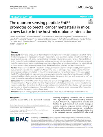
(2022) 20:151Wynendaele et al. BMC ESEARCH ARTICLEOpen AccessThe quorum sensing peptide EntF*promotes colorectal cancer metastasis in mice:a new factor in the host‑microbiome interactionEvelien Wynendaele1†, Nathan Debunne1†, Yorick Janssens1, Anton De Spiegeleer1,2, Frederick Verbeke1,Liesa Tack1, Sophie Van Welden2, Evy Goossens3, Daniel Knappe4, Ralf Hoffmann4, Christophe Van De Wiele5,Debby Laukens2, Peter Van Eenoo5, Lars Vereecke6, Filip Van Immerseel3, Olivier De Wever7 andBart De Spiegeleer1*AbstractBackground: Colorectal cancer, one of the most common malignancies worldwide, is associated with a high mortality rate, mainly caused by metastasis. Comparative metagenome-wide association analyses of healthy individuals andcancer patients suggest a role for the human intestinal microbiota in tumor progression. However, the microbial molecules involved in host-microbe communication are largely unknown, with current studies mainly focusing on shortchain fatty acids and amino acid metabolites as potential mediators. Quorum sensing peptides are not yet consideredin this context since their presence in vivo and their ability to affect host cells have not been reported so far.Results: Here, we show that EntF*, a metabolite of the quorum sensing peptide EntF produced by Enterococcus faecium, is naturally present in mice bloodstream. Moreover, by using an orthotopic mouse model, we show that EntF*promotes colorectal cancer metastasis in vivo, with metastatic lesions in liver and lung tissues. In vitro tests suggestthat EntF* regulates E-cadherin expression and consequently the epithelial-mesenchymal transition, via the CXCR4receptor. In addition, alanine-scanning analysis indicates that the first, second, sixth, and tenth amino acid of EntF* arecritical for epithelial-mesenchymal transition and tumor metastasis.Conclusion: Our work identifies a new class of molecules, quorum sensing peptides, as potential regulators of hostmicrobe interactions. We prove, for the first time, the presence of a selected quorum sensing peptide metabolite in amouse model, and we demonstrate its effects on colorectal cancer metastasis. We believe that our work represents astarting point for future investigations on the role of microbiome in colorectal cancer metastasis and for the development of novel bio-therapeutics in other disease areas.Keywords: Quorum sensing peptides, Microbiota, Colorectal cancer metastasis, Orthotopic mice model, LC-MSBackgroundColorectal cancer (CRC) is the third most commonly†Evelien Wynendaele and Nathan Debunne contributed equally to this work.*Correspondence: Bart.DeSpiegeleer@UGent.be1Drug Quality and Registration Group, Faculty of Pharmaceutical Sciences,Ghent University, Ghent, BelgiumFull list of author information is available at the end of the articleoccurring malignancy worldwide and is associatedwith a high mortality rate, mainly caused by metastasis(mCRC). Primary CRC originates from epithelial cellsthat line the gastrointestinal tract, usually through anadenoma-carcinoma sequence: normal colorectal epithelium transforms into an adenoma and eventually intoan invasive and metastatic tumor. During the initial stepsof the metastatic process, epithelial CRC cells switch The Author(s) 2022. Open Access This article is licensed under a Creative Commons Attribution 4.0 International License, whichpermits use, sharing, adaptation, distribution and reproduction in any medium or format, as long as you give appropriate credit to theoriginal author(s) and the source, provide a link to the Creative Commons licence, and indicate if changes were made. The images orother third party material in this article are included in the article’s Creative Commons licence, unless indicated otherwise in a credit lineto the material. If material is not included in the article’s Creative Commons licence and your intended use is not permitted by statutoryregulation or exceeds the permitted use, you will need to obtain permission directly from the copyright holder. To view a copy of thislicence, visit http:// creat iveco mmons. org/ licen ses/ by/4. 0/. The Creative Commons Public Domain Dedication waiver (http:// creat iveco mmons. org/ publi cdoma in/ zero/1. 0/) applies to the data made available in this article, unless otherwise stated in a credit line to the data.
Wynendaele et al. BMC Biology(2022) 20:151towards a mesenchymal phenotype, known as epithelialto-mesenchymal transition (EMT) [1, 2]. Although therole of genetic predisposition is clear in some cases, astrong association of CRC with diet and lifestyle has beendemonstrated [3]. Moreover, inflammation is believedto play a role in CRC development either as a promoting condition, as shown by colitis-associated colorectalcancers (CAC) in patients with inflammatory bowel disease (IBD), or as a consequence, in patients with sporadicCRC [4].Over the last decade, an increasing amount of evidence suggested that human intestinal microbiota mayplay a role in CRC [5–7]. For example, a high abundanceof Enterococcus, Escherichia, and Fusobacterium species was observed in the feces of patients suffering fromintestinal disorders, including CRC and Crohn’s disease[8–11]. The causative links between gut microbiota andCRC development/progression are not clear to date, andcurrent investigations focus on bacterial-derived compounds such as short-chain fatty acids and amino acidderived amines [12].Quorum sensing peptides are traditionally regardedas intra- and inter-bacterial communication molecules.However, given their structural variety and co-evolutionwith the host, these bacterial metabolites may also affecthost physiology. Indeed, different quorum sensing peptides were previously found to influence the behavior ofhost cells, including brain, muscle, and cancer cells (CRCand breast cancer) [13–16]. In CRC, specific microbialquorum sensing peptides were found to promote tumorcell invasion and angiogenesis in vitro, suggesting potential pro-metastatic effects of these peptides. However, thepresence of quorum sensing peptides in biofluids has notbeen unambiguously demonstrated. In fact, only indirectevidence of the presence of an uncharacterized quorumPage 2 of 16sensing peptide in the stool of patients with Clostridiumdifficile infection was reported so far [17].In this study, we focus on Enterococcus faecium, one ofthe most abundant Enterococcus species in the humanintestinal microbiota [18–21], which synthesizes theenterocin induction factor, i.e., the precursor of the EntFquorum sensing peptide (AGTKPQGKPASNLVECVFSLFKKCN). This peptide acts as a communication signal,regulating the production of enterocin A and B toxins,which inhibit the growth of similar or closely relatedbacterial strains [22–27]. Interestingly, it was previouslydiscovered that EntF*, a metabolite of the quorum sensing peptide EntF, could promote CRC tumor cell invasionin vitro [13].Here, we demonstrate the presence of the EntF* peptide in mice biofluids, along with the EntF*-induced prometastatic effect in vivo. Together, these results indicatea potential role of quorum sensing peptides in the hostmicrobiome interaction and CRC tumorigenesis.ResultsPresence of EntF* in mice biofluidsTo demonstrate that the native EntF peptide can bemetabolized into EntF* in mice, we carried out in vitrometabolization studies (Fig. 1a) on feces and colonictissue homogenates. Our experiments show that theformation rate of the 15-mer peptide EntF* (SNLVECVFSLFKKCN) is 1.71 0.27 % min 1 (mean SEM; n 4)and 0.11 0.01 % m in 1 (mean SEM; n 7) in fecesand colonic tissues, respectively (Fig. 1b, Additional files1 and 2). Similarly to other quorum sensing peptides [14],EntF* is able to cross the intestinal barrier in vitro withan apparent permeability coefficient of 3.67 0.22 10 9 cm s 1 (mean SEM; n 6) in Caco-2 monolayerpermeability assays (Fig. 1c, Additional files 1 and 3).(See figure on next page.)Fig. 1 In vitro formation and in vivo presence of the EntF* metabolite. a Sequences of the enterocin induction factor pro-peptide, mature quorumsensing peptide EntF, and its metabolite EntF*. b In vitro formation rate of EntF* from EntF in colon (n 7) and feces (n 4) homogenates.Bars represent the mean formation rate SEM from independent experiments. Statistically significant differences were determined by aMann-Whitney U test with indicated p-values. c Apparent permeability coefficients (Papp) of PapRIV, EntF*, and EDF-analog in Caco-2 cells. Barsrepresent mean Papp values SEM (n 6 independent experiments); the shaded area represents the limit of detection. d Flow chart displayingthe experimental design stages, from serum sampling to peptide detection and further confirmation of EntF* presence in vivo. Different LC-MSmethods: LC1-MS1, reversed-phase ultra-high-performance liquid chromatography (RP-UPLC) using triple quadrupole (TQ) in MRM mode; L C1-MS2,high-resolution quadrupole time-of-flight; LC1-MS3, high-resolution quadrupole-orbitrap; LC2-MS1, HILIC-amide UPLC using TQ in MRM mode.qPCR was performed on feces sample of mice from the same set to demonstrate the presence of EntF-encoding DNA sequences from E. faecium.e Chromatographic profiles of (1) negative serum sample, (2) positive serum sample, (3) serum sample from EntF*-treated mice. Chromatographicprofiles were obtained using RP-UPLC with detection by electrospray ionization mass spectrometry (ESI-MS) using TQ in MRM mode (m/z 865 202.08 315.17). f Chromatographic profiles of (1) negative serum sample, (2) positive serum sample, (3) serum sample from EntF*-treatedmice. Chromatographic profiles were obtained using HILIC amide UPLC with detection by ESI-MS using TQ in MRM mode (m/z 865 202.08 315.17). g Isotopic distribution of the double charged EntF* measured in a positive serum sample using RP-UPLC with detection by ESI-MSusing quadrupole-orbitrap. h High-resolution tandem mass spectrum of EntF* with characteristic fragments, using RP-UPLC with detection byQ-TOF. i In vivo presence of EntF* in gnotobiotic mice treated with EntF-producing bacterial strains. Number of EntF DNA copies per gram of fecesmeasured four days after treatment with placebo (300 μL BHI medium) (limit of detection: 105 copies/g) (left). EntF* concentration in colon content.No EntF* was detected in the placebo group (the red line indicates the limit of detection) (middle). EntF* concentration in serum content. No EntF*was detected in the placebo group (the red line indicates the limit of detection) (right)
Wynendaele et al. BMC Biology(2022) 20:151Fig. 1 (See legend on previous page.)Page 3 of 16
Wynendaele et al. BMC Biology(2022) 20:151To prove the presence of EntF* in vivo, serum samples from 35 healthy, non-manipulated C57BL/6mice (age 5–18 months) were collected and analyzed(Fig. 1d, Additional files 1 and 4) by reverse phaseultra-high performance liquid chromatography tandem quadrupole mass spectrometry (RP-UPLC-TQMS; LC1-MS1). Notably, we improved the detectionprotocol to avoid carry-over and adsorption and tomaximize selectivity and sensitivity. To achieve thesegoals, we (1) developed an adequate sample preparation protocol using a novel bovine serum albumin(BSA)-based anti-adsorption solution [28] togetherwith a combination of solvent/acid/heat sample treatment followed by solid phase extraction and (2) setup optimal MS detection parameters, including theselection of quantifier (b2: m/z 202.08) and qualifier (b3: m/z 315.17) ions. This method was thoroughly tested and validated (Additional file 5: Fig. S1,Additional file 6). The amount of EntF* detected in theserum of six mice was above 100 pM (limit of quantification, LOQ; Fig. 1e). Taking into account all samples,including those with EntF* values LOQ (consideredas zero), we obtained an EntF* mean value of 329 150pM (mean SEM; n 35; Additional file 5: Table S1).Serum samples from four EntF*-positive and fourEntF*-negative samples (i.e., above and below LOQ,respectively) were analyzed using additional methods(Fig. 1f–h, Additional files 7, 8 and 9). The presenceand identity of EntF* were confirmed through the isotopic distribution of the doubly charged EntF* precursor ion (Fig. 1 g) and by the presence of fragment ions y11 (m/z 1315.61) and y 12 (m/z 1414.69) in thefour positive serum samples (Fig. 1 h, Additional file 5:Table S1). To demonstrate the presence of EntF-encoding bacterial DNA, quantitative real-time PCR wasperformed on fecal samples obtained from the samemice used in the biochemical analysis: EntF-encodingDNA sequences were found in all positive samples(Additional file 5: Fig. S2a, Table S1, Additional file 1).To confirm the bacterial origin of EntF*, EntF-producing E. faecium strains were administered via oralgavage to germ-free mice. Four days after the colonization of the mice gastrointestinal system, EntF* wasobserved in the colon content of the conditioned miceat a concentration of 10 3.2 pmol/g (mean SEM; n 4), while it was below the limit of detection ( LoDEntF* 1.9 pmol/g) in the placebo and control groups. EntF*was also detected in the serum of the test group (Fig. 1i,Additional file 10). In addition, standard BLAST proteinsearches indicated the absence of peptide sequences similar to EntF* in the murine genome (maximum sequencealignment of 67%), supporting the microbial origin of theEntF* peptide found in vivo.Page 4 of 16In vitro activity and molecular targets of EntF*Western blot analysis showed that EntF* and some alanine- or D-amino acid derived analogs affect the expression of E-cadherin, which is linked to the EMT of cancercells (Fig. 2a, Additional file 5: Fig. S3-S4, Additional files11 and 12). Western blot quantifications showed a significant decrease in E-cadherin expression following EntF*treatment ( 38% with respect to control). When thefirst, second, or tenth amino acid of EntF* was replacedby an alanine residue, the reduction in E-cadherin expression was significantly flattened out. These results indicatethat amino acids in position 1, 2, and 10 are important forthe EMT-promoting effects of EntF*. The other residuescontribute to a much lesser extent, as determined by theFisher’s LSD test, and further confirmed using the Jenksnatural breaks algorithm. Replacing the sixth amino acidof EntF* with the corresponding D-amino acid isomerrestored E-cadherin expression to placebo levels. Theseresults suggest that the stereochemical configuration ofthe sixth amino acid of EntF* is important for its EMTpromoting effects.To further investigate the mechanism by which EntF*promotes metastasis, we reasoned that EntF*, by mimicking endogenous ligands of the E-cadherin-regulating pathway, may interact with these receptors, therebyprovoking signaling pathways that lead to an increasedexpression of EMT transcription factors; this is then followed by an upregulation of N-cadherin and subsequentdownregulation of E-cadherin expression. To this end,we searched for similarities between EntF* and activedomains of 13 different natural ligands (Additional file 5:Fig. S5 and Table S2); the selected ligands are knownto trigger signaling pathways that lead to an increasedexpression of EMT transcription factors, thus resulting in the transcriptional downregulation of E-cadherin(Additional file 5: Fig. S5 and Table S2). According tothe respective alignment with the activating domain ofreceptors’ natural ligands and to the relative importanceof the aligned amino acids, three receptors were selectedas the most likely targets of EntF*: C-X-C chemokinereceptor type 4 (CXCR4; alignment score 27), interleukin 6 (IL-6; alignment score 21), and vascularendothelial growth factor (VEGF; alignment score 19).Interestingly, for the activation of the CXCR4 receptor, anine N-terminal amino acid stretch (KPVSLSYRC; aminoacids 22–30) of the endogenous ligand C-X-C motifchemokine 12 (CXCL12) is required [29]; alignment ofthis CXCL12 activating domain and EntF* also shows agood similarity (Fig. 2b).To investigate whether EntF* interacts with the CXCR4receptor, we analyzed by Western blot the effect of theNef-M1 (an antagonist of CXCL12) on the EntF*-mediated E-cadherin downregulation in HCT-8 CRC cells.
Wynendaele et al. BMC Biology(2022) 20:151Page 5 of 16Fig. 2 In vitro activity of the EntF* peptide. a The relative importance of the amino acids of EntF* on E-cadherin expression ranked in five differentclasses. Ranking (blue to red: increasing significance) was performed using the Fisher’s LSD p-values, which was confirmed using the Jenks naturalbreaks algorithm. Based on ranking, it is likely that the first, second and tenth amino acid of EntF* are the most important residues for EntF* activity.b Alignment between EntF* and the active domain of CXCL12. Black amino acids with a line indicate a match. Dark grey amino acids with two dotsmark a similarity between the two residues. Light grey amino acids with one dot are not similar, but no gap is formed. c Antagonistic effects ofNef-M1 on EntF*-mediated E-cadherin downregulation in HCT-8 cells (EntF* n 15; Nef-M1 n 6; EntF* Nef-M1 n 6). Statistically significantdifferences were determined by a one-sided student’s t test. d Antagonistic effects of EntF*1A on EntF*-mediated E-cadherin downregulation inHCT-8 cells (EntF* n 15; EntF*1A n 18; EntF* EntF*1A n 6). Statistically significant differences were determined by a one-sided Student’s ttest. e Effect of EntF* on E-cadherin expression. A significant decrease in E-cadherin levels following EntF* or CXCL12 treatments was observed inHT-29 (n 12), Caco-2 (n 6) and HCT-8 (n 15) cells. Statistically significant differences were determined by one-way ANOVA test. f ProposedE-cadherin-regulating pathway for EntF*. Schematic representation of the CXCR4 receptor and its signaling pathways, leading to the activation ofEMT transcription factors, followed by the downregulation of E-cadherin expression (see also Additional file 5: Fig. S5)
Wynendaele et al. BMC Biology(2022) 20:151Interestingly, co-treatment of HCT-8 CRC cells withEntF* and Nef-M1 restored E-cadherin expression tocontrol levels (Fig. 2c, Additional file 13): E-cadherinlevels increased from 62% (EntF* only) to 94% (EntF* Nef-M1) (Cohen’s d 1.4). These results suggest aninteraction between EntF* and the CXCL12/CXCR4pathway in EMT promotion and tumor metastasis (basedon the E-cadherin-regulating pathway, Additional file 5:Fig. S5).Interestingly, the modified peptide EntF*1A, where theserine amino acid at position 1 is replaced by an alanineresidue, also displayed antagonistic activity towards theEntF*/CXCR4 interaction: E-cadherin relative expressionlevels increased from 62% to 92% (Cohen’s d 0.9) whenEntF*1A was added to EntF*-treated cells (Fig. 2d).Additionally, treatment of HT-29 and Caco-2 CRCcells with EntF* also resulted in a statistically significantreduction of E-cadherin expression. Similar effects onE-cadherin expression were also observed when HT-29and Caco-2 CRC cells were treated with the endogenousCXCL12 peptide. Interestingly, when CXCR4-negative HCT-116 cells were treated with EntF* or with theCXCL-12 ligand, no decrease in E-cadherin expressionwas observed in response to either peptide (Fig. 2e, Additional file 14). Together, these data suggest that EntF*exerts its functions by interacting with the CXCR4-mediated signaling pathway, ultimately leading to E-cadherindownregulation (Fig. 2f ). The lack of response in HCT116 cells suggests that CXCR4 receptor is likely the solemolecular target of the EntF* peptide.In vivo pro‑metastatic properties of EntF*Pro-metastatic properties of EntF* were determinedusing an orthotopic mouse model of colorectal cancer. Luciferase-transfected HCT-8 cells were treatedfor 5 days with phosphate-buffered saline (PBS) vehicle, 102 nM EntF*, or 0.1 μg mL 1 transforming growth factor α (TGFα, positive control). On the sixth day, luciferase-transfected CRC cells were orthotopically injectedinto the cecal wall of 6-week-old female Swiss nu/numice. Subsequently mice were treated once a day intraperitoneally with PBS vehicle (n 17), 102 nmol kg 12 1EntF* (n 38), or 10 μg kg Epidermal Growth Factor (EGF, positive control; n 18) (Fig. 3a). To calculateEntF* daily exposure after intraperitoneal (i.p.) injections (102 nmol kg 1; n 14), in vivo serum kinetic profilesof EntF* were determined (see Materials and Methods). Additionally, endogenous EntF* levels in untreatedfemale Swiss nu/nu mice (n 65) were assessed. Theseanalyses concluded that daily EntF* injections resultedin five times higher EntF* serum levels compared tothe endogenous (natural) exposure in untreated mice,thus demonstrating the biological relevance of thisPage 6 of 16experimental set-up (Fig. 3b, Additional file 15). Bioluminescent imaging of mice was performed weekly tomonitor tumor growth (Fig. 3c, Additional file 16). Overa period of 6 weeks, EntF* caused a statistically significantincrease in luciferase activity compared to the vehicle (p 0.003). This increase was similar to what was observedin EGF-treated controls (p 0.432; Fig. 3d). Our resultsshow, after a 6-week treatment, an effect size of 79%increase in bioluminescence for EntF* compared to theplacebo, ranging from -92%, a relatively small negativeassociation, to a 417% increase, a substantial positiveassociation (Fig. 4a). For the EGF control, a median effectsize of 316% was scored, ranging from 145 to 826%(Fig. 4a). The tumor-promoting activity of EntF* was further confirmed by macroscopically assessing the numberof nodules on the cecum of treated mice: EntF*-treatedsamples showed a 3-fold increase in the number of nodules compared to PBS vehicle (p 0.036) (Fig. 3e, f ),whereas EGF-treated samples showed a 4.5-fold increasecompared to control (Fig. 4b).Histopathological analyses also showed a significantlyhigher number of tumor nodules in the liver (p 0.014)and lungs (p 0.026) of mice after 6 weeks of treatmentwith EntF*, compared to the vehicle (Fig. 5a–d; Additional file 17).Another quorum sensing peptide, Phr0662 from Bacillus species, which also promoted in vitro cell invasion inour initial screening experiments [13], showed no prometastatic effects in the orthotopic mouse model of colorectal cancer used in this work (Additional file 5: Fig. S6).These findings indicate a certain degree of selectivity ofquorum sensing peptides in metastasis promotion in vivo.DiscussionThe in vitro metabolization analysis and the Caco-2 permeabilization assays suggest that EntF* can be present inthe blood circulation of the host. This is likely to occurafter the degradation of the bacterial EntF peptide inthe colon or feces of the host and subsequent intestinal absorption of the resulting EntF* peptide. To unambiguously demonstrate the presence of EntF* in vivo, wedeveloped and optimized a bioanalytical method basedon RP-UPLC-TQ-MS in multiple reaction monitoring(MRM)-mode. Using this method, we detected the presence of EntF* in the serum of six mice. Also, four EntF*positive and four EntF*-negative samples were analyzedthrough additional chromatographic methods: hydrophilic interaction liquid chromatography (HILIC)-UPLCTQ-MS (LC2-MS1) as an orthogonal separation systemand RP-UPLC-QTOF-MS (LC1-MS2) and RP-UPLCQOrbitrap-MS (LC1-MS3) as high-resolution mass spectrometers. All methods confirmed the presence andidentity of EntF* in the positive serum samples.
Wynendaele et al. BMC Biology(2022) 20:151Page 7 of 16Fig. 3 Metastasis-inducing effect of EntF* in an orthotopic mouse model of colorectal cancer. a Schematic timeline of the experimental setup.Female Swiss nu/nu mice were orthotopically injected with 1 106 luciferase-transfected HCT-8 cells at the age of 5 weeks. During the following6 weeks, mice were daily i.p. injected with 102 nmol kg 1 EntF* (n 38), PBS control (vehicle, n 17), or 0.1 mg kg 1 EGF positive control (n 18). Bioluminescent imaging was performed weekly to determine cancer progression. After 6 weeks, mice were euthanized and the cecum, liverand lungs collected. b Graph representing the average daily exposure of placebo-treated (black; n 65) and EntF*-treated (gray; n 14) femaleSwiss nu/nu mice. In the placebo group, mice with an EntF* serum concentration below the limit of detection (100 pM) were counted as zero.The daily exposure after i.p. injection of 102 nmol kg 1 EntF* is 5 times higher than the naturally occurring EntF* levels. c Representative images ofbioluminescence activity in vehicle, EntF*-treated, and EGF-treated mice. Mice were i.p. injected with 150 mg kg-1 luciferin and imaged after 10 minin supine position. d Tumor growth curves derived from the quantification of bioluminescence in vehicle, EntF*-treated, and EGF-treated mice.Based on linear regression slope comparison, EntF* and EGF treatment resulted in a significant increase in tumor growth compared to the vehicle.Data represent the mean relative increase in bioluminescence SEM. e Representative pictures of vehicle, EntF*-treated, and EGF-treated ceca atthe end of the experiment. f Number of tumor nodules in untreated (vehicle), EntF*-treated, and EGF-treated ceca determined by macroscopicinspection of tissues. Data represent the mean nodule number SEM. Statistically significant differences were determined by a Mann-Whitney Utest with indicated p-valuesTo confirm the bacterial origin of the peptide, fecesfrom four EntF*-positive and -negative mice weretested for the presence of E. faecium-specific EntF DNAcoding sequences. All EntF*-positive mice possessedthe EntF coding sequences. In a few cases, UPLC-MScould not detect the EntF* peptide in the serum ofmouse samples that tested positive for the presence ofEntF DNA in the feces (e.g. sample 20181011S8). Thesenegative samples could derive from specific E. faeciumstrains with reduced translational efficiency. Thesedata are presented in Additional file 5: Table S3: out ofthree E. faecium strains containing the EntF gene, onlyone produced measurable EntF levels in vitro (LoD 1.5 nM). Additionally, variability in peptide degradation rates (EntF to EntF*) and/or differences in gastrointestinal absorption of EntF* could further explainthe weak correlation between EntF* levels in the serumand EntF DNA copy number in the feces. The presenceof EntF* in the serum of germ-free mice treated withEntF-producing E. faecium strains represents an additional proof that the detected EntF* is indeed of microbial origin.
Wynendaele et al. BMC Biology(2022) 20:151Page 8 of 16Fig. 4 Effect size for bioluminescence and number of nodules in the cecum of the orthotopic mouse model after a 6-week treatment. a After a6-week treatment, an effect size of 79% increase in bioluminescence for EntF* compared to the placebo was observed, while for the positive controlEGF, a median effect size of 316% was obtained. When calculating the effect size according to Hedges’ G values, a medium to large effect wasobserved for both EntF* and EGF treatment groups, compared to the placebo group. b After a 6-week treatment, a 3-fold increase in the numberof nodules on the cecum was observed for EntF* compared to the placebo PBS, while for the positive control EGF, a 4.5-fold increase was obtained.When calculating the effect size according to Hedges’ G values, a medium and large effect was observed for the EntF* and EGF treatment groups,respectively, compared to the placebo group [Fig. prepared in R-script: https:// github. com/ Joach imGoe dhart/ Plots OfDif f eren ces]EntF* was previously identified by our group in anin vitro screen for molecules that selectively promoteangiogenesis and tumor cell invasion in HCT-8 CRCcells [13]. In this work, we confirm these in vitro effectsby proving that EntF* significantly decreases E-cadherinexpression in three different colorectal cell lines. Additionally, we found that amino acids in the first (S), second (N), tenth (L) position, as well as the stereochemicalconfiguration of the sixth residue, are important for theEMT-promoting effects. Our findings suggest that EntF*acts as an agonist-ligand in the CXCR4/EGFR phosphorylation/ERK activation/E-cadherin pathway. Indeed,when EntF* is administered together with a CXCR4antagonist (Nef-M1), or to a CXCR4-negative colorectalcell line (HCT-116), it shows reduced or no activity.Furthermore, we evaluated EntF* metastasis-promoting activities in vivo using an orthotopic mouse model ofcolorectal cancer [30, 31]. Since we used a single mousestrain and identical housing conditions, we could directlycompare different treatment groups to find causal associations, as only treatments were different among groups(i.e., placebo, peptide or positive control). This experiment demonstrates that the quorum sensing peptidemetabolite EntF* can promote CRC metastasis in vivo.
Wynendaele et al. BMC Biology(2022) 20:151Page 9 of 16Fig. 5 Histopathological evaluation of CRC metastasis after EntF* treatment. a Microscopic images of liver tissue sections from untreated (vehicle),EntF*-treated, and EGF-treated mice. Tissues were stained with Hematoxylin and Eosin (H&E) and imaged at different magnifications (10x upper row,40x lower row). b Histopathological scores with statistically significant differences determined by a Mann-Whitney U test (n 8 for PBS, n 30 forEntF*, n 9 for EGF) with indicated p-values. c Microscopic images of lung tissue sections from untreated (vehicle), EntF*-treated, and EGF-treatedmice. Tissues were stained with H&E and imaged at different magnifications (10x upper row, 40x lower row). d Histopathological scores withstatistically significant differences determined by a Mann-Whitney U test (n 8 for PBS, n 30 for EntF*, n 9 for EGF) with indicated p-valuesThis effect seems quite specific to EntF* as Phr0662,another quorum sensing peptide, did not
Wynendaele et al. BMC Biology Page 5 of 16 Fig. 2 In vitro activity of the EntF* peptide. a The relative importance of the amino acids of EntF* on E-cadherin expression ranked in ve dierent classes. Ranking (blue to red: increasing signicance) was performed using the Fisher's LSD p-values, which was conrmed using the Jenks natural .