Transcription
Hartmann and Seiler BMC Oral Health(2020) ARCH ARTICLEOpen AccessMinimizing risk of customized titaniummesh exposures – a retrospective analysisAmely Hartmann1,2*and Marcus Seiler2AbstractBackground: Recommendations for soft tissue management associated with customized bone regeneration shouldbe developed. The aim of this study was to evaluate a new protocol for customized bone augmentation in a digitalworkflow.Methods: The investigators implemented a treatment of three-dimensional bone defects based on a customizedtitanium mesh (Yxoss CBR , ReOSS, Filderstadt, Germany). Patients and augmentation sites were retrospectivelyanalysed focussing on defect regions, demographic factors, healing difficulties and potential risk factors. Anexposure rate was investigated concerning surgical splint application, A - PRF and flap design.Results: In total, 98 implants could be placed. Yxoss CBR was removed after mean time of 6.53 2.7 months. Flapdesign was performed as full flap preparation (27.9%), full flap and periosteal incision (39.7%), periosteal incision(1.5%), poncho/split flap (27.9%) and rotation flap (2.9%). In 25% of the cases, exposures of the meshes weredocumented. Within this exposure rate, most of them were slight and only punctual (A 16.2%), like one toothwidth (B 1.5%) and complete (C 7.4%). A - PRF provided significantly less exposures of the titanium meshes(76.5% no exposure vs. 23.5% yes, p 0.029). Other parameters like tobacco abuse (p 0.669), diabetes (p 0.568) orsurgical parameters (mesh size, defect region, flap design) did not influence the exposure rate. Surgical splints werenot evaluated to reduce the exposure rate (p 0.239). Gender (female) was significantly associated with lessexposure rate (78,4% female vs. 21.6% male, p 0.043).Conclusions: The results of this study suggest that the new digital protocol including patient-specific titaniummeshes, resorbable membranes and bone grafting materials was proven to be a promising technique. To improvesoft tissue healing, especially A -PRF should be recommended.Keywords: Customized titanium mesh, Digital dentistry, Mesh exposure, Risk parameters, Platelet rich fibrinBackgroundGuided Bone Regeneration (GBR) technique offers thepossibility to reconstruct bony alveolar defects [1] preceding implant placement [2]. A barrier membrane separates the surrounding connective tissue from the bonydefect [3–7].Large defects of the jaws exhibit both hard and softtissue shortages. Titanium meshes are well-known towork as a mechanical scaffold and create stability forbone healing in large three-dimensional defects [8].* Correspondence: amelyhartmann@web.de1Private Practitioner, Affiliate to the Department of Oral and MaxillofacialSurgery, University Medical Centre of the Johannes Gutenberg University ofMainz, Augustusplatz 2, 55131 Mainz, Germany2Department Head, Private Dental Practice, Echterdinger Str. 7, 70794Filderstadt, GermanyTherefore, rebuilding such a complex defect also implicates a detailed focus on soft tissue management. Membrane and graft exposures are frequent complicationsassociated with non-resorbable membranes [9–12].Based on the principles of the GBR technique, individualized titanium meshes are proposed to overcome theproblems of the conventional titanium meshes [13]. Literature [14–16] reveals the advantages of this technologysuch as a shortened and facilitated surgery time in senseof a modern digital work-flow. Although Sumida et al.evaluated less exposures, but not statistically significant,in patient-specific titanium meshes [17], soft tissue management remains one of the most challenging targets incustomized bone regeneration [18]. The Author(s). 2020 Open Access This article is distributed under the terms of the Creative Commons Attribution 4.0International License (http://creativecommons.org/licenses/by/4.0/), which permits unrestricted use, distribution, andreproduction in any medium, provided you give appropriate credit to the original author(s) and the source, provide a link tothe Creative Commons license, and indicate if changes were made. The Creative Commons Public Domain Dedication o/1.0/) applies to the data made available in this article, unless otherwise stated.
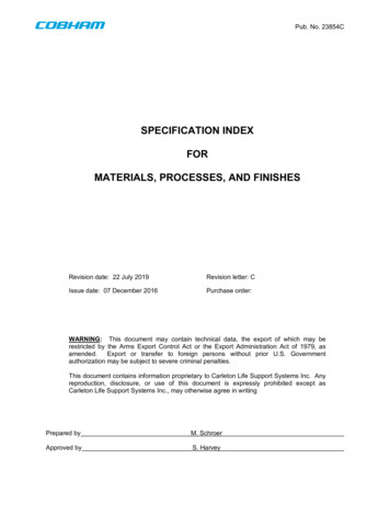
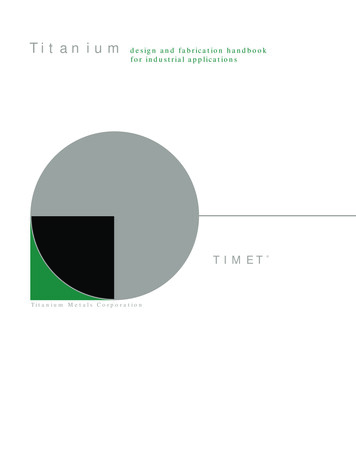
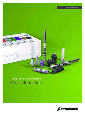
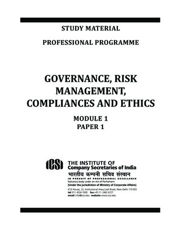
Hartmann and Seiler BMC Oral Health(2020) 20:36Treatment opportunities like self-inflating soft tissue expanders [19] or free fat grafts (FFG) from the buccal fatpad [20] aiming to stabilize the soft tissue healing processincrease comorbidity of the patient and need additionalsurgical skills. A promising solution may be the enhancement of soft tissue wound healing by Platelet-Rich Fibrin(PRF) as shown in literature [21–23].So far, there is no clear recommendation in literatureto reduce this exposure rate in customized boneregeneration.The aim of this study was to describe a new surgicalprotocol in customized bone regeneration and to evaluate parameters that minimize the risk of customized titanium mesh exposures. The influence of variousdemographic, local and systematic factors were assessed.MethodsThis is a clinical non-interventional monocentre study.It was performed retrospectively during the clinical routine without any further consequences for the patient.Data were anonymized and processed in accordancewith the 2013 Declaration of Helsinki on medical protocol and ethics (Declaration of Taipei on Ethical Considerations regarding Health Databases and Biobanks2016). Due to the character of the study no approval bythe local ethics committee was necessary (Regulatory ofthe ethic committee of Rhineland-Palatinate and described in Gaus et al. [24]). No administrative permissions and/or licenses were acquired by the team toaccess the data used in the research due to the characterof the study.Study populationThis study included 55 patients with 68 grafting procedures for consecutive dental implant placement. Patientswith three-dimensional bony defects were included. Allthree-dimensional grafting procedures had to bePage 2 of 9performed by the same trained surgeon (MS; PrivateDental Practice, Filderstadt, Germany) by using apatient-specific titanium lattice structure (Yxoss CBR ,ReOss, Filderstadt, Germany). Female and male patients 18 years were included.Exclusion criteria were mentally disabled patients,pregnant women and patients 18 years. Local exclusioncriteria were horizontal or vertical bony defects. Patientswith three-dimensional defects treated with other boneaugmentation procedures like distraction osteogenesis,block graft or onlay-technique were excluded as well. Ingeneral, patients with systemic or local diseases and malignancies were excluded before enrolment.Workflow and surgeryAfter acquisition of a Cone Beam Computed Tomography (CBCT) dataset, a 3D-projection of the atrophiedsegment was obtained by using a reverse engineeringsoftware. The necessary bone volume was digitallyadded, and the individualized titanium meshes were designed. The inner contour of the lattice structure represented the desired augmentation volume. By usingComputer-Aided Design/Computer-Aided Manufacturing (CAD/CAM) procedures and rapid prototyping thefinal design was achieved and confirmed interactively bythe surgeon (Figs. 1 and 2). After a 3D-Printing Process(Fig. 3), the titanium mesh was sent to the surgeon andsterilized before use.Surgery was performed under local anesthesia. Theopening incision was carried out in accordance with thedefect size and location of the neighboring anatomicalstructures of the region. Flap design was performed asappropriate in each case (full flap preparation, full flapand periosteal incision, periosteal incision, poncho/splitflap or rotation flap). After preparation of the defect,scar tissue was removed. In the lower jaw, some cortexperforations were performed to boost the blood supply.Fig. 1 Design-example. The inner contour of the mesh represents the desired augmentation volume
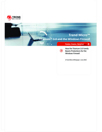
Hartmann and Seiler BMC Oral Health(2020) 20:36Page 3 of 9Fig. 2 By using CAD/CAM-technology, the technician is able to design an individualized titanium meshThe meshes were installed by using a mixture of autologous bone graft and bone substitute biomaterial (BioOss , Geistlich, Wolhusen, Switzerland) in a 1:1 ratio(Fig. 4). Autologous bone was harvested from the conventional intraoral donor sites (n 59, external obliqueline (n 50) and operation site (n 9)) or from the iliaccrest (n 8). In one patient, only bone substitute (BioOss ) was used. Each mesh was fixed to the residualbone with titanium osteosynthesis screws. A resorbablemembrane (Bio-Gide , Geistlich, Wolhusen, Switzerland)was placed on top. In some cases (n 12), an AdvancedPlatelet Rich Fibrin (A-PRF ) clot was applied accordingto manufacturers protocol following the in vitro-protocol of Choukroun [25] (Figs. 5 and 6). Flaps wereadopted without tension by using deep mattress and single interrupted sutures (Seralon5/0). Vacuum formsplints were adjusted to enhance soft tissue healing inn 22 (Fig. 7). Patients received instructions concerningFig. 3 After an additive 3-D- printing processa proper oral hygiene. They had to avoid brushing at thegrafting area and to wear removable dentures. All patients underwent an oral antibiotic therapy (Amoxicillin 1000 mg 1–1-1 or Clindamycin 600 mg 1–1-1) for 5–7days starting at time of the surgery.After a two-week healing period, sutures were removed. Outcomes were also assessed one week aftersurgery and during follow-up each month. In summary, they were controlled each month for 4 to 8months depending on the defect. The Re-openingand removal of the titanium mesh was after approximately 4–8 months depending on size of the defect.Implant placement subgroups (Camlog Screw Line ,Camlog, Wimsheim, Germany) were equally distributed (implantation performed either simultaneouslywith mesh insertion (44.1%) or after a healing periodof 4–8 months combined with the removal of themesh (44.1%)).
Hartmann and Seiler BMC Oral Health(2020) 20:36Page 4 of 9defined as complete loss of the graft. Outcomes and possible healing difficulties were assessed one week aftersurgery and during follow-up each month. Patients wereinstructed to contact the surgeon if any disturbancesoccurred.Secondary aim of the study was to assess possible riskfactors (defect regions, defect and mesh sizes, smoking, tissue phenotype (thin and fragile phenotype, thick phenotype[26]), diabetes) for grafting success and developing an exposure. Exposure rate and impact of such factors as Vacuum form splint, A-PRF and flap design on the exposurerate should be assessed. Exposures of the titanium meshwere classified concerning their size. “A” was a punctual exposure, “B” an exposure like one premolar size and “C” acomplete one whereas “D” was no exposure [27]. Mesh sizewas defined according to the missing teeth.Data evaluationFig. 4 Individualized mesh in situ. Titanium mesh filled with graft;there is a slot for the easy removal function on top and centralOutcome assessmentPrimary outcome was the grafting success defined as thefeasibility of implant placement in the planned positionand achievement of an adequate primary stability(15Ncm-35Ncm) until the re-entry and to finalize withthe individual prosthetic supraconstruction. Failure wasFig. 5 The A-PRF clots according to the protocol of ChoukrounStatistical assessment was done using IBM SPSS Statistics version 22.0 for Windows. Level of significance wasset to p 0.05. Data presentation was performed byusing JMP 10.0 statistical software (SAS Institute, Cary,NC, USA). Grafting success, exposure rate and impact ofsuch factors as Vacuum form splint, A-PRF and flap design on the exposure rate.For secondary outcome parameters possible risk factors (defect regions, defect and mesh sizes, smoking,tissue phenotype (thin and fragile phenotype, thickphenotype [25]), diabetes) for developing an exposurewere defined. Statistical analyses were performed usingChi-Quadrat-Test and Fisher’s Exact-Test as appropriatefor qualitative parameters, T-Test or Mann-Whitney-UTest for quantitative parameters.ResultsA group of 38 female (69.1%) and 17 male (30.9%) patients with 68 three-dimensional defects and a mean age
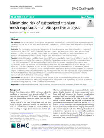
Hartmann and Seiler BMC Oral Health(2020) 20:36Fig. 6 A-PRF clot covering a titanium mesh in situof 58.1 (range of age 18 to 81 years with SD 15.6years) were enrolled. Augmentation site was in the upperjaw (n 35, 51.5%) and in the lower jaw (n 33, 48.5%).No case failed. In total, 98 implants could be placed asplanned. Yxoss CBR was removed after mean time of6.53 2.7 months. Tobacco abuse was documented inn 6 (10.9%) and stable diabetes mellitus (HbA1c 5%)in n 3 (5.5%).Bone quality was found to be D1 (n 12, 17.6%), D2(n 36, 52.9%), D3 (n 13, 19.1%) and D4 (n 7, 10.3%)according to Misch’s classification. Tissue Phenotypewas classified as “thin and fragile” (n 53, 77.9%) and“thick” (n 15, 22.1%). Flap design was performed as fullflap preparation (n 19, 27.9%), full flap and periostealincision (n 27, 39.7%), periosteal incision (n 1, 1.5%),poncho/split flap (n 19, 27.9%) and rotation flap (n 2,2.9%). Implant placement was performed either simultaneously with mesh insertion (n 30, 44.1%) or after ahealing period of 4–8 months (n 30, 44.1%). In n 8Page 5 of 9(11.8%) implant placement was not performed whiledata evaluation. A surgical splint was used in n 22(32.4%) and A - PRF in n 12 (17.6%).In 25% of the cases (n 17), exposures of the mesheswere documented. Within this exposure rate, most ofthem were slight and only punctual (A 16.2%, n 11).Exposure like one tooth width (B 1.5%, n 1) and acomplete (C 7.4%, n 5) occurred as well. Associatedwith these exposures, no loss of grafting material (86.8%,n 59), partial (11.8%, n 8) and complete in 1.5% (onecase) was evaluated. A therapy according to the plannedtreatment protocol was possible in all the cases.A -PRF provided significantly less exposures of the titanium meshes (76.5% no exposure vs. 23.5% yes, p 0.029)(Fig. 8). Other parameters like tobacco abuse (p 0.669),diabetes (p 0.568) or surgical parameters (mesh size, defect region, flap design p 0.368) did not influence the exposure rate. Surgical splints were not found to reduce theexposure rate (p 0.239). Gender (female) was significantlyassociated with less exposure rate (78,4% female vs. 21.6%male, p 0.043, Fig. 9).DiscussionThis study shows that treatment with customized titanium meshes offers the opportunity to provide highquality work in large three-dimensional bony defects.The benefits like precise fit, shorter time of surgery, predictable outcome and good acceptance of the surgicalprocedure were already described in recent literature[27, 28]. Although exposures are a common complication associated with titanium mesh technique, graftingoutcome was not affected. This is according to literaturewhere exposure does not necessarily compromise thefinal treatment outcome [29–31]. Literature aims to reduce exposure rates. Interpreting the results from thepresent study (25% exposure rate), all patients presentedlarge defects which would have made a conventionalFig. 7 Surgical splint application after suturing without tension and a healing process of 10 days
Hartmann and Seiler BMC Oral Health(2020) 20:36Fig. 8 A -PRF provided significantly less exposures of thetitanium meshesGBR technique with resorbable membranes impossibleand would have required block augmentation from otherintra- or extraoral donor sites, onlay technqiues or distraction osteogenesis.Comparing mesh exposure (A-C) of this study to literature, heterogenous data are documented. In customized bone regeneration, Sagheb et al. reported 33%exposures [16]. Other studies range from uneventfulhealing with no [32–34] and from 14.8 to 59% [29, 30,35–38] of membrane exposure. These different resultsmay be caused by a lack in literature to precisely describe the exposures according to size. By distinguishingin severity of the exposure [27], we were able to showFig. 9 In female patients, significantly less exposureswere consideredPage 6 of 9that most of them are only slight and only punctual. Another point is the various titanium mesh techniques,various surgical protocols and skills. Sumida et al. evaluated less exposure rates for customized meshes compared to the conventional titanium mesh technique [17].Evaluating the grafting protocol without a control groupis a shortcoming of this study due to its retrospectivecharacter. In general, there are two major limitations inthis study that could be addressed in future research.First, the study focused on augmentation procedures inthree-dimensional defects. No measurements of the defect (in mm3) were assessed. A more objective way todescribe them clinically and radiographically by CBCTwould be in mm3. This shortcoming is due to the retrospective character of the study and the next prospectivestudy will define the three-dimensional defects in mm3and according to a new classification.Second, grafting outcome should be defined in mm3after another CBCT and matching the two radiographs.According to our best knowledge, and lots of own tests,matching will not work exactly because of X-ray scattered radiation of the titanium lattice structure. Futureresearch should aim to develop a new software excludingthese effects.Within the limitations of this study being retrospectivewithout a control group it was possible to show that anexposure does not necessarily lead to grafting failure asdefined above. A prospective study should compare different methods for bone augmentation in similar defectsizes in comparable study groups to present a superiorityof one method.Analyzing risk factors for soft tissue healing, the results of this study show that tobacco abuse had no influence on the exposure rate. This is surprising becausemany studies describe the negative effects of tobaccoabuse in healing processes because it hinders revascularization and enforces soft tissue inflammation [30] [39].Our findings are supported by Lindfors et al. [30], whostated no correlation between smoking and the development of exposures. The results may be due to a properinformed consent of the patients directly and their acceptance to reduce/avoid smoking after surgery. Additionally, the results may also be associated with thelimited number of patient subgroups in this study.The same goes for the findings concerning Diabetes.Diabetes as another risk factor in wound healing processes [40] was not proven to have an influence on developing exposures. It is tempting to speculate that allthe included patients suffering from diabetes are wellcontrolled type 2 diabetic patients and Erdogan et al.[41] have found GBR technique in such cases being aproper treatment opportunity.One might assume that a thin tissue phenotype [26, 42]is much more difficult to handle than a thick one. Scar
Hartmann and Seiler BMC Oral Health(2020) 20:36tissue will develop easily because of an earlier mucosalrupture. An adequate aesthetic outcome may be difficultto achieve. This was evaluated in previous studies [43].They described thick gingival phenotypes associated withadditional blood support during wound healing because ofthe missing periodontal ligament support in implant therapies. These findings are contrary to the results at hand. Apossible reason for this may be in the patient group itself.Large three-dimensional defects like described in thisstudy, are the result of numerous preceding surgeries liketeeth extraction, inflammation processes or others. According to authors opinion, scar tissue as a result of theseformer interventions – in thin and thick tissue phenotype– might be the real reason for developing an exposure.In this study, neither age nor periodontitis influencethe exposure rate. These findings are according toSagheb et al. [16].In their study, they also evaluated no relationshipbetween gender and the risk to develop an exposure.This goes along with other studies [44] but is not inaccordance with findings of this study where we wereable to find a significant relationship between malegender and the development of exposures. Male patients were already described to suffer from an increased potential infection rate after implantplacement by Figueiredo et al. [45]. This is in accordance with Kim et al. [46] who evaluated male genderto have the highest risk of wound dehiscence inguided bone regeneration. These results are in linewith our findings. The same observation was also described in dermatology. Dao et al. [47] reported aboutgender differences in skin regeneration. They described modified immunological processes in elderlymen caused by a decreasing testosterone level. Malespecific instructions on postoperative care may helpto overcome these healing difficulties. On the otherhand one might assume that especially steroid hormones in female patients appear capable of influencing the normal bacterial flora and the subgingivalecology [48, 49]. Consecutively, healing difficultiesmay arise. This is not according to the results of thisstudy. Female gender was significantly associated withless exposures. Further gender-specific studies inintraoral healing processes are needed.Localization or sizes of meshes were not relevant forthe occurrence of dehiscence. This is contrary to thefindings of Uehara et al. [50], wo found a significant correlation between the success rate of a bone grafting andthe extension of the augmentation site. This may be dueto different techniques in flap management or other patient recruitment. In the study at hand, there were nodifferences between the upper and lower jaw concerningan exposure rate as already described in Her et al. [31]and Louis et al. [37].Page 7 of 9Although flap design did not affect the outcome of thetreatment, according to authors opinion, a proper flapmanagement will remain a key point in working with individualized titanium meshes. This includes planning ofthe incision in advance according to Kleinheinz et al.[51] and adopted to the kind of defect, a careful flap elevation and a tension-free primary wound closure [11].This will avoid necrosis of the flap and a premature exposure. The risk of a consecutive inflammation and possible loss of the graft will be decreased [10, 52].This study evaluated a significant improvement oftherapy outcome if customized titanium mesh techniquewas applied together with A-PRF . This is according to arecent study which found the combination of PRF andFFG to be successful during the soft tissue healingprocess preceding implant placement [53]. Torres et al.[54] also reported 28.5% of their cases in a control groupsuffering from mesh exposure while in a PRP group, noexposures were registered. The success of PRF may bedue to the role of fibrin in initial clot stabilization [22].PRF as a biodegradable scaffold consisting of stem cells,fibrin, platelets and leucocytes boosts microvascularization and epithelial cell migration [55, 56]. This may prevent mesh exposure by using it to cover meshes asapplied in this study. In a recent systematic review, therewas limited evidence on the effects of Lykocite PlateletRich Fibrin (L-PRF) in intraoral bone grafting procedures [55]. They concluded the need for further studieswith special emphasis on the standardized surgical procedures [55]. Therefore, and supported by this study, aproper soft tissue healing is boosted using (A -) PRF .The influence on bone healing processes must beassessed in further randomized, control studies, althougha recent study described positive effects in postextraction sockets [23].The use of surgical splints must be evaluated in prospective studies with more patients. According to authorsopinion, these surgical splints may provide a better woundhealing although the results being not significant in thisstudy. As a part of everyday routine together with soft tissue surgery, it is not a standard procedure applied together with customized mesh technique or titaniummeshes in general. Another study agreed that – especiallyin male patients – the application of surgical pack andsurgical splint would provide a better wound healing [46].ConclusionIn complex bone reconstruction, the new surgical protocol in customized bone regeneration was proven to be apromising technique.Interpreting the primary outcome of this study, exposures are proven to be complications that did not affectthe defined outcome and success of the grafting procedure. To improve soft tissue healing, especially A - PRF
Hartmann and Seiler BMC Oral Health(2020) 20:36should be recommended. A tension-free wound closureseems to be more important than a specific flap design.Concerning the secondary aim of the study, future prospective research should aim to evaluate the gender specific risk of developing exposures and in general fordeveloping healing difficulties in augmentationprocedures.Abbreviations3D-Printing Process: 3Dimensional-Printing Process; 3Dprojection: 3Dimensional-projection; A-PRF : Advanced-Platelet Rich Fibrin;CAD/CAM: Computer-Aided Design/Computer-Aided Manufacturing;CBCT: Cone Beam Computed Tomography; CBR : Customized BoneRegeneration; FFG: free fat grafts; GBR: Guided Bone Regeneration; IBMSPSS : Statistics software; JMP : Statistics software; L-PRF: Lykocite PlateletRich Fibrin; Mann-Whitney-U-Test: Statistical test; mm3: cubic millimetres;MS: Marcus Seiler; N: number; PRF: Platelet-Rich Fibrin; T-Test: Statistical testAcknowledgementsThe authors wish to thank patients, nurses and physicians for their supportwith patients material and reports. Moreover, we thank all our colleagues forhelpful discussions. We thank Klaus Hartmann, M.D. for language proof.Page 8 of 93.4.5.6.7.8.9.Authors’ contributionsA.H. analysing data, article draft, M.S. data generation. Both authors read andapproved the final manuscript.10.FundingWe received a financial support from the Geistlich Company for statisticalanalyses and we will receive a funding for open access publication. Thefunding body did not influence the design of the study, the collection,analysis, and interpretation of data or helped writing the manuscript.11.Availability of data and materialsThe datasets used and/or analysed during the current study are availablefrom the corresponding author on reasonable request.13.Ethics approval and consent to participateThis is a clinical non-interventional monocentre study. It was performedretrospectively during the clinical routine without any further consequencesfor the patient. Data were anonymized and processed in accordance withthe 2013 Declaration of Helsinki on medical protocol and ethics (Declarationof Taipei on Ethical Considerations regarding Health Databases and Biobanks2016). Due to the character of the study no approval by the local ethicscommittee was necessary (Regulatory of the ethic committee of RhinelandPalatinate and described in Gaus et al. [24]). No administrative permissionsand/or licenses were acquired by the team to access the data used in the research due to the character of the study. Using a human study design, written informed consent was obtained from patients.14.Consent for publicationUsing a human study design, written informed consent was obtained frompatients.Competing interestsA.H. is working at the private practice of M.S., M.S. is the creator of YxossCBR.12.15.16.17.18.19.20.Received: 13 September 2019 Accepted: 27 January 2020References1. Boyne PJ, Mikels TE. Restoration of alveolar ridges by intramandibulartransposition osseous grafting. J Oral Surg. 1968;26(9):569–76.2. Dahlin C, Sennerby L, Lekholm U, Linde A, Nyman S. Generation of newbone around titanium implants using a membrane technique: anexperimental study in rabbits. Int J Oral Maxillofac Implants. 1989;4(1):19–25.21.22.Crump TB, Rivera-Hidalgo F, Harrison JW, Williams FE, Guo IY. Influence ofthree membrane types on healing of bone defects. Oral Surg Oral Med OralPathol Oral Radiol Endod. 1996;82(4):365–74.Amano Y, Ota M, Sekiguchi K, Shibukawa Y, Yamada S. Evaluation of a polyl-lactic acid membrane and membrane fixing pin for guided tissueregeneration on bone defects in dogs. Oral Surg Oral Med Oral Pathol OralRadiol Endod. 2004;97(2):155–63.Marouf HA, El-Guindi HM. Efficacy of high-density versus semipermeablePTFE membranes in an elderly experimental model. Oral Surg Oral Med OralPathol Oral Radiol Endod. 2000;89(2):164–70.Merli M, Migani M, Bernardelli F, Esposito M. Vertical bone augmentationwith dental implant placement: efficacy and complications associated with2 different techniques. A retrospective cohort study. Int J Oral MaxillofacImplants. 2006;21(4):600–6.Merli M, Lombardini F, Esposito M. Vertical ridge augmentation withautogenous bone grafts 3 years after loading: resorbable barriers versustitanium-reinforced barriers. A randomized controlled clinical trial. Int J OralMaxillofac Implants. 2010;25(4):801–7.Merli M, Mariotti G, Moscatelli M, Motroni A, Mazzoni A, Mazzoni S, Nieri M.Fence technique for localized three-dimensional bone augmentation: atechnical description and case reports. Int J Periodontics Restorative Dent.2015;35(1):57–64.Aloy-Prosper A, Penarrocha-Oltra D, Penarrocha-Diago M, Penarrocha-DiagoM. The outcome of intraoral onlay block bone grafts on alveolar ridgeaugmentations: a systematic review. Med Oral Patol Oral Cir Bucal. 2015;20(2):e251–8.Friedmann A, Gissel K, Konermann A, Gotz W. Tissue reactions aftersimultaneous alveolar ridge augmentation with biphasic calcium phosphateand implant insertion--histological and immunohistochemical evaluation inhumans. Clin Oral Investig. 2015;19(7):1595–603.Kaner D, Zhao H, Arnold W, Terheyden H, Friedmann A. Pre-augmentationsoft tissue expansion improves scaffold-based vertical bone regeneration - arandomized study in dogs. Clin Oral Implants Res. 2016.Soldatos NK, St
Results: In total, 98 implants could be placed. Yxoss CBR was removed after mean time of 6.53 2.7months. Flap design was performed as full flap preparation (27.9%), full flap and periosteal incision (39.7%), periosteal incision (1.5%), poncho/split flap (27.9%) and rotation flap (2.9%). In 25% of the cases, exposures of the meshes were .