Transcription
Walley et al. Critical Care 2014, 18:681http://ccforum.com/content/18/6/681REVIEWA practical approach to goal-directedechocardiography in the critical care settingPatricia E Walley1*, Keith R Walley2,3, Ben Goodgame4, Vivek Punjabi4 and Demetrios Sirounis2AbstractUrgent cardiac ultrasound examination in the critical care setting is clinically useful. Application of goal-directedechocardiography in this setting is quite distinct from typical exploratory diagnostic comprehensive echocardiography,because the urgent critical care setting mandates a goal-directed approach. Goal-directed echocardiography mostfrequently aims to rapidly identify and differentiate the cause(s) of hemodynamic instability and/or the cause(s) ofacute respiratory failure. Accordingly, this paper highlights 1) indications, 2) an easily memorized differential diagnosticframework for goal-directed echocardiography, 3) clinical questions that must be asked and answered, 4) practicalissues to allow optimal image capture, 5) primary echocardiographic views, 6) key issues addressed in each view,and 7) interpretation of findings within the differential diagnostic framework. The most frequent indications forgoal-directed echocardiography include 1) the spectrum of hemodynamic instability, shock, and pulseless electricalactivity arrest and 2) acute respiratory failure. The differential diagnostic categories for hemodynamic instabilitycan be remembered using the mnemonic ‘SHOCK’ (for Septic, Hypovolemic, Obstructive, Cardiogenic, and (K)combinations/other kinds of shock). RESP-F (for exacerbation of chronic Respiratory disease, pulmonary Embolism, STchanges associated with cardiac or pericardial disease, Pneumonia, and heart Failure) can be used for acute respiratoryfailure. The goals of goal-directed echocardiography in the unstable patient are: assessing global ventricular systolicfunction, identifying marked right ventricular and left ventricular enlargement, assessing intravascular volume, and thepresence of a pericardial effusion. In an urgent or emergent setting, it is recommended to go directly to the best view,which is frequently the subcostal or apical view. The five views are the subcostal four-chamber view, subcostal inferiorvena cava view, parasternal long axis view, parasternal short axis view, and the apical four chamber view. Alwaysinterpret goal-directed echocardiographic findings in the context of clinically available hemodynamic information.When goal-directed echocardiography is insufficient or when additional abnormalities are appreciated, order acomprehensive echocardiogram. Goal-directed echocardiography and comprehensive echocardiography are notto be used in conflict with each other.IntroductionImportant guidelines have recently been published forthe use of echocardiography in the critical care setting[1-6] and a large number of publications have investigated important aspects of echocardiography in criticallyill patients, in particular the goal-directed approach[1-6]. Here we review the current use of, and a problemoriented practical guide to, goal-directed echocardiography in the critical care setting. Effective managementof critically ill patients requires rapid assessment and* Correspondence: pat.walley@gmail.com1Cardiac Echo Laboratory, St Paul’s Hospital, University of British Columbia,Vancouver V6Z 1Y6, CanadaFull list of author information is available at the end of the articleclear goals. Even the expert practitioner, with knowledgeand skills that allow an extensive and comprehensiveexamination, must focus on the urgent problem at handand may benefit from a readily remembered diagnosticframework and a goal-directed approach [7,8]. Compared with a conventional comprehensive echocardiogram [9], goal-directed echocardiography has a morelimited scope [10] that specifically addresses the potential causes of 1) the spectrum of hemodynamic instability, shock, and pulseless electrical activity (PEA) arrestand 2) acute respiratory failure. Portability, the timeneeded to acquire data, ease of use, relatively low cost,and the fact that it is used as an adjunct to the physicalexamination are major benefits of it [11]. Goal-directedechocardiography can be used instantly for goal-directed 2014 Walley et al.; licensee BioMed Central Ltd. The licensee has exclusive rights to distribute this article, in any medium, for12 months following its publication. After this time, the article is available under the terms of the Creative CommonsAttribution License (http://creativecommons.org/licenses/by/4.0), which permits unrestricted use, distribution, andreproduction in any medium, provided the original work is properly credited. The Creative Commons Public DomainDedication waiver ) applies to the data made available in this article,unless otherwise stated.
Walley et al. Critical Care 2014, 18:681http://ccforum.com/content/18/6/681therapy in the setting of a hemodynamically unstable patient [12]. Another major benefit of goal-directed echocardiography is that it can be used serially [13] to assessresponse to interventions in a ‘real-time’ manner andthus guide ongoing therapies [8,14]. Importantly, weemphasize that goal-directed echocardiography shouldalways be interpreted in view of clinically availablehemodynamic information (arterial pressures, centralvenous pressure, use of vasopressor/inotropic drugs,ventilator settings, urine output, and so on) and respiratory data (chest radiography, arterial and venousoxygen saturation, shunt fraction, dead space, acid–basestatus, and so on).Definition and scope of goal-directedechocardiographyGoal-directed echocardiography is different from the comprehensive echocardiograms performed by fully trained,experienced, and certified cardiac sonographers, cardiologist echocardiographers, and advanced critical care echocardiographers [3-5,9]. Comprehensive echocardiogramsare beyond the scope of beginners, who will use only afew echocardiographic views. A key challenge for intensivists is to know their (narrow) field of competencewhile using transthoracic echocardiography in the ICUsetting. Advanced practitioners are capable of measuring many more parameters designed to assess manyadditional aspects of cardiac structure and function andrequiring more time, additional views, additional tools(intravenous contrast, maneuvers), and a higher level ofqualifications and training in the acquisition and interpretation of images [3-5,9,15,16].In conducting goal-directed echocardiography, themost technically difficult patients are imaged in the mostchallenging situations; hence, diagnostic shortcomingsare exaggerated in this population because of, for example, lung interference, mechanical ventilation, positioning limitations, and the urgency to acquire theinformation for immediate treatment. Current researchsuggests that training internists and residents with limitedprevious ultrasound skills is feasible and can result in accurate assessments when using goal-directed echocardiography [11,17-19]. It is important to recognize, however,when the necessary images cannot be acquired or are insufficient for accurate interpretation. It is then necessaryto order a comprehensive echocardiogram to further assess potential findings and findings that fall outside thescope of goal-directed echocardiography [1] (for example,valvular abnormalities, wall motion abnormalities, diastolic dysfunction, abnormal anatomy or other abnormalechocardiographic features). An ‘incidental’ finding of asignificantly thickened aortic valve should be noted due topossible implications during resuscitation attempts [20].When these additional observations are appreciated orPage 2 of 11suspected, a full comprehensive echocardiographic examination is indicated as soon as feasible.The issues of how goal-directed echocardiography isdifferent from conventional echocardiography and whengoal-directed echocardiography alone is sufficient arethe subject of careful discussion in the recent publicationby Spencer and colleagues [1]. The American Society ofEchocardiography Expert Consensus Statement specifically discusses the role of ‘goal-directed echocardiographywhen echocardiography is not promptly available’. Weaddress a narrow aspect of these guidelines in detail,specifically the use of goal-directed echocardiographyto distinguish between differential diagnostic categoriescontributing to 1) hemodynamic instability, shock, andPEA arrest requiring immediate therapeutic decisionsand interventions - the central indication for goal-directedechocardiography in the critical care setting - and 2) evaluation of acute respiratory failure. In every case the operatorshould consider whether a subsequent comprehensiveechocardiographic examination would contribute additional information necessary for ongoing care.Guidelines for trainingAn expert round table led by the European Society ofIntensive Care Medicine has recently developed an international consensus statement on guidelines for training and accreditation which have been widely adopted[3]. Earlier published statements [15,21], the CanadianSociety of Echocardiography [22], and the AmericanSociety of Echocardiography [1] have also addressed aspects of training.While cardiologists and cardiac sonographers are generally the most highly trained and experienced operators,there are emerging important roles for different kinds ofproviders. Goal-directed echocardiography is often performed by physicians who are not trained in acquisitionand interpretation of comprehensive echocardiogramsand who come from diverse backgrounds and different clinical experiences. For these physicians guidelines for goal-directed echocardiography training inthe ICU are much more focused and are currently indevelopment [1,15,19,23]. Transesophageal echocardiography also plays a role in the ICU but is not explored in this review as it is appropriately performedby skilled and highly knowledgeable operators conducting sufficient examinations every year to maintain competency [2]. Training guidelines for transesophagealechocardiography in the critical care setting have recently been published [3].Developing an efficient training programApplication of ultrasonography by intensivists and otherclinicians is increasing and widespread use is inevitable[10,24,25]. Technical training for performing goal-directed
Walley et al. Critical Care 2014, ography and interpretation takes time, practice,and supervision [11,26,27]. It is imperative to recognizetechnical and interpretive limitations. The ‘trainees’ arefrequently physicians with very busy schedules who are accustomed to being experts [28]. The recent InternationalConsensus Statement on training standards for advancedcritical care echocardiography are clear that a trainingprogram should be rigorous and include competencebased testing [3]. Other organizations have endorsed concordant views [1]. For example, the American College ofEmergency Physicians suggests that didactic training, extensive hands-on experience, and expert review should beincluded in every case [29] and partnership with a comprehensive echocardiography laboratory is required [1].This should be supplemented with a continuing medicaleducation program [2] and there should be a formal certification process to ensure competence in technical skillsand interpretation [3,30] as poor training may result inadverse consequences [31].IndicationsGoal-directed echocardiography has now become a reliable tool and can be performed within minutes [17] andis useful for assessing the spectrum of hemodynamic instability, shock, and PEA arrest [2,32-34]. The differential diagnostic categories for hemodynamic instabilitycan be remembered using the mnemonic ‘SHOCK’ (forSeptic, Hypovolemic, Obstructive, Cardiogenic, and (‘K’)combinations or other kinds of shock) . ‘S’ stands for thebroad category of septic shock due to infection or similar distributive shock from non-infectious causes. Hypovolemia (H) is identified by changes in inferior vena cava(IVC) diameter and by small, underfilled ventricles. Obstructive shock (O) is most frequently due to pericardialtamponade or pulmonary embolism, which manifest differently (see below). Cardiogenic shock (C) is usuallycharacterized by dilated and/or poorly contractile ventricles or marked valvular dysfunction recognized by usingcolor Doppler imaging [5]. When the clinical pictureand goal-directed echocardiography do not fit clearlyinto one of these major categories, other possibilitiesmust be considered - ‘K’, remembered phonetically, for‘combinations’ and other ‘kinds’. That is, shock may bedue to combinations of the above etiologies and rarelydue to other disease processes such as adrenal insufficiency, neurogenic shock, and so on.Goal-directed echocardiography can be combined withthoracic ultrasound imaging [35] and improves diagnostic accuracy over conventional imaging for patients presenting with acute respiratory failure [36]. The mostcommon causes are captured by the mnemonic RESP-F exacerbation of chronic Respiratory disease, pulmonaryEmbolism, ST changes associated with acute cardiac orpericardial disease, Pneumonia, and heart Failure. LungPage 3 of 11and thoracic imaging (beyond the scope of this review) arean essential component of this examination to additionallyidentify pleural effusion, pneumothorax, and consolidatedor edematous lung [35,36].Specific goalsThe goal of goal-directed echocardiography is to identifythe cause (mnemonic SHOCK) of hemodynamic instability, shock, or PEA arrest and to expedite directed therapies[1,32-34]. To accomplish this, the specific goals are thesystematic assessment of 1) left ventricule (LV) systolicfunction (cardiogenic) [2,17,37], 2) right ventricule (RV)size and function [38] (hypovolemia, obstruction [39,40],cardiogenic), 3) pericardial effusion, potential signs ofhemodynamic compromise (obstruction), and 4) size anddistensibilty of the IVC for evaluation of volume status(hypovolemia) [14].A structured approach to accomplishing the goals ofgoal-directed echocardiography involves specifically asking and addressing key practical questions, as follows.First, does the LV appear significantly dilated or not?Does the LV function appear ‘significantly’ impaired ornot? Second, is the RV dilated or not? Third, is there evidence of hypovolemia (small LV, narrow IVC ( 21 mm)and collapsing 50% with spontaneous respiration). Inthe more severe cases of hypovolemia, and in a nonmechanically ventilated patient, an IVC diameter of 1 cmusually indicates preload responsiveness [41]. Fourth, ispericardial effusion present (moderate to large)?Goal-directed echocardiography should note the presence of additional abnormalities but the diagnostic pursuit of these additional findings is accomplished by acomprehensive echocardiographic examination. Specifically, the presence of significant valvular abnormalities,wall motion abnormalities, LV aneurysm, RV hypertrophy, cardiac masses, thrombus, diastolic dysfunction,or a dilated ascending aorta with potential dissectionshould be pursued with a comprehensive echocardiographic examination, when recognized [1].Equipment set upTransducerUse a low-frequency transducer for adequate penetrationto image cardiac structures. Transducers with a higherfrequency have better resolution at the expense of deeper tissue penetration. Transducers with a lower frequency have better penetration at the expense of higherresolution. The ‘footprint’ or size of the low-frequencytransducer head is designed to be small enough to betterfit between the intercostal spaces.Initial settingsDepth is increased to 20 to 24 cm to identify pericardialeffusion and the incidental finding of pleural effusion.
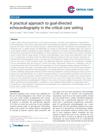
Walley et al. Critical Care 2014, 18:681http://ccforum.com/content/18/6/681Decrease ‘depth’ to 14 to 16 cm for a full screen cardiacimage. Continually adjust the ‘focus’ control to the anatomy imaged for better resolution. Gain should be adjusted to see endocardial borders clearly (Figure 1A).Be careful not to ‘over gain’ (too bright, washes outimage) or ‘under gain’ (weak echocardiographic images)(Figure 1B,C).ViewsGoal-directed echocardiography comprises four mainviews. In an urgent or emergent setting, ‘go for the gold’.Apical and subcostal views are frequently most useful[42]. That is, if unable to obtain an adequate image inone view, move on to the next view, when necessary. Incritical care settings, you may not always have the electrocardiogram on the echocardiographic monitor to provide a timing marker. For timing of the cardiac phase,pay attention to whether the mitral/tricuspid valves areopen (diastole) or closed (systole). Remember, you mustPage 4 of 11see endocardium to accurately assess ventricular function for all the views listed.Subcostal viewThe subcostal view (Table 1 and Figure 2) provides moreinformation than any other individual view and, as a result, addresses more of the key questions. In particular,volemia is best addressed in this view and multiple aspects (IVC collapse [14,43], RV and LV diastolic size) aresometimes evident from this one window. The IVC canbe used for estimating central venous pressure, reflectiveof right atrial pressure [14,43,44]. The ratio of inspiratory and expiratory changes in diameter is very useful inevaluating volume status [45-47]. The subcostal view, aswith the apical four-chamber (A-4) view, is frequentlythe best view to appreciate the presence of a pericardialeffusion and associated echocardiographic evidence ofhemodynamic compromise [2]. This view is frequentlyused for a pericardiocentesis. It is sometimes difficultFigure 1 The parasternal long axis (PLAX) view. (A) Normal gain. (B) Gain too high; with the gain too high, echocardiographic anatomy datamay be obliterated. (C) Gain too low; with the gain too low, echocardiographic anatomy data may be incomplete. LA, left atrium; LV, leftventricle; RV, right ventricle.
Walley et al. Critical Care 2014, 18:681http://ccforum.com/content/18/6/681Page 5 of 11Table 1 Subcostal and apical four-chamber viewSubcostal viewApical four-chamber viewGoalVolume status - IVC collapse. LV and RV size and function.Pericardial effusion. Right pleural effusionLV size and function, RV size and function, and pericardium.RV/LV ratio. Pericardial effusion. Left pleural effusion.Aortic and mitral valve thickening. Significant valvularregurgitation - color DopplerPatient positionSupine with pillow under knees to relax abdominal wallFar left lateral, when possibleInitial transducer placementTransducer at the subxyphoid position and angled slightlymedially, pointing the transducer (soundwave beam) tothe patient’s left side. Transducer marker at approximately3 o’clock. Transducer almost flat against the abdominalwall, approximately at a 15 degree angleStart at the apex. Rotate the transducer clockwise with thetranducer marker at approximately 3 o’clock and angle thetransducer or sound wave beam up through the apex ofthe heart or right shoulderSearch for the best windowTransducer angled laterally to image the apex of the RV,upper right side of screen, LV on the lower right side ofscreen, right atrium on the upper left side of screen, andleft atrium on the lower right side of screenThe right and left apex will be at the top of the screenwith the atria belowFor IVC, angle the transducer medially or towards the patient’sright shoulder to view the right atrium. Slightly rotate thetransducer counter clockwise. Tilt the transducer andsoundwave beam slightly inferiorly or down to ‘open up’or image the IVC in the long axis plane for measurementsand IVC collapse/distensibility calculation. The measurementshould be taken 1 to 2 cm in from the IVC/right atriumconnectionThe LV on the upper right side of the screen and the RVon the upper left side of the screen. If not sure which isthe RV or LV, the tricuspid valve leaflets are always insertedcloser to the apex than the mitral valveIf the apex is leftward on the screen, angle or slide thetransducer and soundwave beam mediallyIf the apex is tilted rightward on the screen, angle or slidethe transducer and soundwave beam laterally or rightwardIVC, inferior vena cava; LV, left ventricle; RV, right ventricle.to differentiate pericardial fluid, fat, clot, or thrombus.A small, focal or loculated pericardial effusion maynot be visualized nor easily ‘tapped’. When resultsare in question, order a comprehensive echocardiographicexamination.An incidental finding of ascites or a right pleural effusion may be visualized in this view. A left-sided pleuraleffusion, visualized in other views, is more frequently associated with a pericardial effusion.Parasternal long-axis viewThe parasternal long-axis view (Table 2 and Figure 2) isused in the assessment of LV systolic function (normal,decreased, or severely decreased) [17]. This view is oftentechnically easier to obtain due to patient positioning,reliable landmarks, and ease of maintaining a steadytransducer position [1]. A small and hyperdynamic LVmay indicate hypovolemia. A pericardial effusion may beappreciated. An ‘incidental’ finding of a left pleural effusion may be noted.Parasternal short-axis viewThe parasternal short-axis (PSAX) view (also known asthe transverse view; Table 2) is used for the visual assessment of LV function at the mid-LV. The mid-LV (Figure 2)is identified when the tips of the papillary muscles are visualized as just ‘flickers’ of the mitral valve apparatus. The LVshould appear round, with RV visualization at the mid-LV.The main pulmonary artery with the bifurcation may bevisualized.In the setting of hypovolemia or volume depletion, theLV may appear small and mitral valve leaflets may bevisualized. The presence of regional wall motion abnormalities may be noted, though a comprehensive echocardiographic examination is indicated for evaluation ofregional wall motion abnormalities. The RV may be evaluated for the presence of abnormal or paradoxical septalmotion and the ‘D’ sign: flattening of the interventricularseptum (RV pressure overload). This view is also usedfor presence of pericardial effusion. Incidental findingsof clot/thrombus in the main pulmonary artery may benoted.Apical four-chamber viewSome authors caution about the use of A-4 views sinceoff-axis views lead to erroneous interpretation of the LVand, particularly, RV size and function [1]. This view canbe useful for identifying circumferential pericardial effusion. Assessment of RV size should be carefully made[38]. The RV/LV ratio may be used for evaluating RVsize [48]: a RV/LV ratio 0.6 indicates normal RV size, aRV/LV ratio of 0.6 to 1.0 indicates moderate RV dilatation and an RV/LV ratio 1.0 indicates severe dilation[31,49,50]. The RV apex and LV apex must be at the topof the screen with the atria visualized directly below. Anoff-axis view may inaccurately cause the RV to appeardilated. This view is used for the assessment of pericardial effusion and for evidence of hemodynamic compromise. Circumferential pericardial effusion is oftenassociated with a large pericardial effusion. Right atrial
Walley et al. Critical Care 2014, 18:681http://ccforum.com/content/18/6/681Page 6 of 11Figure 2 Standard four views of goal-directed echocardiography. (A) Parasternal long-axis view. Horizontal view of the heart, including theascending aorta, the aortic valve (AV), the right ventricle (RV), the left ventricle (LV), the left atrium (LA), frequently excluding the apex, and thepericardium. (B) Parasternal short-axis view. A transverse view of the mid-LV at the level of the papillary muscles, including the RV, and thepericardium. (C) Apical four-chamber view, including the LV, RV at the RV inlet, trabeculated apical, and the infundibulum or smooth myocardialoutflow regions, LA, right atrium (RA), mitral valve, tricuspid valve, and pericardium. (D) Subcostal four-chamber view. A more perpendicularorientation of the LV, RV, LA, RA, mitral and tricuspid valves, interatrial septum, pericardium, inferior vena cava (IVC), and liver.and/or ventricular diastolic collapse/buckling may beseen [2]. An incidental finding of a left pleural effusionmay be visualized.InterpretationGoal-directed echocardiography should always be interpreted in the context of clinically available hemodynamicand other clinically relevant information. Accordingly, agoal-directed echocardiography is not an anatomic investigation; it is a physiologic study or hemodynamic assessment (Table 3). Full interpretation involves linkinggoal-directed echocardiography findings to hemodynamicand other clinical information. We provide a practical‘primer’ approach while full discussion of all aspects ofhemodynamic instability, shock, and PEA arrest is beyond the scope of this review [51].Septic or distributive shock that is less severe or in theearlier stages may show a hyperdynamic LV and asmaller to normal size IVC with 50% collapsibility because septic shock is often initially associated with a degree of hypovolemia. More severe distributive shock maydemonstrate impaired LV function [31].Hypovolemic shock may demonstrate small ventricles,sometimes ‘near cavity obliteration’, due to a small andhyperdynamic LV, and a small IVC with wide respiratory variation which collapses 50% on inspiration ina spontaneously breathing patient [47,52,53]. If thepatient is fully mechanically ventilated on positive pressure breathing, variation of the IVC diameter is associated with volemia [43,54]. Variation by more than 12%(the difference been maximal IVC diameter and minimalIVC diameter divided by maximal IVC diameter) suggests
Walley et al. Critical Care 2014, 18:681http://ccforum.com/content/18/6/681Page 7 of 11Table 2 Parasternal viewsParasternal long-axis viewParasternal short-axis viewGoalLV size and function, RV size, RV wall motion, pericardialeffusion, left pleural effusion. Mitral and aortic valvethickening. Significant valvular regurgitation - colorDopplerLV size, wall thickness, and function. RV size and function.Main pulmonary artery (dilated or not). Pericardialeffusion. Aortic valve thickening. Significant valvularregurgitation - color DopplerPatient positionFar left lateral, when possibleFar left lateral, when possibleInitial transducer placementSecond or third ICS, as close to the sternum as possible,with the transducer ‘marker’ directed at approximately11 o’clock (towards right shoulder)Second or third ICS, as close to the sternum as possible.From the PLAX view, rotate the transducer clockwiseor to your right until the transducer ‘marker’ is atapproximately 2 o’clockSearch for the best imaging ‘window’ Move the transducer up or down an ICS to searchfor the best view. Structures should appear horizontalon the screen. Image in the middle of the screen byadjusting transducer positionMid-LV: angle the transducer slightly laterally andinferiorly or towards the patient’s left hip to image themid-LV. Visualize the tips of the papillary muscles withinthe LV, below the level of the mitral valve. LV shouldappear round with RV clearly visualized. If the LV appears‘egg-shaped’ or oblong and the RV is not visualized,slide the transducer ‘up’ an ICSIf the apex appears to be positioned ‘uphill’ on thescreen, slide the transducer ‘up’ an ICSMain pulmonary artery: angle the transducer slightlylaterally and slightly superiorly or toward the patient’sleft shoulder with a slight clockwise rotation. Youmay need to move up an ICS, but in doing so maynot be able to visualize the pulmonary artery bifurcationIncrease the ‘depth’ to 20 to 24 cm to image thepresence of a possible pleural effusionDecrease the depth to approximately 14 to 16 cm,depending on the size of the heart, to fill the screenwith cardiac structureICS, intercostal space; LV, left ventricle; PLAX, parasternal long axis; RV, right ventricle.that cardiac output may increase significantly following volume infusion [55]. Whether fluid should begiven, even when IVC diameter variation is large, is aclinical judgment that depends on other clinical variables [56]. Goal-directed echocardiography only supplements other clinical information. Patients who areresponsive to volume loading can increase their cardiacoutput by 15% [31,54].Table 3 SHOCK: a practical mnemonic for the differentialdiagnosis of the spectrum of hemodynamic instability,shock, and pulseless electrical activity arrestType of shockGoal-directed echocardiographic featuresSeptic/distributiveLess severe shock - hyperdynamic. Severeshock - can be hypodynamicHypovolemicCollapsing IVC, small ventricles, may be hyperdynamicObstructiveTamponade: pericardial effusion, tamponadephysiologyPulmonary embolism: large RV, septal shift/diastolicflatteningCardiogenicDecreased LV ( RV) function, dilated ventriclesKCombinations (for example, septic plus hypovolemic,septic plus cardiogenic, and so on)Other kinds (adrenal insufficiency, neurogenic, andso on). This broader category is raised by thequestion ‘What does not fit?’IVC, inferior vena cava; LV, left ventricle; RV, right ventricle.Obstructive shock due to tamponade (evidence ofhemodynamic compromise) may be observed in the A-4view (Figure 3A) and the subcostal view (Figure 3B).These views will demonstrate a significant pericardialeffusion (usually encompassing the heart) with rightatrial wall or RV diastolic collapse. Delayed RV diastolicexpansion may also be noted. These findings may indicate increased pericardial pressure [2]. The IVC isdilated indicating high right-sided filling pressures.‘Swinging heart’ motion always indicates a large amountof pericardial effusion, but is not always associated withhemodynamic compromise. A patient may have tamponade physiology at times with a smaller amount of pericardial effusion. This is sometimes associated with fasterpericardial fluid accumulation. It is beneficial to detectpericardial effusion in patients with PEA an
the use of echocardiography in the critical care setting [1-6] and a large number of publications have investi-gated important aspects of echocardiography in critically ill patients, in particular the goal-directed approach [1-6]. Here we review the current use of, and a problem-oriented practi