Transcription
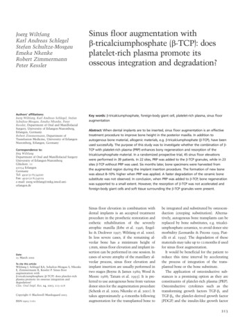
case reportSinus elevationwith short implantAuthors: Prof. Dr Mauro Marincola, Prof. Dr Dr Rolf Ewers, Prof. Giorgio Lombardo (University of Verona) &Prof. Miguel Simancas Pallares (University of Cartagena), Austria/Italy/ColombiaIntroductionAn upper posterior edentulous maxilla with diminished bone height usually represents a challengingsituation for clinicians when dental implantation isthe solution for tooth absence. Bone decrease is theresult of a reduction in both height and width of thealveolar process due to the maxillary sinus pneumatisation.1Fig. 1: Pre-operative patientassessment: X-ray of the edentulousspace showing maxillary sinus pneumatisation.Two main options have been proposed to lift theSchneiderian membrane and then create somespace to place implants: the lateral approach thatincreases treatment-related costs, postoperativemorbidity and also surgical time, and the osteotomesinus floor elevation (OSFE) firstly introduced in1986 by Tatum. Then in 1994 Summers introducedthe maxillary sinus lifting internal sinus lift (ISL)technique by the use of osteotomes, in which boneis added to the apical part of the implant to improveimplant primary stability.2,3 This technique wasshown to be less invasive, less time consuming andreduces postoperative discomfort to the patient.ISL is indicated when residual bone height (RBH) isbetween 5 and 7 mm. Other authors perform ISL inRBH extreme conditions such as 4 mm.4There’s an increasing debate whether a bone graftis needed into the elevated area to maintain the spacefor new bone formation in the future. According toSummers’ original report, autogenous, allogenic orxenogeneic grafting materials are often placed.5 Recently Nedir et al. showed no differences (P 0.05)between ISL procedures performed with or withoutbone graft on the apical portion.6 However, the mainclinical challenge arises when a bone graft is placedand the bone around the implant-abutment interfaceneeds to be maintained.Rammelsberg et al. performed ISL but withoutbone graft in a retrospective study in 66 patientswith 101 dental implants in 2015. By using X-rays,they determined bone changes over the time.7 Theyfurther obtained results that mesial and distal meanapical bone gain (initial-final bone height) were0.5 mm and 0.4 mm respectively, thus recommending that implants placed in combination with ISLwithout graft material would have bone gain.Likewise, Nedir et al. compared dental implantsplus ISL placed in combination with and withoutbone graft in 2013. They concluded that, althoughmore bone is observed when the grafting material isused (5 mm) in comparison with non-grafted sites(3 mm, P 0.05), this may not be required to promoteendo-sinus gain.Although there’s no consensus whether bonegraft should be placed via ISL procedure or not, thisoption is highly recommended due to the benefitsregarding osseous level maintenance. The aim of thisstudy was to describe radiographic parameters ofminimally-invasive internal sinus elevation in combination with plateau-form short implants. Thus thispaper is intended to describe the surgical techniqueFig. 1a20implants2 2017
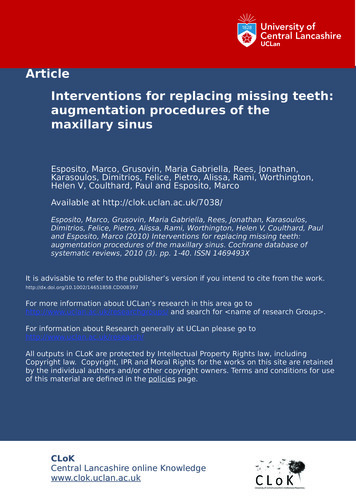
case reportFig. 2aFig. 2bFig. 2cFig. 2dFig. 2eFig. 2fof a predictable procedure we developed during our20 years of practical hands-on training with students and course participants.Case reportA female 58-year-old patient consulted us becauseof her desire of functional and aesthetic repairment.The patient did not relate any medical background ofclinical interest. She also signed informed consentprior the beginning of study, held ASA I status and required dental implantation in the upper posteriormaxilla with at least 4 mm on RBH and measuredthrough a digital periapical radiograph by a single operator calibrated for this (Intraclass Correlation Coefficient: 0.83).Amoxicillin 500 mg was prescribed to the patienttwo days prior the surgical procedure, once everyeight hours in order to avoid infections. Surgical procedure was performed by a different trained clinicianwith more than 25 years of experience in the field.Surgical TechniqueInfiltrative anaesthesia was used in the procedure.Initially, a non-epinephrine anaesthesia was used Fig. 2: Description of the surgicalprocedure: a) clinical condition ofthe alveolar ridge that shows theedentulous area of teeth 14, 15 and16 prior the beginning of the surgicalprocedure; b) crestal incision andblood collection with a sterile syringefor further bone graft; c) pilot drillingat the site of dental implantation withsimultaneous ISL; d) 2.5 mm latchreamer with no cutting edge to startosteotomy widening at thecrestal bone; e) 2.5 mm latch reamer perforation in the bone startingthe osseous widening; f) X-ray showing RBH (4.24 mm) and also 2.5 mmlatch reamer insertion into the bone(4.29 mm).(PRICANEST 4 %, Ropsohn Therapeutics, Bogotá,Colombia) in order to collect blood (Fig. 2b) to mixwith the grafting material (50–500 μm Synthograft,Bicon Dental Implants, Boston, USA). Then 2 % Xylocaine (Dentsply Pharmaceutical, York, USA) was usedto complete the surgical procedure.Intrasulcular incision was performed by using a size15 blade in a Bard-Parker scalpel. Full thickness flapwas obtained in the area and then a previously published protocol8 was followed to perform ISL: pilot(2 mm diameter) drilling to achieve cortical perforation was started. Pilot drilling length (1–2 mm) wasdetermined upon residual bone height prior measurement (Fig. 1b). This high-speed 1,100 rpm drill is usedwith external water irrigation and has a cutting edgeat the apical portion. The pilot osteotomy should be1 to 2 mm shorter than the calculated RBH measuredon the periapical xR. The following steps are achievedwith latch reamers at 50 rpm without water irrigation. The reamer consists of two vertical cuttingedges, which stops 2 mm before the apical portion.The apex is tapered and without a cutting edge toavoid Schneiderian membrane perforation. A 2.5 mmlatch (mechanical) reamer was inserted to start thewidening of the crestal cortical bone and to deepenimplants2 201721
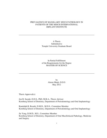
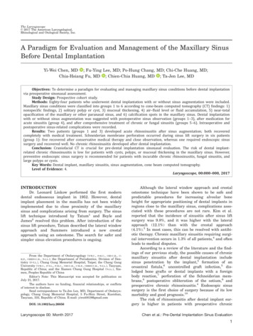
case reportFig. 3: Microfracture points alongthe cortical zone performed withthe 3.5 mm hand reamer. a) Firstmicrofracture at distal, b) third microfracture at buccal and thefourth microfracture was performedat distal. c) Clinical picture of thegrafting material being injectedand d) the 3.5 mm osteotome firstinsertion into the cavity.Fig. 3aFig. 3bFig. 3cFig. 3dthe bur with finger pressure towards the corticalbone of the sinus floor. The pressure allows thenon-cutting edge to be pressed through the smoothcancellous bone but stops at the hard tissue of thesinus floor (Fig. 2d). With this 2.5 mm latch reamer, anX-ray was taken to determine the reaming finallength before sinus floor (Fig. 2f). The RBH was measured to determine the final drilling length and thelatch reamer series with 0.5 mm diameter improvement were inserted until the 4.5 mm implant diameter was reached.The following step describes the microfracturetechnique of the sinus floor. With the 3.5 mm handreamer that has a single vertical cutting edge andends with a knife-edge at the apex of the reamer, wetapped the sharp tip of the hand reamer at four different points along the buccopalatal and mesiodistalaxis to facilitate the microfracture of the sinus floorcortical bone. The first fracture point was the lowestRBH on the periapical. We started the fracture at thedistal aspect of the osteotomy (Fig. 3a). The secondand fourth fracture points are always the buccal(Fig. 3b) and the palatal because of their higher pneumatisation towards the buckle. The third point inthis case is the mesial aspect.22implants2 2017A synthetic and bacteriostatic grafting material(Synthograft, -TCP, size 50–500 m) was mixedwith the collected blood until getting a putty consistency and no liquid was obvious in the mixture.A 4.0 mm bone graft syringe was used to place abone graft material into the apical portion of the oste o tomy (Figs. 3c). Once resistance against the Schneiderian membrane was detected, a slow retraction of the syringe while continuously injectingwas done. After bone graft material was injected, a3.5 mm Summer osteotome was used to gently pushthe material into the osteotomy. With the graft material in place, the osteotome was advanced via gentle tapping until the cortical bone was fully fractured,and lifted with the sinus mucosa (Figs. 3d, 4a–c).A 4.5 x 6.0 mm Implant (Bicon Dental Implants,Boston, USA) was inserted into the grafted oste otomy using first an implant inserter and retrievermounted in a straight handle and then by gently tapping with a seating tip (Figs. 4d–f).If the remaining RBH is more than 3 mm, the firstplateaus following the sloping shoulder will be engaged against the osteotomy walls and this press fitimplant will not move during the healing because a
case report Fig. 4: a) The 3.5 mm osteotomeinserted into the osteotomy pushingthe material against the Schneiderianmembrane; b) X-ray image showingthe 3.5 mm osteotome position andalso the grafting material; c) 4 mmosteotome performing the greenstick fracture; d–f) implant insertioninto the osteotomy with the straighthandle and the 3 mm seating tip.Fig. 4aFig. 4bFig. 4cFig. 4dFig. 4eFig. 4fprimary stability is achieved. When a RBH 3 mm ispresent, a sinus lift abutment (Bicon Dental Implants, Boston, USA) needs to be placed in order toavoid implant displacement into the lifted sinus. Thisimplant design will not have a primary stability alongthe osteotomy walls because it is placed 2 mm underthe crest and the implant body would be fully immerged into the grafting material. Plateau-formedimplants with healing chambers between the plateaus do not need a primary stability but the internalsinus abutment stabilises the implant into its finalprosthetic position. Single suture with polyglycolidacid (PGA) was used to close the mesial anddistal relieving incisions (ACE Surgical Supply CO, Brockton, USA). After implant insertion, immediatepost-op X-ray was performed (Fig. 5). The Patient received post-op instructions and homecare instructions. Antibiotics (Amoxicillin) and analgesics(Nimesulide) were prescribed to avoid infections andpain/swelling.DiscussionDental implantation is still the most effective approach to replace a missing tooth due to the observed survival rates over the time. However, some-times the anatomic conditions could restrict implantpositioning into the ideal space, thus limiting theprosthetic options.9 Maxillary sinus pneumatisationoccurs as the result of the upper posterior tooth loss.As a consequence, internal sinus lift (ISL) has beendocumented as one of the surgical approaches toaccomplish implant placement in the same surgicalprocedure.10 Results of this case report suggest thatISL and simultaneous implantation were successfully performed on the patient with no intraoperative or early postoperative complications.The employment of sinus grafting in conjunctionwith the ISL procedure is still open to debate. According to Summers’ recommendation, autogenous, allogenic or xenogenic grafting materials are often inserted into the elevated area to maintain the space fornew bone formation. Moreover, several studies havesuggested that the Schneiderian membrane elevationby itself promotes bone regeneration by means of theformation of a fibrin clot in the created space. Thisclot, which is stabilised and protected from externaltrauma and intra-sinus pressure, would have the potential to stimulate bone formation.11,12 However, thisoption is highly susceptible to membrane perforationor membrane invagination around the implant apex.implants2 201723
case reportFig. 5: a) Clinical picture showingthe implant position after its placement via gentle tapping; b) placedimplant with the healing plug; c) cutted view of the healing plug for theplaced implants in the zone; d) autologous bone graft over the implantwith the collected bone during theosteotomy; e) immediate final X-rayviews of the placed implant togetherthe internal sinus lifts procedure. Onthe apical portion of the implant thegrafting material is observable.Fig. 5aFig. 5bFig. 5cFig. 5dchanical stress in the crestal alveolar bone area andassists in enhancing the vascular supply to hard- andsoft-tissue in case of reduced interdental space.16,17Fig. 5eWe then decided to use pure-phase tricalcium phosphate, a synthetic material into the created space toavoid the collapse of the Schneiderian membranearound the internal sinus implant portion and promote bone formation during the osseointegrationperiod also around the implant apex.Associated complications with the sinus augmentation procedures are well described in the literature.The most common complication is membrane perforation during procedure and its prevalence is between7 and 44 %. Haemorrhage, infection and rhinosinusitis are also described as expected complications.13However, none of them were present in our patient,indicating an immediately successful surgical procedure which was specially developed for clinicians withno experience in sinus lift elevation.Implant survival placed in conjunction with ISL hasbeen also well reported in the literature ranging from94 % to 100 %.14,15 Nevertheless, the most critical issue is the crestal bone level maintenance over thetime. This is achieved by placing the implant in a subcrestal (submerged) fashion and by the usage of animplant with convergent crest module, representedby the sloping shoulder geometry, which enhancesthe platform switching (PS) to occur. This PS allows anincrease in residual crestal alveolar bone volumearound the neck of the implant, repositions the papillato a more aesthetic and apposite level, reduces me-24implants2 2017ISL is reliable if used with the proper protocol, lesstime consuming procedure, with lower rates of complications that can be considered in patients with upper posterior decreased alveolar ridge. In a non-traumatic way and during the same surgical procedure itallows implant placement with no immediate complications during the procedure nor a short postoperative time period.Conflict of interest statement: Authors declarethey do not have any conflict of interest.Source of funding: NoneAuthor detailsLiteraturecontactMiguel Simancas PallaresAvenue Del Consulado 48-152University of CartagenaHealth Sciences CampusFaculty of Dentistry, Research Department, Suite 301Cra. 6 #36, Cartagena, Bolívar, ColombiaTel.: 57 5 6698172-110msimancasp@unicartagena.edu.co
1986 by Tatum. Then in 1994 Summers introduced the maxillary sinus lifting internal sinus lift (ISL) technique by the use of osteotomes, in which bone is added to the apical part of the implant to improve implant primary stability.2,3 This technique was shown to be less invasive, less time consuming and