Transcription
Revista Odontológica MexicanaVol. 17, No. 3Facultad de OdontologíaJuly-September 2013pp 173-178CASE REPORTMaxillary sinus elevation and simultaneous implant placementusing PRGF (plasma rich in growth factors), hydroxyapatite andallogenic graft. Seven year case reportElevación de seno maxilar y colocación simultánea de implantes utilizandoplasma rico en factores de crecimiento (PRFC), hidroxiapatita y aloinjerto.Reporte de un caso de siete añosNayibe Hernández Tejeda,* Ma. del Carmen López Buendía§ABSTRACTRESUMENMaxillary sinus floor elevation is a predictable surgical proceduremeant to vertically increase the amount of bone in the posteriorregion of the upper jaw to enable placement of a prostheticrehabilitation device supported by implants. The aim of the presentarticle was to describe elevation of the maxillary sinus floor usingplasma rich in growth factors, absorbable hydroxyapatite and boneallograft as sub-antral graft materials with simultaneous placementof two surface treated implants (Osseotite, 3i). The present articlealso reported clinical and radiographic results obtained at sevendays, six months and seven years after the surgery. From the clinicaland radiographic standpoint suitable healing was observed. Thesurgical procedure used in the present clinical case was considereda suitable option to place implants in atrophic maxillary areas.La elevación de piso de seno maxilar es un procedimiento quirúrgico predecible que se realiza con la finalidad de aumentar verticalmente la cantidad de hueso en la región posterior del maxilarpara poder realizar una rehabilitación protésica implantosoportada.El propósito de este trabajo es describir un caso clínico donde serealizó elevación de piso de seno maxilar utilizando plasma rico enfactores de crecimiento, hidroxiapatita absorbible y aloinjerto óseocomo materiales de injerto subantral y la colocación simultánea dedos implantes de superficie tratada (Osseotite, 3i) y reportar los resultados clínicos y radiográficos obtenidos siete días, seis meses ysiete años después de la cirugía, observando una cicatrización adecuada tanto clínica como radiográficamente. El procedimiento quirúrgico utilizado en este caso clínico resultó una buena opción parapoder colocar implantes en áreas maxilares posteriores atróficas.Key words: Maxillary sinus elevation, sub-antral graft, dental implants, plasma rich in growth factors.Palabras clave: Elevación de seno maxilar, injerto subantral, implantes dentales, plasma rico en factores de crecimiento.INTRODUCTIONBone-integrated oral implants have shown longterm predictable results. The greatest failure ratein implants was found among those placed in theposterior region of the upper jaw. This is due to theanatomical characteristics of the region which includequality and amount of present bone.1 Bone availabilityin this area is reduced. This is due to several causes,which can include, among others: premature toothloss causing thus sinus pneumatization, periodontaldisease, iatrogenic or physiological bone resorption.All the aforementioned would preclude treatment ofprosthetic rehabilitation supported by implants.2Elevation of the maxillary sinus floor is a surgicalprocedure which consists on vertically increasing theamount of bone found in that location. This procedurewas first designed and described by Hilt Tatum in 1976,at the Dental Implant Meeting held in Birmingham,Alabama. Nevertheless, the first published data on thissubject matter were released by Boyne ad James in1980.3*§Graduate, Periodontics Specialty, Graduate School, NationalSchool of Dentistry, National University of Mexico (UNAM).Professor, Periodontics Specialty, Graduate School, NationalSchool of Dentistry, National University of Mexico (UNAM).This article can be read in its full version in the following aunam
174Elevation of the maxillary sinus is a recommendedtechnique for edentulous areas of the maxillaryposterior region lacking adequate bone. In that region,placement of dental implants is required in order toachieve successful prosthetic treatment. 4 Differentanatomical situations and different topography ofthe sinus with respect to the maxillary ridge give riseto establishing a classification with respect to thepneumatization and atrophy or resorption of the subantral maxillary area. Four grades are identified:Grade I. The height of the sub-antral maxillarysegment is 10 mm or more, thus allowing implantplacement without having to elevate the sinus floor.Grade II. Sub-antral maxillary segment height islesser than 10 mm and over 8 mm; these cases canbe treated with osteotomes.Grade III. The height of the sub-antral segmentis between 4 and 8 mm. In this case it is neededto increase the vertical volume of the bone. Thisis achieved by surgically elevating the floor of themaxillary sinus through placement of a sub-antral graftas well as implants.Grade IV. The height of the sub-antral segmentis under 4 mm. With these dimensions, obtainingacceptable implant primary stability is very risky.Therefore, a two-stage surgical technique wasdesigned: the first involved elevation of the maxillarysinus and the second entailed implant placement.5Contraindications to this procedure are: inadequatesinus transversal dimension, ostium location atthe surgical site, excessive or inadequate interocclusal space, sinus disease, as well as all generalcontraindications to dental implant placement.6The first grafting material used in sinus elevationprocedure was autologous bone harvested from theiliac crest. 7 Commonly used donor sites within themouth are: maxillary tuberosity, mandibular retromolararea as well as mandibular ramus. Additionaly toauto-grafts, allografts, xenografts, alloplastic graftsor combinations of these have been used. Variedresults of these techniques have been reported at theconsensus of the 1996 Conference.4Plasma rich in platelets (PRP) is an autologoushuman platelet (above normal) concentrate. It isconsidered a source rich in growth factors. It wasintroduced in 1998 by Marx et al. Combined with anautologous bone graft, it was used to reconstructmandibular defects. Their research showed thatPRP addition to bone grafts accelerated the rateof bone maturation, and radiographically increasedbone density when compared to bone graft by itself.8Hernández TN et al. Maxillary sinus elevation and implantsSince that point in time, it has been used in differentclinical procedures such as, among others, sinusfloor elevation, ridge increase, periodontal defecttreatments, alveolus preservation.9At a later point, plasma rich in growth factors (PRGF)would emerge. This technique was proposed byEduardo Anitua, and several studies informed of theiruse as an additional option within the scope of severalmaterials to achieve a sub-antral graft. This techniquepurported the aim of compacting particulate grafts andimproving conditions for bone regeneration.10-12The aim of the present research paper was todescribe this technique and report clinical andradiographic results achieved in a patient after havingconducted the surgical procedure entailing elevationof the maxillary sinus floor mucosa and simultaneousimplant placement, with usage of PRGF, absorbableHA as well as FDBA as sub-antral bone graft.CASE REPORT43 year old female patient attended the GraduateSchool, National School of Dentistry, NationalUniversity of Mexico (UNAM) seeking periodontaltreatment. The patient exhibited good general healthand had been diagnosed with moderate generalizedchronic periodontitis.Initial periodontal therapy was undertaken: itentailed personal control of oral hygiene, scalingand root planning as well as consultation with OralProsthesis Department to launch a comprehensivetreatment.Teeth 16 and 17 were deemed hopeless. Therefore,surgical phase consisted on extractions with alveoluspreservation by placing 0.5 g of allograft (DFDBA) andusing a collagen membrane. Tooth 15 was missing.Teeth 44 and 45, 24, 25 and 27 were subjected todebridement surgery. Teeth 36 and 37 were subjectedto regenerative periodontal surgery (Enamel-DerivedProtein Matrix).Eleven months after performing alveoluspreservation surgery in the upper right posteriorarea, bone amount was radiographically andclinically assessed. According to the aforementionedclassification presence of Grade III residual bonewas established. Therefore, maxillary sinus elevationwas programmed with simultaneous placement oftwo implants. Sub-antral graft was composed byPlasma Rich in Growth Factors (PRGF), absorbablehydroxyapatite (absorbable HA, Osteogen ) as well asmineralized lyophilized bone allograft MLBAG.Before undertaking surgical procedure, andaccording to PRGF protocol established by Anitua in
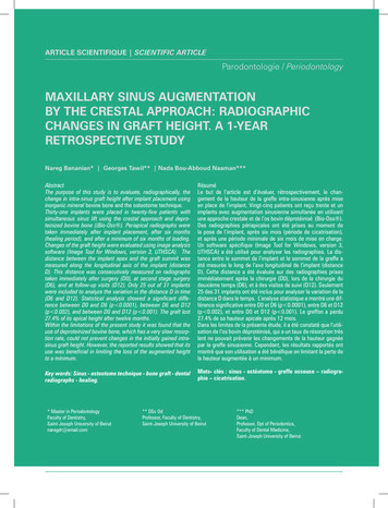
Revista Odontológica Mexicana 2013;17 (3): 173-1781966, 30 cm3 of the patient’s blood were obtained.10Sterile tubes with 3.8% sodium citrate were usedas anticoagulant. Blood was centrifuged for sevenminutes at 1,700 rpm. After this, plasma wasseparated into fractions by means of meticulouspipetting to avoid creation or turbulence in obtainedfractions. The first 500 µL (0.5 cm3, fraction 1) wasa platelet-poor plasma, it was consequently poorin growth factors. The next 500 µL (fraction 2)corresponded to plasma containing similar number ofplatelets to those found in peripheral blood. Finally,the plasma fraction richest in growth factors was the500 µL fraction, found to be immediately above thered series (fraction 3). 1 cm3 of PRGF was found toevery 4.5 cm3 tube. Total was 6 cm3 which were thenactivated with 10% calcium chloride (50 µL per eachPRGF cm 3) as activator and platelet aggregationmeans. Obtained PRGF was combined with bonegraft materials, 1 g of mineralized bone allograft(Pacific Coast Tissue Bank ) and 1 g of absorbablehydroxyapatite (Osteogen ).Ad interim, at the surgical site, after havingadministered local anesthesia, a liberating mesialincision was performed on the alveolar ridge in orderto gain improved surgical field visibility, lifting thus afull-thickness graft.An antrostomy was performed following thetechnique described by Tatum, known as incompletefenestration technique, in which approach is tracedon the maxillary external or lateral side according totopographic projections of the radiographic study aswell as sinus intra-oral transillumination.7 This type ofosteotomy, due to the elevation of the bone segment,allows the transformation of a new maxillary sinusfloor. To this effect a low speed, number six, ballshaped, carbide burr was used.Once the access was elevated, the sinus membranewas detached (Schneider membrane) with specificcurettes (Figure 1). The next step, with the assistanceof the surgical guide, was to prepare the bed whereinto place the implants. Two 4 x 15 mm Osseotite 3 were used.The graft was placed into the sinus cavity followinga two-stage technique. The first stage involved placingthe graft before the implants, so as to be able toreach the medial wall and thus easily compact thegraft material. The remaining graft was placed aftersituating the implants in their final position (Figure 2).Using the plasma’s fraction 2, a fibrin clot wasformed to be placed as biological membrane on themaxillary lateral wall and thus achieve sinus cavitysealing (Figure 3). The consistency of the membranewas achieved after placing this plasma fraction in a175tube containing calcium chloride. This procedure wassimilar to that used when using the fraction rich ingrowth factors, with the difference that in this case, athermal block was used to accelerate clotting, whichtook approximately 15 minutes.The flap was repositioned and secured withhorizontal mattress sutures as well as simplesutures, using 3-0 silk. Finally, the surgical site wasdampened with the fraction of plasma poor in growthFigure 1. Lateral antrostomy (incomplete fenestrationtechnique) and elevation of sinus membrane.Figure 2. Graft compacted within the sinus and implantsplacement.
Hernández TN et al. Maxillary sinus elevation and implants176factors (fraction 1) so as to improve and accelerateepithelialization process in the area.The patient was thoroughly informed onpostoperative care to be observed. Antibiotic coveragewas achieved with amoxicillin, 750 mg every 12 hoursfor seven days. Ibuprofen was equally prescribed tocontrol pain and inflammation. 0.12% chlorhexidrinegluconate, 15 mL twice a day for two weeks was usedas local antiseptic.RESULTSObtained results were assessed at the time whensutures were removed, that is, seven days aftersurgery. Suitable soft tissue healing was observed.14 days after surgery, the wound epithelializationwas completed (Figure 4). No implant exposition wasobserved. The patient reported little post-operativediscomfort.Six months after surgery results were deemedfavorable (Figures 5 and 6). Radiographically, the areaof the sub-antral graft appeared dense and compact,lacking radiolucent areas or bone sequestrations. Theimplants were uncovered ten months after surgicalprocedure Two months later prosthetic rehabilitationwas undertaken. Presently, seven years after the sinuselevation surgical procedure, upon clinical assessment,the implants appear stable, without radiographicchanges at the level of the bone crest (Figures 7 and 8).DISCUSSIONMaxillary sinus elevation with sub-antral graft is asurgical procedure currently considered one of theFigure 3. Fibrin clot over sinus approach site.Figure 5. Initial X-ray with surgical guide.Figure 4. Soft tissue healing 14 days after surgical procedure.Figure 6. X-ray taken six months after surgical procedure.
Revista Odontológica Mexicana 2013;17 (3): 173-178most common and accepted methods to increasebone volume in the posterior lower jaw area in orderto place implants. Reports in scientific literatureshow high success rates when using this technique.This technique is a safe, effective and predictableprocedure.13In some cases, guided bone regeneration (GBR)procedures can be undertaken to increase verticalridge height, whenever the inter-occlusal space mightallow it. GBR and maxillary sinus elevation techniquecan even be combined in cases of very severe boneloss in the region.On the other hand, use of short implants in theposterior upper region can solve the problem.Nevertheless, this can result in a poor diagnosis, incases of low bone density, as is often the case inthat anatomical region.14 Due to the aforementionedFigure 7. Clinical results seven years after surgical procedure.Figure 8. Radiographic image seven years after surgicalprocedure.177reasons, use of implants exhibiting suitable lengthand diameter might require elevation of the sinusmembrane and placement of a sub-antral graft.15Suitable treatment plan, step-by step observanceof one or two-stage surgical protocol, appropriateselection of sub-antral graft as well as propermedication and pre- and postoperative care are someof the factors essential for achievement of successfullong and short term results of this procedure.16For this technique, there are many surgical protocolsas well as modifications.17 Nevertheless, when sinuselevation is conducted simultaneously to implantplacement (one stage) there is a great advantage withrespect to total restoration time, although it is importantto achieve primary stability of the implant. This implieshaving appropriate amounts of remaining bone, if thiswere not to be the case failure rates would increase.Increase of dental implants use and therefore,the need to improve bone conditions for successfulplacement, has driven clinicians in the search of betteralternatives in the field of bone implants. There is widevariety of materials used for sub-antral bone grafts.To this day, the best option still is autologous bone,since, it not only provides osteoblastic cells, it alsoconfers osteo-induction and osteo-conduction, offeringorganic and inorganic matrixes and viable bone cells,without incurring in antigenicity risks. Nevertheless,obtaining autologous grafts from extraoral donor sitesinvolves longer recovery time, the need for generalanesthesia and hospitalization of the patient, besideshigher treatment cost. Reports indicate that intraoralsites yield suitable results, nevertheless, limitations inavailability of donor sites constitutes a disadvantage,as well as increased procedure time, morbidity ofanother surgical site and increased patient discomfort.The amount of bone harvested intra-orally is generallyinsufficient for a complete increase of bilateralmaxillary sinus floor.18,19Therefore, many authors support the use of materials obtained from other sourcces (allografts, autografts xeno-grafts and alloplastic) in combination withautologous bone. They further argue that with thesetechniques a more suitable resorption time is generated. Choice of one or more materials often depends onthe amount of requered bone.4,13,16,20-22Plasma rich in growth factors is one of the materialsdeveloped with the purpose of improving grafthandling. This material improves soft tissue healing.10Incorporation of this technique can bring benefits forthe patient without incurring in any risk of spread ordisease transmission. PRGF use entails no secondaryeffect. Use of PRGF allows simplification of thesubantral graft compaction technique. Allowing thus
Hernández TN et al. Maxillary sinus elevation and implants178to improve consistency and facilitating handling aswell as increasing the amount of graft. Recent studiesresults have shown that PRGF as well as PRP (plasmarich in platelets) do not provide significant differencesin the procedures final results.23,24With respect to the implants’ inherent characteristics,it has been well established that there is greatersurvival rate whenever implants placed within themaxillary sinus come from a treated surface.13Knowledge of complications which can ariseas a result of a sinus elevation procedure such asmembrane perforation or maxillary sinus infection willhelp us to avoid them, or, in case they were to occur,to then properly treat them.It is important to take into consideration the factthat this surgical procedure requires a thoroughtreatment plan as well as a knowledgeable, skillful andexperienced surgeon.25,26CONCLUSIONSIn the present clinical case and based upon clinicaland radiographic results obtained seven years aftercompleting the surgical procedure, we could concludethat elevation of maxillary sinus using as sub-antralgraft a combination of PRGF, absorbable HA andallograft with a simultaneous placement of dentalimplants provided good results. This proceduredecreased as well total treatment time for thepatient, and proved to be a predictable and effectiveprocedure, in cases where a thorough treatment planhad been previously designed.REFERENCES1. Albrektsson T, Dahl E, Enbom L, Engevall S, Engquist B,Erikksson AR et al. Osseointegrated oral implants. A Swedishmulticenter study of 8,139 consecutively inserted implants.Journal of Periodontology. 1988; 59: 287-296.2. Bruggenkate C, Bergh J. Maxillary sinus floor elevation: avaluable pre-prosthetic procedure. Periodontology. 2000; 1998;17: 176-182.3. Boyne P, James R. Grafting of the maxillary sinus floor withautogenous marrow and bone. J Oral Surgery. 1980; 38: 613616.4. Jensen O, Shulman L, Block M, Iacono V. Report of the sinusconsensus conference of 1996. Int J of Oral and MaxillofacialImplants. 1998; 13: 11-55.5. Salagaray V, Lozada J. Técnica de elevación sinusal. Unidad deImplantología Oral y Prótesis Biointegrada. España, 1993.6. Baladrón J, Colmenero C, Elizondo J. Cirugía avanzada enimplantes. Ed. Ergon. España 2000.7. Tatum H. Maxillary and sinus implant reconstruction. Dent ClinNorth Am. 1986; 30: 227-229.8. Marx R, Carlson E, Eichstaedt R. Platelet-rich plasma. Growthfactor enhancement for bone grafts. Oral Surg Oral Med OralPathol. 1998; 85: 638-646.9. Marx R. Platelet-rich plasma: evidence to support its use. J OralMaxillofac Surg. 2004; 62: 489-496.10. Anitua E. Un nuevo enfoque en la regeneración ósea. PRFC.España: Puesta al día publicaciones; 2000.11. Anitua E. Plasma rich in growth factors: preliminary results ofuse in the preparation of future sites of implants. Int J Oral andMaxillofacial Implants. 1999; 4: 529-535.12. Anitua E. La utilización de los factores de crecimientoplasmáticos en cirugía oral, maxilofacial y periodoncia. Revistadel Ilustre Consejo General de Colegios de Odontólogos yEstomatólogos de España. 2001; 3: 305-315.13. Wallace S, Froum S. Effect of maxillary sinus augmentation onthe survival of endosseous dental implants. A systematic review.Annals of Periodontology. 2003; 8: 328-343.14. van den Bergh J et al. Anatomical aspects of sinus floorelevations. Clin Oral Impl Res. 2000; 11: 256-265.15. Yamamichi N, Itose T, Neiva R, Wang H. Long-term evaluationof implant survival in augmented sinuses: a case series. Int JPeriodontics Restorative Dent. 2008; 28: 163-169.16. Del Fabbro M, Testori T, Francetti L, Weinstein R. Sistematicreview of survival rates for implants placed in the graftedmaxillary sinus. Int J Periodontics Restorative Dent. 2004; 24:565-577.17. Smiler Dg. The sinus lift graft: basic technique and variations.PP&AD. 1997; 9 (8): 885-893.18. Mellonig J. Autogenous and allogenic bone grafts in periodontaltherapy. Critical Reviews in Oral Biology and Medicine. 1992; 3(4): 333-352.19. Lang N, Karring T. Proceedings of the 3rd European Workshopon Periodontology Implant Dentistry. Ed. Quintessence Books;Switzerland. 1999; 535-543.20. Kassolis J, Rosen P, Reynolds M. Alveolar ridge and sinusaugmentation utilizing platelet-rich plasma in combination withfreeze-dried bone allograft: case series. J Periodontol. 2000; 71:1654-1661.21. Jensen O. The sinus bone graft. Second Ed. QuintessenceBooks. 2006.22. Gonshor A. Technique for producing platelet-rich plasmaand concentrate: background and process. Int J PeriodonticsRestorative Dent. 2002; 22: 547-557.23. Plachokova A, Nikolidakis D, Mulder J, Jansen J, Creugers N.Effect of platelet-rich plasma on bone regeneration in dentistry: asystematic review. Clin Oral Impl Res. 2008; 19: 539-545.24. Torres et al. Effect of platelet-rich plasma on sinus lifting: arandomized-controlled clinical trial. J Clin Periodontol. 2009; 36:677-687.25. Kan J et al. Factors affecting the survival of implants placedin grafted maxillary sinuses: a clinical report. J Prosthet Dent.2002; 87: 485-489.26. Pjetursson B, Ching Tan W, Zwahlen M, Lang N. A systematicreview of the success of sinus floor elevation and survival ofimplants inserted in combination with sinus floor elevation. J ClinPeriodontol. 2008; 35 (Suppl. 8): 216-240.Mailing address:Nayibe Hernández TejedaE-mail: nayiht@hotmail.com
acceptable implant primary stability is very risky. Therefore, a two-stage surgical technique was designed: the first involved elevation of the maxillary sinus and the second entailed implant placement.5 Contraindications to this procedure are: inadequate sinus transversal dimension, ostium location at