Transcription
Molecular Biology Reports (2019) 4713-xMETHODS PAPERIdentification of reference genes for quantitative PCRduring C3H10T1/2 chondrogenic differentiationSerena Cappato1· Francesca Giacopelli1 · Laura Tonachini1 · Roberto Ravazzolo1,2 · Renata Bocciardi1,2Received: 25 October 2018 / Accepted: 20 February 2019 / Published online: 7 March 2019 The Author(s) 2019AbstractC3H10T1/2, a mouse mesenchymal stem cell line, is a well-known in vitro model of chondrogenesis that can be easilyemployed to recapitulate some of the mechanisms intervening in this process. Moreover, these cells can be used to validatethe effect of candidate molecules identified by high throughput screening approaches applied to the development of targetedtherapy for human disorders in which chondrogenic differentiation may be involved, as in conditions characterized by heterotopic endochondral bone formation. Chondrogenic differentiation of C3H10T1/2 cells can be monitored by applying quantitative polymerase chain reaction (qPCR), one of the most sensitive methods that allows detection of small dynamic changesin gene expression between samples obtained under different experimental conditions. In this work, we have used qPCR tomonitor the expression of specific markers during chondrogenic differentiation of C3H10T1/2 cells in micromass cultures.Then we have applied the geNorm approach to identify the most stable reference genes suitable to get a robust normalizationof the obtained expression data. Among 12 candidate reference genes (Ap3d1, Csnk2a2, Cdc40, Fbxw2, Fbxo38, Htatsf1,Mon2, Pak1ip1, Zfp91, 18S, ActB, GAPDH) we identified Mon2 and Ap3d1 as the most stable ones during chondrogenesis.ActB, GAPDH and 18S, the most commonly used in the literature, resulted to have an expression level too high comparedto the differentiation markers (Sox9, Collagen type 2a1, Collagen type 10a1 and Collagen type 1a1), therefore are actuallyless recommended for these experimental conditions. In conclusion, we identified nine reference genes that can be equallyused to obtain a robust normalization of the gene expression variation during the C3H10T1/2 chondrogenic differentiation.Keywords qPCR · C3H10T1/2 · Reference genes · Gene expression · Chondrogenesis · GeNormIntroductionC3H10T1/2 cells are a mouse cell line functionally similar tomesenchymal stem cells [1] that can be induced to differentiate towards chondrocyte, osteoblast and adipocyte lineageswhen cultured in presence of specific culture media [2–4].In particular, these cells represent an established cellularElectronic supplementary material The online version of thisarticle (https ://doi.org/10.1007/s1103 3-019-04713 -x) containssupplementary material, which is available to authorized users.* Renata Bocciardibocciardi@unige.it1Department of Neurosciences, Rehabilitation,Ophthalmology, Genetics, Maternal and Child Health(DINOGMI), Università degli Studi di Genova,16132 Genova, Italy2Medical Genetics Unit, IRCCS Istituto Giannina Gaslini,16147 Genova, Italymodel to simulate in vitro chondrogenesis, because they donot spontaneously differentiate under normal culture conditions, but can be induced by cellular condensation (highdensity micromass culture) and treatment with bone morphogenetic protein 2 (BMP2) [3, 5–8].Due to limitations of primary cell lines and complexity of animal models, in the last decades, various in vitrocellular models with chondrogenic capabilities have beenestablished to have cost effective, reproducible and simpleexperimental systems available to study the mechanisms thatunderlie chondrogenesis [8]. These in vitro models can besuccessfully applied also to recapitulate some of the crucialevents that might be deregulated in pathological conditions.Fibrodysplasia Ossif icans Progressiva (FOP,MIM135100) is one of the most severe and disabling disorder due to extra-skeletal ossification (heterotopic ossification, HO). The disease is caused by mutations of theACVR1 gene encoding a type I receptor for bone morphogenetic proteins (BMPs), and is characterized by ectopic13Vol.:(0123456789)
3478bone formation affecting skeletal muscles, ligaments andtendons [9, 10]. Early FOP lesions are characterized by localinflammatory response, muscle degeneration and fibro-proliferative reaction, followed by mesenchymal condensation,chondrogenesis and bone neoformation. Therefore, cartilage differentiation can be considered a crucial step in FOPpathogenesis. Regarding the origin of cells undergoing theaberrant differentiation process, the source of the progenitors involved is still actively investigated and debated, andaccording to recent studies is likely to be heterogeneous[11–15].So far, no specific treatment is available to cure FOP andprevent the occurrence of the ossification flares. However,in the last years many effort have been devoted to study thecellular and molecular mechanisms altered in the disease andthe advancements of the research in the field has providedtargets to develop specific therapies by both drug discoveryor drug repositioning approaches (for a revision see [11,16]).In vitro cellular models like C3H10T1/2 cells representan essential tool to evaluate compounds identified by highthroughput screening (HTS) of large collections of smallmolecules to find out candidate “hits” to be further developed as targeted therapy in diseases in which chondrogenesisrepresents a crucial step as in FOP. Indeed, primary screenings provide a list of candidate molecules that need to bevalidated in reliable secondary assays to confirm their effecton defined targets and to investigate pathways involved intheir activity.The progression of chondrogenic differentiation inC3H10T1/2 micromass cultures can be monitored by applying specific histological stainings such as Alcian Blue orToluidin Blue, able to detect the deposition of extra-cellularmatrix glycosaminoglycans; or by verifying the expressionof differentiation markers through immunohistochemicalassays with specific antibodies; by using quantitative realtime PCR (qPCR) to evaluate changes in the expression ofdifferentiation markers at different stages of the process.qPCR is definitely one of the most sensitive methods thatallows detection of small dynamic changes in gene expression between samples, therefore it is mandatory to proceedwith extreme care in every step of the process, from samplepreparation to data interpretation.MIQE (minimum information for publication of quantitative real-time PCR experiments) guidelines help to definethe most important steps from RNA to qPCR to minimizeerrors and the minimum set of information required to evaluate the reliability of qPCR data [17]. According to theseguidelines, after accurate evaluation of RNA quality andcDNA synthesis, it is essential to rely on the use of solidreference genes. Moreover, it is highly recommended to useat least two different reference genes in order to normalizethe expression of mRNA under analysis.13Molecular Biology Reports (2019) 46:3477–3485Nevertheless, few studies report a systematic screeningof panels of reference genes before the choice of the bestcandidates to be used in relationship to the cell type, differentiation stage and experimental conditions [18]. Very frequently, only a single gene is applied to normalize, and themost commonly used remain Glyceraldehyde 3-phosphatedehydrogenase (GAPDH), β-actin (ActB) and 18S ribosomalRNA also in C3H10T1/2 differentiation studies [19–21].Reference genes stability can be evaluated by severalmethods. The most popular rely on the use of dedicatedprograms such as geNorm [22], BestKeeper [23], and NormFinder [24] that are based on different algorithms, but allevaluate the variance in Cq values (quantification cycle,the cycle at which fluorescence from amplification exceedsthe background fluorescence) of candidate reference genesacross different samples, cell types, experimental conditions,etc.In the current work, PrimerDesign g eNormPLUS kit wasemployed to screen a panel of 12 reference genes in orderto select the best candidates to be applied in monitoring thechondrogenic differentiation process in C3H10T1/2 cells.To this aim, C3H10T1/2 were cultured in the appropriateconditions to induce chondrogenesis and used to select thebest reference genes to obtain a reliable expression profile ofdifferentiation markers which could be applied in differentexperimental settings.Materials and methodsCell cultureC3H10T1/2 cells were purchased from ATCC cell biologycollection (C3H/10T1/2, Clone 8, ATCC CCL-226 ).Cells were used between the 5th and 15th passage and wereroutinely cultured in complete medium consisting of MEMwith Earle’s Salts (Euroclone S.p.a) supplemented with10% fetal bovine serum (FBS, Gibco, ThermoFisher Scientific), 2 mM Glutamine, 100 U/ml Penicillin, 0.1 mg/mlStreptomycin (Euroclone S.p.a). Cells were cultured at37 C in a humidified atmosphere with 5% CO2.C3H10T1/2 and chondrocyte‑like differentiationIn order to induce chondrocyte-like differentiation, micromass cultures were obtained as described by Denker et al.,1999 [3]. Briefly, cells were trypsinized and 1 0 7 cells/ml were resuspended in Ham’s F12 medium with 10%fetal bovine serum. 10 µl drop of cell suspension wasplaced in the center of a well in a standard 24-well plate(Euroclone S.p.a). The cells were allowed to adhere for2–3 h at 37 C and 5% C O2, and then 500 µl of medium
Molecular Biology Reports (2019) 46:3477–3485containing 100 ng/ml of BMP2 (Peprotech) was added toeach well. Medium was replaced every 3 days.To check chondrocyte differentiation, cells were eitherprocessed for RNA extraction or for Alcian Blue staining to verify glycosaminoglycans deposition. To this aim,micromass cultures were rinsed with Dulbecco’s phosphate-buffered saline (DPBS without Ca and Mg, Euroclone S.p.a), fixed for 10 min in 4% Paraformaldehydesolution (Santa Cruz Biotechnology, Inc.) and stainedovernight with a mixture containing 0.05% w/v AlcianBlue 8GX (Sigma-Aldrich), 0.2 M sodium acetate buffer,pH5.8 and 0.5 M MgCl2.Quantitative PCR primersIn this study, we used the Mouse geNormPLUS kit (PrimerDesign) that includes a list of 12 reference genes (Ap3d1,Csnk2a2, Cdc40, Fbxw2, Fbxo38, Htatsf1, Mon2, Pak1ip1,Zfp91, 18S, ActB, GAPDH) selected for their high stability level in different biological samples and treatmentconditions. Oligonucleotides detect all the transcript variants and are PrimerDesign proprietary information (SeeTable S1 for accession numbers and anchor nucleotide ofthe assays). Primers sequences of the other genes used inthis work are shown below and were selected from theliterature [25, 26] to produce mouse-specific amplicons.Primer sequences: Sox9, Fwd GAG C CC GAT C TG A AG AAG GA, Rev GCT TGA CGT GCG GCT TGT TC (151 bp,[25]); Collagen type 2a1, (Col2a1) Fwd CAC CAA ATT CCT G TT CAG C C, Rev TGC ACG A AA CAC ACT G GT AAG (124 bp, [25]); Collagen type 10a1, (Col10a1) FwdTTC TGC TGC TAA TGT TCT TGACC, Rev GGG ATG AAG TAT TGT GTC TTGGG (115 bp, [26]); Collagen type 1a1,(Col1a1) Fwd ATG CCG CGA CCT CAA GAT G, Rev TGA GGC ACA GAC GGC TGA GTA (153 bp, [26]).RNA extraction and cDNA synthesisTotal RNA was isolated from T0, T1, T3, T6 and T13time points by using the RNeasy Plus Mini kit (Qiagen)according to the manufacturers’ protocol. RNA quality andconcentration were established by electrophoresis on 1%Agarose gel (Fig S1) and by nanodrop spectrophotometer(ThermoFisher Scientific). All RNA samples displayed260/280 ratios 2.0 and first strand cDNA was obtainedfrom 1 µg RNA, using the iScript reverse transcriptionsupermix for RT-qPCR (Bio-Rad), according to the manufacturers’ instructions. cDNA preparations were dilutedsix-fold prior to quantitative PCR analysis.3479Quantitative PCR (qPCR)Gene expression profiles were evaluated through qPCRusing the SYBER Green system, (IQ SYBER GreenSupermix, Bio-Rad). Reactions were prepared in duplicatein 20 µl volumes using 5 µl of diluted cDNA (around 25 ngcDNA assuming 1:1 synthesis) per well. qPCR was performed on the IQ5 instrument from Bio-Rad with the following protocol: 95 C for 3 min, 40 amplification cycles: denaturation at 95 C for 30 s, annealing at 60 C for 30 s andextension at 72 C for 40 s. The presence of single specificamplification product was checked by melting curve analysisand template-free controls in all runs performed. All sampleswere quantified in the same run for a given reference geneand reaction efficiencies were estimated by standard dilutioncurve and analysis of individual traces.Quantification cycle (Cq) values for each replicate reaction were extracted from IQ5 analysis and analyzed throughthe qBasePLUS software (Biogazelle, https: //www.qbasep lus.com/) to calculate geNorm values. Details about geNormalgorithm are described in Vandesompele et al., 2002 [22].Relative expression of differentiation markers was calculatedby the Ct Method, derived as a modification of the 2 Ct(Livak) approach [27]. The applied Ct Method uses thedifference between reference and target Ct values for eachsample, (Ratio (reference/target) 2 Ct(reference) Ct(target)).Statistical analysisThe results obtained from reference genes study were analyzed by the q BasePLUS software (Biogazelle). Experimentsto evaluate gene expression profile by qPCR were performedfrom three independent biological replicates and the resultswere represented as average Standard Deviation of generelative expression. The unpaired two-tailed Student’s t-test(GraphPad, https ://www.graph pad.com/quick calcs /ttest 1)was applied to verify statistical significance of the observedvariation. Significant variations were defined as *P 0.05.ResultsChondrogenic differentiation of C3H10T1/2C3H10T1/2 micromass cell cultures were induced to differentiate with BMP2, starting from day 1 to day 13. In orderto evaluate the differentiation process, micromass cultureswere stained with Alcian Blue, specific for the deposition ofthe matrix glycosaminoglycans, after 1, 3, 6, and 13 days ofculture in inductive medium (Fig. 1). The expression levelof markers associated with chondro-differentiation and cartilage formation (Sox9, Col2a1, Col10a1 and Col1a1) wasalso monitored (Fig. S2).13
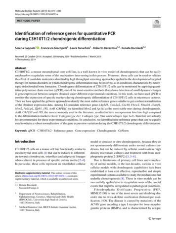
3480Molecular Biology Reports (2019) 46:3477–3485Fig. 1 Chondrogenic differentiation of C3H10T1/2 cells.Representative C3H10T1/2micromass cultures after 1, 3, 6and 13 days of BMP2 treatment.Cells were fixed and stainedwith Alcian blue. DM, differentiation mediumStudy of 12 candidate reference genesIn parallel, cells cultured in the same conditions and blockedat the different time points were used to identify the bestreference genes suitable to study gene expression variation during differentiation of C3H10T1/2. RNA quality andconcentration (Fig. S1) were checked for all the samplesderiving from two different groups of experiments and theexpression of a panel of 12 reference genes was evaluated.qPCR results were processed with the IQ5 software and Cqvalues were exported to be analysed with q base software(Table S2). Amplification efficiency, calculated from standard curves was between 1.9 and 2.10 for all genes, corresponding to 90–110% which represents the recommendedrange (Table S1).GeNorm study was applied following the instruction ofthe software. GeNorm M value represents reference genestability and according to the geNorm manual, candidategenes with lower M value have to be considered more stable.The graph starts with the least stable gene on the left sideand ends with the most stable gene on the right part, with athreshold of 0.5 between stable and unstable genes. Therefore, all the tested reference genes appeared to be stable uponthe different conditions and thus suitable for gene expressionanalysis at different time points of C3H10T1/2 chondrogenicdifferentiation (Fig. 2a). However, the best candidate reference genes identified were Mon2 and Ap3d1, whereas theless stable were ActB and GAPDH, curiously among themost commonly used in the literature. The geNorm V valuesuggests the minimum number of reference genes requiredfor normalization. The threshold value is 0.15, below whichthe inclusion of an additional control is not required [22].As shown in Fig. 2b, all the reference genes combinationhas Vn/n 1 0.15 (n optimal number of reference genes),suggesting that a combination of two reference genes wassufficient for a correct normalization.after 6 days of chondrogenic differentiation and processed forqPCR assay.The expression level of markers associated with chondro-differentiation and cartilage formation (Sox9, Col2a1,Col10a1) was obtained by the Ct Method. In this evaluation,various reference genes were employed, including all singlecandidate reference genes, a combination of the most stableones identified in this study, calculated by the geometric average of Mon2&Ap3d1, Ap3d1&Fbxw2, Mon2&Ap3d1&Fbxw2(indicated as M&A, A&F, M&A&F), and the geometric average of all 12 reference genes Ct values, (designed as ‘All’).As shown in Fig. 3 up-regulation of the chondrogenic genesSox9, Col2a1, Col10a1 was demonstrated by all normalizationstrategies. As a control, we evaluated also the expression ofCol1a1, a gene not associated with cartilage differentiation andexpected to be downregulated in micromass cultures inducedby BMP2 [28]. As shown in Fig. 3, the decrease in Col1a1expression upon differentiating conditions does not appear significant when ActB is applied as normalizing gene, comparedto ‘All’ and to other single or combined reference genes.Comparison of relative expression of 12 candidatereference genesIn order to select the proper reference genes with transcriptionlevel similar to that of target genes, Ct values deriving fromqPCR assay on RNA samples at T0 and T6, were normalizedagainst Fbxo38, which showed the lowest expression level.As reported in Fig. 4, relative expression of most of the reference genes have similar magnitude, whereas ActB, GAPDHand 18S showed the highest expression in C3H10T1/2 cells.This analysis suggested the exclusion of ActB, GAPDHand 18S because their transcription levels were too far fromthat of target genes and this might affect the accuracy of thenormalization process.DiscussionExpression profile of the differentiation markersnormalized against the different reference genesTo validate geNorm analysis and reference genes selection,C3H10T1/2 micromass cultures were harvested at time 0 and13qPCR is one of the most sensitive method to evaluate theexpression level of genes of interest under different experimental conditions, and thus in order to obtain reliable resultsit is crucial to select the suitable reference genes [26, 29–34].
Molecular Biology Reports (2019) 46:3477–34853481Fig. 2 GeNorm analysis of12 candidate reference genes.a geNorm M value indicatesreference genes stability. Thegenes with geNorm M valuebelow 0.5 can be considered forthe normalization and the loweris the value, the more stable arethe genes. b GeNorm V valuerepresents the pairwise variation (n/n 1) and indicates theminimum number of referencegenes required for normalization. Values below 0.15 indicatethe optimal number (n) of reference genes to be considered. Inthis study, a combination of twogenes was sufficient for a correct normalization, V2/3 0.082In the last years, several algorithms were developed toidentify the optimal reference genes in different cell types/tissues or treatment conditions. GeNorm [22], BestKeeper[23], and NormFinder [24] are the most frequently applied tothis purpose and are reported to usually show similar results[35, 36].To the aim of using C3H10T1/2 cells as an in vitro modelof chondro-like differentiation [3], we applied the geNormsoftware to identify the most stable references genes suitablefor normalization during the in vitro differentiation processof these cells. GeNorm has been developed according to thenormalization strategy described by Vandesompele et al.,2002 [22], conceived to identify the most stably expressedcontrol genes in a given set of tissues, and to determine theminimum number of genes required to calculate a reliablenormalization factor.In our study, we used a commercial kit of 12 murine‘house-keeping’ genes, specifically designed by PrimerDesign for geNorm studies, and we performed two biologicalreplicates of C3H10T1/2 chondrogenic differentiation after1, 3, 6, and 13 days.The first parameter generated by the g eNormPLUS analysisis geNorm M, which evaluates expression stability of candidate reference genes between samples. M value representsthe average pairwise variation of a particular gene with allthe other control genes: the lowest M value indicates themost stable gene expression. The cut-off value is 0.5 andall the genes we analyzed in this study could be consideredpotential reference genes.According to the MIQE guidelines, an accurate normalization process relies on the use of two or more referencegenes and geNorm V value (pairwise variation, V (n/n 1) coefficient) helps to determine the number of them required foroptimal normalization. The suitable number of referencegenes “n” is found when V(n/n 1) drops below 0.15.As shown in Fig. 2, V 2/3 was less than 0.15 (V2/3 0.082)and two reference genes were sufficient for a correct normalization, ideally Mon2 and Ap3d1.13
3482Fig. 3 Fold-change expressionof chondrogenic differentiationmarkers, normalized againstdifferent reference genes after6 days of differentiation. aSox9, b Col2a1, c Col10a1, dCol1a1. Relative expressionwas calculated by Ct Methodand histograms representaverage Standard Deviationof three independent biological replicates. The statisticalsignificance was evaluated byStudent’s T-test analysis with*P 0.0513Molecular Biology Reports (2019) 46:3477–3485
Molecular Biology Reports (2019) 46:3477–34853483Fig. 4 Relative expression ofthe 12 reference genes. Cellswere analysed after 6 days ofdifferentiation. Gene expressionvalues were calculated using Ct Method against Fbxo38,that showed the lowest expression. Histograms representaverage and standard deviationof three independent biologicalreplicates. Y axis was on a l og10scaleTo validate geNorm analysis, we evaluated the expression levels of markers specific for chondrogenic differentiation of C3H10T1/2 cells at T0 and after 6 days of micromass culture in inductive medium containing BMP2. Sox9,Col2a1 and Col10a1 showed high expression level afterBMP2 treatment and all normalization strategies showedcomparable expression level. In particular, the combination of Mon2 and Ap3d1, Ap3d1 and Fbxw2, and Mon2,Ap3d1 and Fbxw2, gave similar results to the normalization obtained with ‘All’, which is considered as the mostaccurate strategy.Furthermore, it is known that for accurate gene expression studies the expression level of reference genes shouldbe similar to that of target genes [26, 37]. Therefore, wecompared the expression profile of all the 12 referencegenes in our cell model. Our analysis suggested that ActB,GAPDH and especially 18S genes should not be considered for normalization because their expression level significantly diverges from that of the selected differentiationmarkers in C3H10T1/2 cells.Moreover, the use of ActB as reference gene failed toefficiently detect the expected down-regulation of Col1a1expression in differentiating conditions [28].This result suggested the exclusion of ActB, one of themost applied in expression studies, from the best reference genes group, because during the evaluation of treatment effects, also small gene expression differences maybecome relevant. All the other reference genes and combination analysed in this work, showed similar expressionlevels and pass our validation.ConclusionsqPCR is a potent method to study gene expression profilesin different experimental settings. Reliability of results isstrongly dependent on the normalization procedure; reference genes have to be carefully selected for every tissue/cell types and for the different experimental conditions.We applied the geNorm analysis to select the best reference genes in a widely used in vitro model of chondrogenesis. According to our analysis, the most commonlyused reference genes, such as ActB, GAPDH and 18S,are not actually the best candidates for normalization.We suggest to use a different combination of at least twogenes selected from the panel we tested, such as Mon2 andAp3d1 when normalizing gene expression profiles duringchondrogenesis in C3H10T1/2 cells.Acknowledgements This study was funded by Fondazione Telethon(Grant No. GGP15196 to RR), by FOP Italia Onlus, “Ricerca corrente”and Italian Ministry of Health.Author contributions SC conceived, designed, performed and analysedqPCR experiments, and wrote the manuscript; FG and LT performeddifferentiation assays; RR and RB supervised the experiments andmanuscript preparation.Compliance with ethical standardsConflict of interest The authors declare that they have no conflict ofinterest.13
3484Open Access This article is distributed under the terms of the Creative Commons Attribution 4.0 International License (http://creat iveco mmons .org/licen ses/by/4.0/), which permits unrestricted use, distribution, and reproduction in any medium, provided you give appropriatecredit to the original author(s) and the source, provide a link to theCreative Commons license, and indicate if changes were made.Molecular Biology Reports (2019) 46:3477–348515.16.17.References1. Reznikoff CA, Brankow DW, Heidelberger C (1973) Establishment and characterization of a cloned line of C3H mouseembryo cells sensitive to postconfluence inhibition of division.Cancer Res 33(12):3231–32382. Ahrens M, Ankenbauer T, Schröder D, Hollnagel A, Mayer H,Gross G (1993) Expression of human bone morphogenetic proteins-2 or -4 in murine mesenchymal progenitor C3H10T1/2cells induces differentiation into distinct mesenchymal cell lineages. DNA Cell Biol 12(10):871–8803. Denker AE, Haas AR, Nicoll SB, Tuan RS (1999) Chondrogenicdifferentiation of murine C3H10T1/2 multipotential mesenchymal cells: I. Stimulation by bone morphogenetic protein-2 inhigh-density micromass cultures. Differentiation 64(2):67–764. Fakhry M, Hamade E, Badran B, Buchet R, Magne D (2013)Molecular mechanisms of mesenchymal stem cell differentiationtowards osteoblasts. World J Stem Cells 5(4):1365. Haas AR, Tuan RS (1999) Chondrogenic differentiation ofmurine C3H10T1/2 multipotential mesenchymal cells: II.Stimulation by bone morphogenetic protein-2 requires modulation of N-cadherin expression and function. Differentiation64(2):77–896. Izzo MW, Pucci B, Tuan RS, Hall DJ (2002) Gene expressionprofiling following BMP-2 induction of mesenchymal chondrogenesis in vitro. Osteoarthritis Cartilage 10(1):23–337. Roy R, Kudryashov V, Doty SB, Binderman I, Boskey AL(2010) Differentiation and mineralization of murine mesenchymal C3H10T1/2 cells in micromass culture. Differentiation79(4–5):211–2178. Takács R, Matta C, Somogyi C, Juhász T, Zákány R (2013) Comparative analysis of osteogenic/chondrogenic differentiation potential in primary limb bud-derived and C3H10T1/2 cell line-basedmouse micromass cultures. Int J Mol Sci 14(8):16141–161679. Shore EM, Xu M, Feldman GJ, Fenstermacher DA, Cho TJ,Choi IH et al (2006) A recurrent mutation in the BMP type Ireceptor ACVR1 causes inherited and sporadic fibrodysplasiaossificans progressiva. Nat Genet 38(5):525-7. Epub 2006 Apr23. Erratum in: Nat Genet. 2007 Feb;39(2):27610. Kaplan FS, Xu M, Seemann P, Connor JM, Glaser DL, Carroll L et al (2009) Classic and atypical fibrodysplasia ossificansprogressiva (FOP) phenotypes are caused by mutations in thebone morphogenetic protein (BMP) type I receptor ACVR1.Hum Mutat 30(3):379–39011. Cappato S, Giacopelli F, Ravazzolo R, Bocciardi R (2018) Thehorizon of a therapy for rare genetic diseases: a “druggable” futurefor fibrodysplasia ossificans progressiva. Int J Mol Sci 19(4):98912. Medici D, Olsen BR (2012) The role of endothelial-mesenchymal transition in heterotopic ossification. J Bone Miner Res27(8):1619–162213. Wosczyna MN, Biswas AA, Cogswell CA, Goldhamer DJ (2012)Multipotent progenitors resident in the skeletal muscle interstitium exhibit robust BMP-dependent osteogenic activity and mediate heterotopic ossification. J Bone Miner Res 27(5):1004–101714. Dey D, Bagarova J, Hatsell SJ, Armstrong KA, Huang L,Ermann J et al (2016) Two tissue-resident progenitor 1.32.drive distinct phenotypes of heterotopic ossification. Sci TranslMed 23(366):366ra163Lees-Shepard JB, Goldhamer DJ (2018) Stem cells and heterotopic ossification: lessons from animal models. Bone 109:178–186Katagiri T, Tsukamoto S, Kuratani M (2018) Heterotopic boneinduction via BMP signaling: potential therapeutic targets forfibrodysplasia ossificans progressiva. Bone 109:241–250Bustin SA, Benes V, Garson JA, Hellemans J, Huggett J, KubistaM et al. (2009) The MIQE guidelines: minimum informationfor publication of quantitative real-time PCR experiments. ClinChem 55(4):611–622Chapman JR, Waldenström J (2015) With reference to reference genes: a systematic review of endogenous controls in geneexpression studies. PLoS ONE 10(11):e0141853Huang X, Xu J, Huang M, Li J, Dai L, Dai K et al. (2014)Histone deacetylase1 promotes TGF-β1-mediated early chondrogenesis through down-regulating canonical Wnt signaling.Biochem Biophys Res Commun 453(4):810–816Howard M, Tuan RS, Wallis GA (2016) The function and interrelationship between GDF5 and ERG-010 during chondrogenesis in vitro. In Vitro Cell Dev Biol Anim 52(2):182–192Dai J, Yu D, Wang Y, Chen Y, Sun H, Zhang X et al (2017)Kdm6b regulates cartilage development and homeostasisthrough anabolic metabolism. Ann Rheum Dis 76(7):1295–1303Vandesompele J, De Preter K, Pattyn F, Poppe B, Van Roy N,De Paepe A et al. (2002) Accurate normalization of real-timequantitative RT-PCR data by geometric averaging of multiplein
C3H10T1/2, a mouse mesenchymal stem cell line, is a well-known in vitro model of chondrogenesis that can be easily . established to have cost effective, reproducible and simple experimental systems available to study the mechanisms that underlie chondrogenesis []. These in vitro models can be 8 . T1, T3, T6 and T13 time points by using the .