Transcription
ORIGINAL ARTICLENovel Impactor and Microsphere-Based Assay Used toMeasure Containment of Aerosols Generated in a FlowCytometer Cell SorterStephen P. Perfetto,1* Phillip J. Hogarth,2 Simon Monard,3 Ben Fontes,4 Kristen M. Reifel,5Brandon K. Swan,5 Jan Baijer,6 Evan R. Jellison,7 Geoffrey Lyon,8 Patty Lovelace,9Richard Nguyen,1 David Ambrozak,1 Kevin L. Holmes101Vaccine Research Center, NIAID, NIH,Bethesda, Maryland2Animal & Plant Health Agency (APHA,Surrey, UK3Walter and Eliza Hall Institute, Victoria,3052, Australia4Yale School of Public Health, NewHaven, Connecticut5National Biodefense Analysis andCountermeasures Center, Frederick,Maryland6CEA-DSV-IRCM, Fontenay-aux-Roses,France7UConn Health, Farmington, Connecticut8Yale University Environmental Health &Safety, New Haven, Connecticut9The Institute for Stem Cell Biology andRegenerative Medicine, Stanford,California10Research Technologies Branch,NIAID, Bethesda, MarylandReceived 29 June 2018; Revised 25September 2018; Accepted 16October 2018Grant sponsor: Department of HomelandSecurity (DHS) Science and TechnologyDirectorate (S&T), Grant numberHSHQDC-15-C-00064Additional Supporting Information may befound in the online version of this article.*Correspondence to: Stephen P. Perfetto,Vaccine Research Center, NIAID, NIHCytometry Part A 2018 AbstractToday’s state-of-the-art cell sorting flow cytometers are equipped with aerosol containment systems designed to evacuate aerosols from the sort chamber during a sort.This biosafety device is especially important when the sort operator is sorting infectious or potentially infections samples. Hence, it is critical to evaluate the performance for this system in normal operation and in “failure” mode to determine theefficacy of containment. In the past decade, the most popular published method forevaluating containment has been the Glo-Germ bead procedure. These highly fluorescent and multisize particles can easily be detected on a microscope slide and enumerated using a fluorescent microscope. Collecting particles on this slide is accomplishedusing an Aerotech impactor. This sampler collects potentially escaping aerosols fromthe sort chamber before enumerating any particles. Although the Glo-Germ procedure has been adopted by many labs, there are several drawbacks with the procedurethat have limited its adoption by cell sorter laboratories: The Aerotech impactor is areusable device that requires rigorous cleaning between measurements. The surfacearea of the collection slide is large and difficult to scan on a fluorescence microscope.These beads produce a wide variation in sizes resulting in inconsistency in flow rates.Here, we describe a novel and replacement method utilizing a Cyclex-d impactor andDragon Green beads. This method was compared for sensitivity of detection ofescaped aerosols with a published method for aerosol detection which utilizes a UVAPS aerodynamic particle sizer and a UV-excitable dye. One of the advantages of theCyclex-d system is the narrow-defined field of collection as compared to the standardGlo-Germ bead procedure, this means a smaller sampling area is used in the Cyclexd impactor as compared to the AeroTech impactor. In addition, the sensitivity ofdetection was found to be better using the Cyclex-d collection device as compared tothe standard Glo-Germ bead procedure. 2018 International Society for Advancement ofCytometry Key termsinfectious cell sorting; biosafety; aerosol measurement; shared resource lab operationsITis well established that stream-in-air cell sorters may produce high concentrations of aerosols during fail mode (i.e., partial nozzle obstruction and with streamdeviations; FM) (1). Due to the possible exposure of the operator to these aerosolswhile sorting potentially infectious samples, cell sorting is considered a high-risk laboratory procedure. Instrument manufacturers have designed aerosol containmentsystems to mitigate this risk. Therefore, as recommended in the latest InternationalSociety for the Advancement of Cytometry (ISAC) Cell Sorter Biosafety Standards(2), aerosol containment testing of these systems must be performed at intervalsdetermined by a risk assessment to validate containment. These published standards
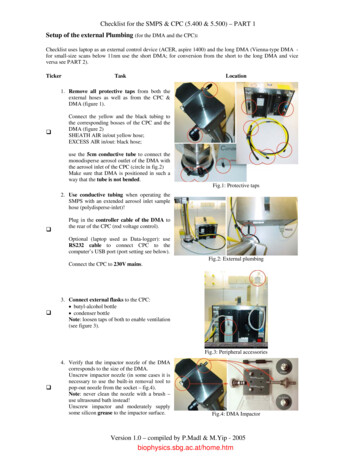
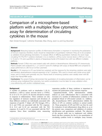
ORIGINAL ARTICLEBethesda, MarylandEmail: sperfetto@mail.nih.govPublished online in Wiley Online Library (wileyonlinelibrary.com)reference a method for containment testing measuring therelease of Glo-Germ beads (melamine copolymer resin beads)collected in an Aerotech impactor (3). However, this methodhas several drawbacks and alternate methods have beenactively pursued with the goal of establishing a sensitive assayfor aerosol containment testing.Several other methods for the evaluation of containmenthave been described previously. These containment assayscan be broadly characterized as utilizing either active(e.g., impactor) or passive deposition methods (3–7) or realtime measurement methods (1,8,9). Real-time measurementmethods offer the advantage of immediate determination ofparticle counts, but require fluorescence detection instrumentsto distinguish ambient air particles from cell sorter derivedaerosols to prevent false positives (1). Such instruments(e.g., UV-APS, TSI, Inc.) represent significant additionalexpense and service considerations. In addition, some of theseinstruments were designed to measure solid particles such asfungal spores, dust particles, and other airborne allergens.Optical particle counters such as the Fluke 985 Air Qualitymeter used by Xie and Waring (9), for example, determineparticle size by measuring light scatter intensity and is calibrated using polystyrene latex beads. This instrument will,therefore, underestimate particle size if used to measure liquidparticles (i.e., aerosolized sheath fluid particles that do notcontain fluorescent beads) due to the lower index of refraction of liquid particles versus polystyrene beads (10,11). Inaddition, this instrument cannot distinguish ambient air particles from cell sorter generated aerosols.Although the capture of aerosols containing highly fluorescent Glo-Germ beads seemed to provide a simple and sensitive assay for containment testing, there are severaldrawbacks with the assay that have forced a re-evaluation ofcurrent containment methods. These include the following:the size distribution of these particles is very large, resultingin inconsistency in cell sorter flow rates; the Aerotech collection device is not used as designed. Specifically, the Aerotechimpactor is a viable impactor, which is designed to capturebio-aerosols onto an agar-coated plate for subsequent growthanalysis, and has a cutoff diameter, or d50, of 0.65 μm (12).However, as used in cell sorter containment testing, the glassslide is positioned on an empty petri dish in the Aerotechimpactor; this setup changes the jet-to-plate distance and willchange the d50, which is related to this distance (13). It is possible that the altered d50 may not be within the desirable aerodynamic diameter (AD) range for cell sorter aerosolmeasurements causing an underestimation of escaped aerosols. In this regard, a preliminary experiment comparingyellow–green (YG) fluorescent beads, collected in a Cyclex-dimpactor with Glo-Germ beads collected in the AeroTech2DOI: 10.1002/cyto.a.23680 2018 International Society for Advancement of Cytometryimpactor showed threefold higher collection for the Cyclex-dat identical flow rates (Fig. 1). Finally, and perhaps the biggestdrawback with the Aerotech impactor is that it must becleaned thoroughly after every test to eliminate carry-overparticles from being detected, which could lead to false positive results if not cleaned properly.This work was undertaken to develop a novel cell sorteraerosol containment assay that met the following criteria:(1) the assay must be performed and results available withinthe same day and before cell sorter operation; (2) the sensitivity must be high, and the assay must be validated using analternative aerosol testing procedure using other establishedinstruments; (3) equipment and supplies must be affordableand utilize commonly available flow cytometry laboratoryequipment; (4) accuracy and specificity must be high, with little false positives or ambient air background readings for bothBiological Safety Cabinet (BSC) enclosed and nonenclosed cellsorters; (5) efficiency of aerosol collection must be highwithin the AD range of cell sorter aerosol production.This article describes the use of a nonviable disposableimpactor and uniform fluorescent microspheres as an alternative to the currently recommended containment assay. Theresults of this work describe the development of a rapid andefficient method for testing containment of aerosols generatedFigure 1. Cyclex-d cassette collection of YG beads (gray bars)compared to the collection of Glo-Germ beads using Aerotechimpactors (black bars). Data shows the increase in collection ofboth particles with increase sample rate. In addition, the datashows the collection of YG beads by the Cyclex-d cassette is 3xmore efficient as compared to the Aerotech impactor and thecollection of Glo-Germ particles.Novel Cell Sorter Containment Assay
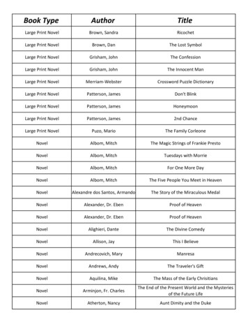
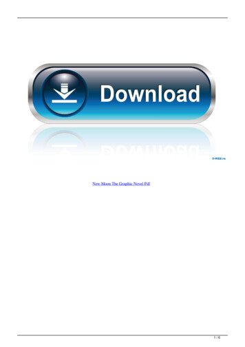
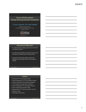
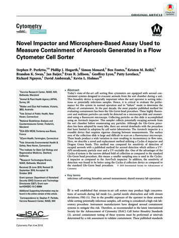
ORIGINAL ARTICLEby cell sorters. The sensitivity of this method was evaluatedby utilizing the UV-APS particle sizer (TSI Inc.) and samplecontaining UV excitable dye.MATERIALS AND METHODSBeads and ImpactorInternally fluorescent 1.0 μm Dragon Green (DG) beads(Excitation (Ex) 480 nm; Emission (Em) 520 nm; Bangs Laboratories) and 0.75, 1.0, and 2.0 μm Fluoresbrite yellow green(YG) beads (Ex 441 nm; Em 486 nm; Polysciences, Inc.)were used.Cyclex-D Setup and Particle DetectionCyclex-d impactor sampling cassettes, MegaLite pump,and Rotameter (Environmental Monitoring Systems, Charleston, SC) were used to collect aerosol samples. Figure 2a showsthe location and distance of the Cyclex-d impactor (15 cm)relative to the sorting chamber of a FACSAria. Note the distance used in the collection for this article is different thanthe collection distance used in the routine procedure (see(a)Appendix: Standard Operating Procedure: Aerosol Containment Measurement), which is set at 5 cm for maximal sensitivity. The impactor was connected to a vacuum regulator(Fig. 2b) and the vacuum set at a constant vacuum equal to20 l/min. Figure 2c show an example of the histograms generated after 10 min of collection of Dragon Green beads at arate of 50,000 beads/s. The Appendix: Standard OperatingProcedure: Aerosol Containment Measurement, outlines acomplete description of the routine procedure for aerosolmeasurement using the Cyclex-d impactor and Dragon Greenbeads.Microscope and Slide MeasurementDragon Green microspheres were collected by Cyclex-dimpactor sampling cassettes and counted using a NikonEclipse E400 Epi-Fluorescent microscope with a 450–490 nmexcitation filter (Fig. 3a). To optimize visualization and quantification of DG beads after collection, the cover slip insidethe cassette was removed and placed (adhesive side down)onto a gridded microscope slide (Electron Microscopy(b)(c)Figure 2. The location and angle of the Cyclex-d impactor in front of the cell sorting chamber of the FACSAria cell sorter was placed at15 cm from the sort chamber as shown in 2a. The tubing from the impactor is attached to the MegaLite pump and rotameter as picturedin 2b. Figure 2c is an example of the collection of dragon green beads after a 10 min collection at 50,000 beads/s as measured in FITCdetector using a 515/20 nm bandpass filter (labeled as the 515-A detector).Cytometry Part A 20183
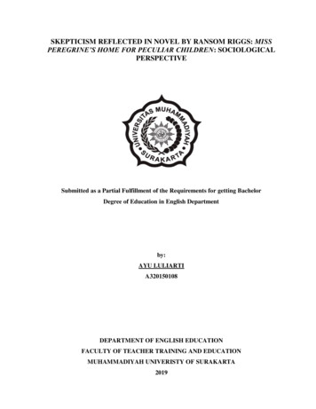
ORIGINAL ARTICLEFigure 3. This figure shows the removal of coverslip from the Cyclex-d impactor (a and b) and the examination of the beads measured inthe fluorescent microscope (c). Note the grid lines on the microscope slide provides a focal plane to ensure the focus of the dragon greenbeads, which is helpful when scanning slides with very few or no beads collected.Sciences #63405–02). As a critical note, it is important to havethe gridded side up before attaching the cover slip from theCyclex-d impactor (Fig. 3b). This technique ensured the focalplane of the DG beads is the same as the grid lines, facilitating viewing of slides with few or no beads (Fig. 3c).UV-APS and Aerosol Concentrations and ADMeasurementsAerosol concentrations and the AD measurements wereconducted on a BD FACSAria II model cell sorter(BD biosciences, San Jose, CA) operating at 70 psi(482,633 Pa), using an Aerosol Particle Sizer (APS: UV-APSModel 3314; TSI, Shoreview, MN) equipped with a UV laser(350 nm). Sorter generated aerosols were distinguished fromambient particles with a UV-excitable dye (Clear Blue Fluorescent Water Tracer Dye [CBD]; Risk Reactor, Santa Anna,CA) that was added to the sample tube. In some experiments,aerosol measurements were conducted on a FACSAriaenclosed in a Class II BSC abrogating the use of the UV dye.Use of the UV dye, methods and analysis of data were performed as previously described (1). For containment testing,large number of aerosols were created by covering the wastetrough with a small piece of tubing while running the wastestream as previously described (14). This was also known asthe Fail Mode (FM) which also simulated the event of cloggedflow cell tip.4Evacuation Airflow Restriction TestsThe Buffalo filter AMS (Medtec Devices Inc, BuffaloNY) was operated at the manufacturer’s recommended settingof 20% of maximum vacuum. AMS airflow reduction experiments utilized a PVC valve connected inline of the AMS hosejust prior to its connection to the sorter AMS port (Fig. 4).Prior to containment tests, airflow (ft/min) was measured atthe end of the AMS hose using a hot wire anemometer(VelociCal Air Velocity Meter Model 9535; TSI, Shoreview,MN). Measurements were in ft/min and then were convertedto CFM (ft3/min) using the standardized airflow calculation,as based on the AMS tubing diameter of 1.25 in. (3.18 cm) atthe point of measurement.RESULTSDetermination of Optimal Microsphere DiameterThe goal of any cell sorter containment assay is to accurately and reliably detect aerosols that have escaped from thepoint of generation, that is, sorting or collection chambers.Since it has been determined that a high concentration ofaerosols with an AD in the range of 1–3 μm are produced bycell sorters in FM (1), detection of aerosols in this range isessential. The initial use of the Cyclex-d cassettes and fluorescent microspheres in a cell sorter containment assay, as firstdescribed (15) utilized YG microspheres of graded sizes 0.5,1.0, 2.0, 6.0, 10.0, and 20.0 μm. Since microspheres can neverNovel Cell Sorter Containment Assay
ORIGINAL ARTICLEFigure 4. This figure shows the installation of the restrictionvalve (yellow arrow) in the vacuum lines of the aerosolmanagement system (FACSAria) used in airflow reductionexperiments. As the valve is turned, the airflow is reduced, whichwas accurately measured at the opened end of the tube attachedto the sort chamber using a hot wire anemometer as described inthe methods section.occupy an aerosol smaller than its own physical size, thephysical size is the starting point for AD and, therefore, itwas reasoned that a smaller, single diameter microspherealone would be sufficient. However, physical size of microspheres does not necessarily equate with AD, since this isdependent on size, shape, and density of the particle. Todetermine the size of the microsphere that occupied the largest range of aerosols generated by a sorter, YG microspheresof 0.75, 1.0, and 2.0 μm were run separately as samples on aFACS Aria enclosed in a BSC in FM with AMS off, andresulting aerosols were measured with the UV-APS.Although, the excitation maximum of the YG microspheres is441 nm, the 351 nm UV laser of the UV-APS was capable ofexcitation of the microspheres due to the very broad excitation spectrum of the microspheres. The results (Fig. 5)showed that the smallest size of YG microsphere tested occupied the largest range of AD aerosols and that the microspheres were always contained in aerosols of AD greater thanthe physical bead size. However, the ability to reliably detectmicrospheres when visualized on a slide is critical and it wasfound that the 0.75 μm microspheres were more difficult todetect at low magnification. Since 1.0 μm microspheres areeasier to detect, and were found in aerosols of AD of interest,that is, 1 to 3 μm, 1.0 μm microspheres were used for subsequent experiments.The frequency of aerosols occupied by the fluorescentmicrospheres was measured to be in the range of 0.13% to0.3% of total (Fig. 5a–c). However, it was hypothesized thatthe frequency of occupied aerosols will vary dependent onPoisson statistics, concentration of microspheres, and thesample flow rate (16). This was verified by measuring aerosolsCytometry Part A 2018with the UV-APS, generated by running YG beads as a sample at two different event rates, with the instrument in FM,aerosol management system off, and sort chamber ajar.Figure 6 shows the frequency of occupied drops ranged froma mean of 0.16% at 75,000 events/s to 0.21% at 82,000 events/s. Also shown are measurements under the same conditions(75,000 events/s) but with the sort chamber door closed.Equally critical to a robust containment assay, is the ability to wash or remove beads subsequent to containment testing. Unfortunately, the ability to easily remove the YG beadsfrom the fluidics of the cytometer proved problematic. Microspheres manufactured by Bangs Laboratories (Fishers, IN),hard dyed with Dragon Green (DG) fluorescent dye werefound to be more hydrophilic and much easier to clean andremove completely from the instrument fluidics system. Inaddition, these were also much brighter than the YG microspheres when viewed under 20x magnification as described inthe methods section. Therefore, due to the brightness factorand the completeness of the wash/removal of these microspheres for containment testing procedures, all subsequentexperiments were performed using 1.0 μm DG microspheres.Development and Validation of Containment AssayAs detailed in the published containment assay usingGlo-Germ beads (3), the procedure for determining the containment efficiency of a cell sorter AMS uses an impactorplaced in close proximity to the sort chamber to collect aerosols during fail mode while running the fluorescent microspheres as a sample. Use of the Cyclex-d cassettes and DGmicrospheres showed that under standard operating procedures (routine method) as described in the methods section,greater than 250 beads were detected when the AMS (positivecontrol test) is off and the instrument is set to FM as compared to zero particles detected under the same conditions(negative control test, data not shown). However, it wasimportant to establish the level of sensitivity of detection inthis assay or the lowest level of concentration of aerosolsescaping from the cell sorter. Experiments were conducted tocorrelate the aerosol concentration (using the UV-APS) withDG microsphere counts, under conditions where the AMSairflow was reduced using a restriction device (see methodssection). In these experiments, at each AMS airflow condition,FM was initiated with CBD (UV tracer dye) as a sample andconcentration of UV (nonambient) aerosols was determinedusing the UV-APS after 10 min. collection time. Subsequently, DG beads were substituted as a sample, and collection with the Cyclex-d cassettes was performed over the sametime period. This permitted the concentration of the escapedaerosols (UV aerosols) from the sort chamber to be correlated with DG bead detection. The results (Fig. 7) showed thatthe lowest aerosol concentration in which DG beads weredetected ranged from 0.026 to 0.04 aerosols/cm3. However,since there were experiments in which no DG beads weredetected within this aerosol concentration range and DGbeads were always detected above 0.04 aerosols/cm3, this concentration was established as the level of sensitivity. It isimportant to note that DG bead-aerosols were measurable5
ORIGINAL ARTICLE(a)(b)(c)Figure 5. UV-APS analysis of YG 0.75 μm (a), 1.0 μm (b), and 2.0 μm (c) microspheres run separately as samples on a FACS Aria enclosedin a Class II biosafety cabinet showing that the smallest microsphere occupied the largest AD range of aerosols. Figure 1a. Represents0.75 μm beads with a minimum AD 0.9 μm; mean AD 3.3 μm and % positive fluorescence 0.20. Figure 1b. Represents 1.0 μm beadswith a minimum AD 1.1 μm; mean AD 3.4 μm; and % positive FL 0.13. Figure 1c. Represents 2.0 μm beads with a minimum AD 2.1;mean AD 3.5; and % positive FL 0.30.only when the AMS airflow was reduced to 4.43 CFM(0.13 m3/M). This represents a 70% reduction in normalAMS airflow (i.e., 15.34 CFM or 0.43 m3/M). In addition, itwas shown that, similar to experiments with YG beads, therewas a direct correlation to event rate and the detection of DGbeads, indicating that at higher flow rates more aerosols containing particles can be captured by the Cyclex-d (Fig. 8).DISCUSSIONcriteria. In brief, these criteria are: (1) Same-day analysis andresults; (2) High sensitivity with independent validation;(3) Affordable equipment and supplies; (4) High accuracyand specificity with low background; (5) High efficiency ofaerosol collection within the desired AD range.An assay to verify cell sorter aerosol containment mayutilize any of the widely available aerosol collection methodologies of impaction, filtration and impingement that meet theabove criteria. It was determined that collection of 1 μm DGmicrospheres provided an appropriate surrogate of aerosolsAs stated earlier, this work was undertaken to develop anovel cell sorter aerosol containment assay that met specificFigure 6. Aerosols containing YG beads were generated whilethe sort chamber door open (ajar) at two event rates; 75,000 and82,000 beads/s. Aerosols containing YG beads were alsogenerated with the sort chamber door closed (CL) as a controltest. The data shows the increase in aerosols containing YGbeads as measured by the UV-APS, as a function of event rate;the higher the rate the greater the number of aerosols containingYG beads.6Figure 7. Dragon green beads were collected with the Cyclex-d,subsequent to aerosol concentration measurements determinedwith the UV-APS and CBD, under the same conditions of air flowrestriction. Data shows that at an aerosol concentration of 0.04number/cm3 (or above), corresponding to an airflow restriction of4.43 CFM (0.13 m3/M), dragon green microspheres were detectedon the slide of the impactor (numbers beside dots; experimentswith no beads detected are indicated in red). This demonstratedthat the sensitivity of the Cyclex-d/DG bead assay to be at anaerosol concentration of 0.04 number/cm3.Novel Cell Sorter Containment Assay
ORIGINAL ARTICLEFigure 8. This figure shows the linear relationship (r 0.99) ofaerosols containing dragon green beads as a function ofincreased event rate. Dragon green microspheres were capturedusing the Cyclex-d cassettes when the cell sorter was placed inFM and the impactor was used under standard collectionprocedures as described in the methods section.within the size range released from the sorter (Fig. 5). Capture of these fluorescent microspheres with a nonviableimpactor, such as the Cyclex-d, in which counting is easilyperformed on a microscope is appropriate since culturabilityand viability are not a concern. Use of fluorescent microspheres and disposable cassettes also meets the requirementfor low background measurements, suitable for both BSCenclosed and nonenclosed cell sorters. In addition, for nonviable impactors of this type, collection efficiency approaches100% when the AD is greater than the impactor d50 (17). Thed50 of the Cyclex-d is 1 μm (18) and the median AD of theaerosols in validation experiments was 1.6 μm (data notshown), in agreement with previously published reports of1.7 μm (1) and meeting the criteria of high collection efficiency of cell sorter derived aerosols.Sequential measurements of cell sorter derived aerosolswith the UV-APS/UV dye and the Cyclex-d/microspheresshowed the lowest level of detection of the Cyclex-d to be0.04 aerosols/cm3 (Fig. 7). This concentration approachesbackground aerosol measurements of 0.022 UV aerosols/cm3 measured in these same experiments (data notshown), demonstrating the high sensitivity of the DG microsphere/Cyclex-d assay.In addition, this high level of sensitivity was measured inspite of the low frequency of microsphere-occupied droplets(Fig. 5). This is most likely due to the higher sampling volume of air (200 l vs. 50 l; Table 1) captured by the Cyclex-dversus the UV-APS over the same collection time. In thisTable 1. Air Sampling Volume: Cyclex-d vs. UV-APSCOLLECTIONDEVICECyclex-dUV-APSRATETIMETOTAL VOLUME SAMPLED20 l/min5 l/min600 s600 s200 l50 lCytometry Part A 2018regard, sensitivity could likely be increased with the Cyclex-dand DG microspheres by increasing the collection time. Forbioaerosols, increasing collection time can be problematic dueto desiccation of the sample, but this is not an issue with theplastic DG microspheres. Figure 6 shows that sensitivity canbe increased by increasing the event rate due to an increase inthe percentage of microsphere-occupied droplets. However,increasing sensitivity by increasing the event rate will eventually plateau due to an increase in coincident events and Poisson distribution statistics.These results also illustrate the large amount of airflowloss (70% reduction of normal) required on this model of cellsorter before containment is compromised. This highlightsthe robustness of the AMS but also suggests that real-timemonitoring of AMS airflow could be an effective method ofmonitoring containment during operation of the cell sorterand could be an important adjunct to containment testing asrecommended by the ISAC Cell Sorter Biosafety Standards (2).The assay described here was validated on a BDFACSAria, but it could be easily adapted to other sorters. Themain considerations in adapting this assay to other instruments are the following: (1) Determine the measurementlocation of the Cyclex-d, which should be as close as possibleto the sort chamber; (2) Determine the optimal method forcreating a FM, mimicking partial nozzle obstruction with subsequent stream deviation; (3) Determine the best procedurefor a positive control; and (4) Perform assay at the highestsheath pressure that will be used, which will result in the largest concentration of aerosols.In summary, the Cyclex-d/DG microsphere assay fulfillsthe criteria for a robust cell sorter aerosol containment assayand is recommended as a replacement assay for the previously published Glo-Germ/Aerotech impactor collectionmethod (2).ACKNOWLEDGMENTSThe views and conclusions contained in this documentare those of the authors and should not be interpreted as necessarily representing the official policies, either expressed orimplied, of the DHS or S&T. In no event shall DHS, NBACC,S&T, or Battelle National Biodefense Institute have anyresponsibility or liability for any use, misuse, inability to use,or reliance on the information contained herein. DHS doesnot endorse any products or commercial services mentionedin this publication.FUNDING STATEMENTThis work was funded under Contract No. HSHQDC15-C-00064 awarded by the Department of Homeland Security (DHS) Science and Technology Directorate (S&T) for theoperation and management of the National Biodefense Analysis and Countermeasures Center (NBACC), a FederallyFunded Research and Development Center. The funders hadno role in study design, data collection and analysis, decisionto publish, or preparation of the manuscript. This work was7
ORIGINAL ARTICLEsupported by the Intramural Research Program of the Vaccine Research Program, NIH.LITERATURE CITED1. Holmes KL. Characterization of aerosols produced by cell sorters and evaluation ofcontainment. Cytometry A 2011;79A:1000–1008.2. Holmes KL, Fontes B, Hogarth P, Konz R, Monard S, Pletcher CH Jr, Wadley RB,Schmid I, Perfetto SP. International society for the advancement of cytometry cellsorter biosafety standards. Cytometry A 2014;85A:434–433.3. Perfetto SP, Ambrozak DR, Koup RA, Roederer M. Measuring containment of viable infectious cell sorting in high-velocity cell sorters. Cytometry A 2003;52A:122–130.4. Merrill JT. Evaluation of selected aerosol-control measures on flow sorters. Cytometry 1981;1:342–345.5. Schmid I, Hultin LE, Ferbas J. Testing the efficiency of aerosol containment duringcell sorting. Curr Protoc Cytom 2001;78–84, Chapter 3:Unit3.3.6. Oberyszyn AS, Robertson FM. Novel rapid method for visualization of extent andlocation of aerosol contamination during high-speed sorting of potentially biohazardous samples. Cytometry 2001;43:217–222.7. Wallace R, Aguila HL, Fomenko J, Price KW. A method to assess leakage from aerosolcontainment systems: Testing a fluorescence-activated cell sorter (FACS) containmentsystem using the radionuclide technetium-99m. Appl Biosaf J 2010;15:77–158.8. Ferbas J, Chadwick KR, Logar A, Patterson AE, Gilpin RW, Margolick JB. Assessmentof aerosol containment on the ELITE flow cytometer. Cytometry 1995;22:45–47.9. Xie M, Waring MT. Evaluation of cell sorting aerosols and containment by an optical airborne particle counter. Cytometry A 2015;87A:784–789.10. Rosenberg PD, Dean AP, Williams PI, Dorsey JR, Minikin A, Pickering MA,Petzoldand A, Petzold A. Particle sizing calibration with refractive index correctionfor light scattering optical particle counters and impacts upon PCASP and CDP datacollected during the Fennec campaign. Atmos Meas Tech 2012;5:1147–1163.11. Liu Y, Daum PH. The effect of refractive index on size distributions and light scattering coefficients derived from optical particle counters. J Aerosol Sci 2000;31:945–957.12. Vincent JH. Aerosol Sampling: Science, Standards, Instrumentation and Applications.Chichester, UK: Wiley & Sons, 2007;p. 480–481.13. Yao M, Mainelis G. Inve
Dragon Green beads. This method was compared for sensitivity of detection of escaped aerosols with a published method for aerosol detection which utilizes a UV-APS aerodynamic particle sizer and a UV-excitable dye. One of the advantages of the Cyclex-d system is the narrow-defined field of collection as compared to the standard