Transcription
General Introduction to ECGReading Assignment (p2-16 in PDF ‘Outline’)Objectives1. Practice the 5-step ‘Method’2. Differential Diagnosis: R & L axis deviation3. Differential Diagnosis: Poor R-wave progression4. Differential Diagnosis: Prominent Anterior Forces
Welcome to the “5-Step Method”ECG #:Mearurements:A Rhythm (s):Conduction:Waveform:Interpretation:V PR QRS QT Axis 1. Compute the 5 basic measurements: HR, PR interval, QRS duration, QT interval, Axis2. What’s the basic rhythm and other rhythm statements (e.g., PACs and PVC’s)3. Any conduction abnormalities (SA blocks, AV blocks (Types I or II), and IV blocks4. Waveform abnormalities beginning with P waves, QRS complexes, ST-T, and U waves5. Final interpretations: Normal ECG or Borderline or Abnormal ECG (list finalconclusions)
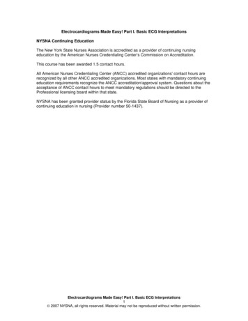
30 year old woman (explain the sequence of activation from sinus node to ventricularmuscle)What are ‘septal’ q-waves?1-1
****Mearurements:A 55PR 140QRS 100QT 4301-1Axis 80V 55Rhythm (s):Normal Sinus rhythmConduction:Normal SA, AV, IV conductionSequence of conduction: SA node (RA LA) AVnode His Bundle RBB &LBB LAF & LPF & LSF Purkinje network leftseptal surface (onset of QRS)Waveform: Normal P, QRS, ST, T; notenormal U waves inprecordial leads (*)Septal q‘s in II, III, aVF(onset of ventricular activationbegins on the left ventricularseptal surface resulting insmall septal q-waves)Interpretation:Normal ECG (septal q-waves normallyseen in II, III, aVF in ECG‘s when the QRSaxis is 60 ; see arrows)
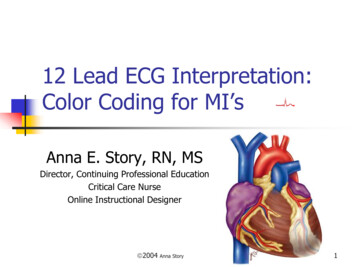
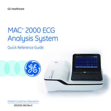
IIIIII65 Year old womanWhere are the ‘septal’ q-waves?1-2
IIIIIIMearurements:A 65V 65Rhythm (s):Sinus RhythmConduction:Normal SA, AV, IVWaveform: Normal P, QRS, ST-TSeptal q-waves I, aVL (arrows)PR 169QRS 70QT 3801-2Axis 30(onset of ventricular activationbegins in the left ventricularseptal surface)Interpretation:Normal ECG (septal q-waves arenormally are seen in leads I, andaVL when the QRS axis is 60 )
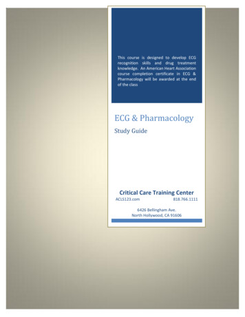
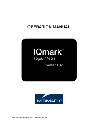
Age 22IIIIII22 year old man; just waking up.1-3
Age 22*I*II*IIIMearurements:A 48V 48PR noneQRS 90QT 4001-3Axis 100Rhythm (s):Junctional escape rhythm(Escape rhythms serve asbackup pacemakers whenthe primary pacemakergets too slow or whenheart block preventsprimary pacemaker fromreaching the ventricles)Conduction: Normal IVWaveform: Normal QRS, ST, T, U (*)Retrograde P waves after the QRS inthe ST segment, best seen in II, III,aVF (arrows); it‘s like someone took abite out of the T wave!Note: normal U waves are best seen inleads V2-5 (*); these are the best leads tosee U waves especially at slow heartrates.Interpretation:Abnormal ECG (likely a normal variant inan athlete)1. Slight right axis deviation (can benormal in 22 year old man)2. Junctional escape rhythm (probablydue to vagal slowing of the sinus ratein a healthy athlete; sinus rhythmwould reappear after light exercise)
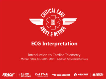
F, Age 2727 year old woman; feeling anxious.1-4
Mearurements:A 110V 110Rhythm (s):Ectopic atrial tachycardiaConduction:Normal AV, IVWaveform: PR 120 Inverted P waves II, III, aVF;upright P waves in lead avR; (lowatrial ectopic pacemaker)Normal QRS, ST, T wavesQRS 80QT 3001-4Axis 10Note: In the horizontal plane (V1-6)ectopic atrial P waves may looknormal in morphology; i.e., upright indirection.Interpretation:Abnormal ECG:1. Rhythm (this rhythm abnormality canbe the result of various internal orexternal stress perturbations; e.g.,hypoxia, stimulants, sepsis, et al.Brief ectopic atrial rhythms (usually 3-6beat runs) are common in otherwisehealthy people (may also occur in sickindividuals)
Just an ordinary guy getting an insurance physical.1-5
Mearurements:A 80PR 120QRS 110QT 3601-5Axis -60V 80Rhythm (s):Sinus rhythmConduction:Normal SA, AV, slight IVconduction delayWaveform: Normal PrS in II, III, aVF (SIII SII)Small q in I, aVLDelayed QRS transition inhorizontal plane (V5); notepersistent S waves in V5-6.Interpretation:Abnormal ECG:1) Left anterior fascicular block (LAFB isthe most common IV conductiondisorder)The left bundle branches into two(sometimes three) fascicles: anterior,(septal), and posterior. (see pp 55-58 inthe Outline)
IIIIIIF, Age 87 (sick and dehydrated)1-6
I**IIIIIMearurements:A 80PR 160QRS 801-6QT 480Axis -20V 80Rhythm (s):Sinus rhythm with 2 PACs (*)Note: The PAC’s are earlybeats with different P wavemorphology; the first PAC isfollowed by a pause (longer RRcycle)Conduction:Normal SA, AV, IV Waveform:Interpretation:Normal P, QRSSlight ST depression V5-6T inversion in III, V2-4Abnormal ECG:1) Prolonged QT (upper limit @ 80 bpmis 380 ms); many etiologies toconsider!2) Nonspecific ST-T abnormalities(consider abnormal electrolytes,drugs, various heart diseases, etc)3) Rhythm: 2 PACs
V1-6: Differential Diagnoses Poor R-wave progression (smallor no r-waves V1-3, R:SV4 1) Normal variant (esp. in women) Misplaced precordial leads Left ventricular hypertrophy Anterior and anteroseptal MI LBBB and incomplete LBBB Left anterior fascicular block Emphysema and COPD Some cases of WPW Diffuse infiltrative diseases Dextrocardia Prominent anterior forces(PAF: R:S V1-2 1) Normal variantMisplaced precordial leadsRight ventricular hypertrophyRBBB and incomplete RBBB‘True Posterior’ (now calledLateral) MI Some cases of WPW Left septal fascicular block Muscular dystrophy
68 y.o. woman (History of hypertension on Rx)1-7
Mearurements:A 85PR 140QRS 90QT 3601-7Axis 15V 85Rhythm (s):Sinus rhythmConduction:Normal SA, AV, IVWaveform: Increase P terminal force V1 (arrow)Multiple voltage criteria for LVHPoor R wave progression V1-4ST depression, T inversion in I, aVL,V5-6Interpretation:Abnormal ECG1. LAE2. LVH with strain pattern (seen in LVpressure overload conditions likeaortic stenosis, hypertensive heartdisease, IHSS)(See p61 in the 2018 pdf Outline forvarious LVH criteria; ECG criteria for LVHhas very poor sensitivity but highspecificity)
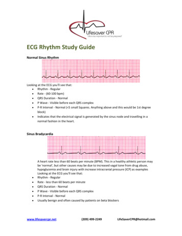
IIIIII18 year old woman who is pretty sick!1-8
IIIIIIMearurements:A 100 V 100Rhythm (s):Sinus tachycardiaConduction:Normal SA, AV, IVWaveform: PR 180 QRS 80QT 3301-8Axis 130PII. V2 2.5 mm (arrows)Prominent anterior forces(PAF) with qR pattern in V1ST depression, T waveinversion multiple leadsInterpretation:Abnormal ECG:1. Right atrial enlargement (RAE)2. RVH with strain pattern3. Heart rate (tachycardia)(This is a patient with primary pulmonaryhypertension; severe right heart disease)(See p64 in the 2018 pdf Outline forvarious RVH criteria)
35 year old woman admitted for acute alcohol intoxicationWhat else went wrong?1-9
Mearurements:A 105V 105Rhythm (s):Sinus tachycardiaConduction:Normal SA, AV, IVWaveform: P, QRS, T in lead I are allinverted (this is a clue!) Minimal signal in lead II (this isclue!)PR 150QRS 80QT 3001-9Axis 150 Poor R wave progression (canbe a normal variant in women)Interpretation:Abnormal ECG:1. Lead reversal error (RA and LA)2. Lead error (RA and right foot)3. Heart rate (tachycardia)Note: lead errors are common (the mostcommon is RA / LA reversal; RA / right legreversal gives no signal in lead II; why is that?).answer: lead II becomes right leg vs. leftleg (i.e., no potential difference).
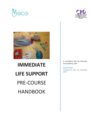
IIIIIIOh, oh . What to do ?1-10
IIIIIIMearurements:A 110PR 140QRS 70QT 3001-10Axis ?V 110Rhythm (s):Sinus tachycardiaConduction:Normal SA, AV, IVWaveform:Much artifact (but you canstill recognize aspects of theECG waveform (see lead III)Interpretation:Artifact precludes accurate ECGinterpretation; sinus tachycardia ispresent.Artifact in this case is from a patient withParkinson‘s disease (skeletal muscle).
Abnormal ECG 1. LAE 2. LVH with strain pattern (seen in LV pressure overload conditions like aortic stenosis, hypertensive heart disease, IHSS) (See p61 in the 2018 pdf Outline for various LVH criteria; ECG criteria for LVH has very poor sensiti