Transcription
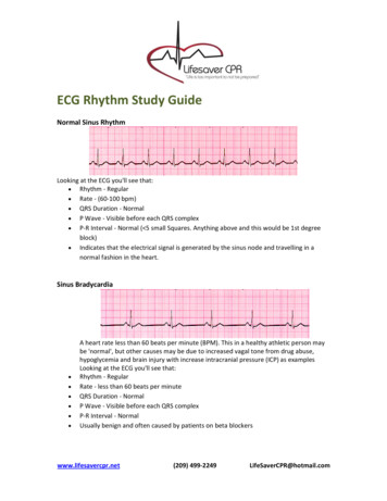
ECG Rhythm Study GuideNormal Sinus RhythmLooking at the ECG you'll see that: Rhythm ‐ Regular Rate ‐ (60‐100 bpm) QRS Duration ‐ Normal P Wave ‐ Visible before each QRS complex P‐R Interval ‐ Normal ( 5 small Squares. Anything above and this would be 1st degreeblock) Indicates that the electrical signal is generated by the sinus node and travelling in anormal fashion in the heart.Sinus Bradycardia A heart rate less than 60 beats per minute (BPM). This in a healthy athletic person maybe 'normal', but other causes may be due to increased vagal tone from drug abuse,hypoglycemia and brain injury with increase intracranial pressure (ICP) as examplesLooking at the ECG you'll see that:Rhythm ‐ RegularRate ‐ less than 60 beats per minuteQRS Duration ‐ NormalP Wave ‐ Visible before each QRS complexP‐R Interval ‐ NormalUsually benign and often caused by patients on beta blockerswww.lifesavercpr.net(209) 499‐2249LifeSaverCPR@hotmail.com
Sinus Tachycardia An excessive heart rate above 100 beats per minute (BPM) which originates from the SAnode. Causes include stress, fright, illness and exercise. Not usually a surprise if it istriggered in response to regulatory changes e.g. shock. But if there is no apparenttrigger then medications may be required to suppress the rhythmLooking at the ECG you'll see that:Rhythm ‐ RegularRate ‐ More than 100 beats per minuteQRS Duration ‐ NormalP Wave ‐ Visible before each QRS complexP‐R Interval ‐ NormalThe impulse generating the heart beats are normal, but they are occurring at a fasterpace than normal. Seen during exerciseSupraventricular Tachycardia (SVT) Abnormal A narrow complex tachycardia or atrial tachycardia which originates in the 'atria' but isnot under direct control from the SA node. SVT can occur in all age groupsLooking at the ECG you'll see that:Rhythm ‐ RegularRate ‐ 140‐220 beats per minuteQRS Duration ‐ Usually normalP Wave ‐ Often buried in preceding T waveP‐R Interval ‐ Depends on site of supraventricular pacemakerImpulses stimulating the heart are not being generated by the sinus node, but insteadare coming from a collection of tissue around and involving the atrioventricular (AV)nodewww.lifesavercpr.net(209) 499‐2249LifeSaverCPR@hotmail.com
Atrial Fibrillation Many sites within the atria are generating their own electrical impulses, leading toirregular conduction of impulses to the ventricles that generate the heartbeat. Thisirregular rhythm can be felt when palpating a pulseLooking at the ECG you'll see that:Rhythm ‐ Irregularly irregularRate ‐ usually 100‐160 beats per minute but slower if on medicationQRS Duration ‐ Usually normalP Wave ‐ Not distinguishable as the atria are firing off all overP‐R Interval ‐ Not measurableThe atria fire electrical impulses in an irregular fashion causing irregular heart rhythmAtrial Flutter Looking at the ECG you'll see that:Rhythm ‐ RegularRate ‐ Around 110 beats per minuteQRS Duration ‐ Usually normalP Wave ‐ Replaced with multiple F (flutter) waves, usually at a ratio of 2:1 (2F ‐ 1QRS)but sometimes 3:1P Wave rate ‐ 300 beats per minuteP‐R Interval ‐ Not measurableAs with SVT the abnormal tissue generating the rapid heart rate is also in the atria,however, the atrioventricular node is not involved in this case.www.lifesavercpr.net(209) 499‐2249LifeSaverCPR@hotmail.com
1st Degree AV Block 1st Degree AV block is caused by a conduction delay through the AV node but allelectrical signals reach the ventricles. This rarely causes any problems by itself and oftentrained athletes can be seen to have it. The normal P‐R interval is between 0.12s to0.20s in length, or 3‐5 small squares on the ECG.Looking at the ECG you'll see that:Rhythm ‐ RegularRate ‐ NormalQRS Duration ‐ NormalP Wave ‐ Ratio 1:1P Wave rate ‐ NormalP‐R Interval ‐ Prolonged ( 5 small squares)2nd Degree Block Type 1 (Wenckebach) Another condition whereby a conduction block of some, but not all atrial beats gettingthrough to the ventricles. There is progressive lengthening of the PR interval and thenfailure of conduction of an atrial beat, this is seen by a dropped QRS complex.Looking at the ECG you'll see that:Rhythm ‐ Regularly irregularRate ‐ Normal or SlowQRS Duration ‐ NormalP Wave ‐ Ratio 1:1 for 2,3 or 4 cycles then 1:0.P Wave rate ‐ Normal but faster than QRS rateP‐R Interval ‐ Progressive lengthening of P‐R interval until a QRS complex is droppedwww.lifesavercpr.net(209) 499‐2249LifeSaverCPR@hotmail.com
2nd Degree Block Type 2 When electrical excitation sometimes fails to pass through the A‐V node or bundle ofHis, this intermittent occurrence is said to be called second degree heart block. Electricalconduction usually has a constant P‐R interval, in the case of type 2 block atrialcontractions are not regularly followed by ventricular contractionLooking at the ECG you'll see that:Rhythm ‐ RegularRate ‐ Normal or SlowQRS Duration ‐ ProlongedP Wave ‐ Ratio 2:1, 3:1P Wave rate ‐ Normal but faster than QRS rateP‐R Interval ‐ Normal or prolonged but constant3rd Degree Block 3rd degree block or complete heart block occurs when atrial contractions are 'normal'but no electrical conduction is conveyed to the ventricles. The ventricles then generatetheir own signal through an 'escape mechanism' from a focus somewhere within theventricle. The ventricular escape beats are usually 'slow'Looking at the ECG you'll see that:Rhythm ‐ RegularRate ‐ SlowQRS Duration ‐ ProlongedP Wave ‐ UnrelatedP Wave rate ‐ Normal but faster than QRS rateP‐R Interval ‐ VariationComplete AV block. No atrial impulses pass through the atrioventricular node and theventricles generate their own rhythmwww.lifesavercpr.net(209) 499‐2249LifeSaverCPR@hotmail.com
Bundle Branch Block Abnormal conduction through the bundle branches will cause a depolarization delaythrough the ventricular muscle, this delay shows as a widening of the QRS complex.Right Bundle Branch Block (RBBB) indicates problems in the right side of the heart.Whereas Left Bundle Branch Block (LBBB) is an indication of heart disease. If LBBB ispresent then further interpretation of the ECG cannot be carried out.Looking at the ECG you'll see that:Rhythm ‐ RegularRate ‐ NormalQRS Duration ‐ ProlongedP Wave ‐ Ratio 1:1P Wave rate ‐ Normal and same as QRS rateP‐R Interval ‐ NormalPremature Ventricular Complexes Due to a part of the heart depolarizing earlier than it shouldLooking at the ECG you'll see that:Rhythm ‐ RegularRate ‐ NormalQRS Duration ‐ NormalP Wave ‐ Ratio 1:1P Wave rate ‐ Normal and same as QRS rateP‐R Interval ‐ Normalwww.lifesavercpr.net(209) 499‐2249LifeSaverCPR@hotmail.com
Also you'll see 2 odd waveforms, these are the ventricles depolarizing prematurely inresponse to a signal within the ventricles.(Above ‐ unifocal PVC's as they look alike ifthey differed in appearance they would be called multifocal PVC's, as below)Junctional Rhythms Looking at the ECG you'll see that:Rhythm ‐ RegularRate ‐ 40‐60 Beats per minuteQRS Duration ‐ NormalP Wave ‐ Ratio 1:1 if visible. Inverted in lead IIP Wave rate ‐ Same as QRS rateP‐R Interval ‐ VariableBelow ‐ Accelerated Junctional Rhythmwww.lifesavercpr.net(209) 499‐2249LifeSaverCPR@hotmail.com
Ventricular Tachycardia (VT) Abnormal Looking at the ECG you'll see that:Rhythm ‐ RegularRate ‐ 180‐190 Beats per minuteQRS Duration ‐ ProlongedP Wave ‐ Not seenResults from abnormal tissues in the ventricles generating a rapid and irregular heartrhythm. Poor cardiac output is usually associated with this rhythm thus causing the pt togo into cardiac arrest. Shock this rhythm if the patient is unconscious and without apulseVentricular Fibrillation (VF) Abnormal Disorganized electrical signals cause the ventricles to quiver instead of contract in arhythmic fashion. A patient will be unconscious as blood is not pumped to the brain.Immediate treatment by defibrillation is indicated. This condition may occur during orafter a myocardial infarct.Looking at the ECG you'll see that:Rhythm ‐ IrregularRate ‐ 300 , disorganizedQRS Duration ‐ Not recognizableP Wave ‐ Not seenThis patient needs to be defibrillated!! QUICKLYwww.lifesavercpr.net(209) 499‐2249LifeSaverCPR@hotmail.com
Asystole ‐ Abnormal Looking at the ECG you'll see that:Rhythm ‐ FlatRate ‐ 0 Beats per minuteQRS Duration ‐ NoneP Wave ‐ NoneCarry out CPR!!Myocardial Infarct (MI) Looking at the ECG you'll see that:Rhythm ‐ Regularwww.lifesavercpr.net(209) 499‐2249LifeSaverCPR@hotmail.com
ECG Rhythm Study Guide Normal Sinus Rhythm Looking at the ECG you'll see that: Rhythm ‐ Regular Rate ‐ (60‐100 bpm) QRS Duration ‐ Normal P Wave ‐ Visible before each QRS complex P‐R Interval ‐ Normal ( 5 small Sq