Transcription
Wang et al. Biological Procedures 19) 21:20RESEARCHOpen AccessComprehensive Transcriptomic Analysis ofMouse Gonadal Development InvolvingSexual Differentiation, Meiosis andGametogenesisJian Wang1†, Geng G. Tian1†, Zhuxia Zheng1†, Bo Li2, Qinghe Xing4* and Ji Wu1,2,3*AbstractBackground: Mammalian gonadal development is crucial for fertility. Sexual differentiation, meiosis and gametogenesis arecritical events in the process of gonadal development. Abnormalities in any of these events may cause infertility. However,owing to the complexity of these developmental events, the underlying molecular mechanisms are not fully understoodand require further research.Results: In this study, we employed RNA sequencing to examine transcriptome profiles of murine female and male gonadsat crucial stages of these developmental events. By bioinformatics analysis, we identified a group of candidate genes thatmay participate in sexual differentiation, including Erbb3, Erbb4, and Prkg2. One hundred and two and 134 candidate genesthat may be important for female and male gonadal development, respectively, were screened by analyzing the global geneexpression patterns of developing female and male gonads. Weighted gene co-expression network analysis was performedon developing female gonads, and we identified a gene co-expression module related to meiosis. By alternativesplicing analysis, we found that cassette-type exon and alternative start sites were the main forms of alternativesplicing in developing gonads. A considerable portion of differentially expressed and alternatively spliced geneswere involved in meiosis.Conclusion: Taken together, our findings have enriched the gonadal transcriptome database and provided novelcandidate genes and avenues to research the molecular mechanisms of sexual differentiation, meiosis, andgametogenesis.Keywords: Gonad, RNA-Seq, Sex-biased expressed genes, Time series cluster, WGCNA, Alternative splicingIntroductionInfertility is estimated to affect as many as 48.5 millioncouples worldwide [1]. Its incidence may be as high as15%. Following cancer, cardiovascular and cerebrovasculardiseases, infertility has become the third major disorderthat seriously affects human health and causes considerable stress to patients and their families.* Correspondence: xingqinghe@hotmail.com; jiwu@sjtu.edu.cn†Jian Wang, Geng G. Tian and Zhuxia Zheng contributed equally to thiswork.4Children’s Hospital & Institutes of Biomedical Sciences, Fudan University, 131Dong-Chuan Road, Shanghai 200032, China1Renji Hospital, Key Laboratory for the Genetics of Developmental &Neuropsychiatric Disorders (Ministry of Education), Bio-X Institutes, School ofMedicine, Shanghai Jiao Tong University, Shanghai 200032, ChinaFull list of author information is available at the end of the articleGonads are the important reproductive organs formammals. The process of gonadal development includescritical developmental events, such as sexual differentiation, meiosis and gametogenesis. Abnormalities in anyof these events may cause infertility. The molecularregulatory mechanisms of these crucial developmentalevents have been the focus of many studies in reproductive biology.Many genes that are important for gonadal developmenthave been discovered. For example, the Sry, Sox9, M33,Dax1, Wt1, Rspo1, Sf1, Dmrt1, Atrx, Wnt4, Amh, Fgf9,Lhr, Dhh, Insl3, Foxl2 and Sox3 genes play important rolesin sexual determination and differentiation [2–5]. TheStra8, Sycp1, Sycp2, Sycp3, Spo11, Rce8, Dmc1, Dazl, Mlh1 The Author(s). 2019 Open Access This article is distributed under the terms of the Creative Commons Attribution 4.0International License (http://creativecommons.org/licenses/by/4.0/), which permits unrestricted use, distribution, andreproduction in any medium, provided you give appropriate credit to the original author(s) and the source, provide a link tothe Creative Commons license, and indicate if changes were made. The Creative Commons Public Domain Dedication o/1.0/) applies to the data made available in this article, unless otherwise stated.
Wang et al. Biological Procedures Online(2019) 21:20and Msh5 genes regulate meiosis [5–9]. The Figla, Lhx8,Nobox, Sohlh1, Sohlh2, Bax, Ahr, Gdf9, Pten, Scf, Bcl2 andRps6 genes have roles in follicular and oocyte development [5, 10]. The Adamts2, Bcl2l2, Cadm1, Ddx25, Piwil1,Prm1, Prm2, Tbpl1, Tlp, Tnp1, Tnp2 and Ube2b genes areinvolved in regulating spermatogenesis [5].Many previous studies of gonadal development usedgene knockout mouse models, and only a small numberof genes could be studied at a time. Gene chips werealso used to research gonadal development [11–14]. Although higher throughput was obtained by gene chipanalysis, this research was typically limited to the morehighly expressed known genes. Classical gene chip analysis was unable to research unknown or low abundancegene transcripts, nor alternative splicing.In recent years, high-throughput RNA sequencing(RNA-Seq) overcame the above technical shortcomings ofgene chips, and also allowed high-throughput investigation of the whole transcriptome [15]. Previous studieshave used RNA-Seq to investigate the development ofmouse gonads. However, these studies either focused onsingle sex gonads at certain developmental stages, or specific cell types in the gonads. Gong et al. investigated thetranscriptome profiling of mouse testes at three postnatalages: 6 days postnatal, 4 weeks old and 10 weeks old,representing infant, juvenile and adult stages, respectively[16]. Pan et al. performed a comparative transcriptomicanalysis using RNA-Seq of ovaries isolated from mice aged1 week and 8 weeks old [17]. McClelland et al. carried outtranscriptomic analysis of mouse fetal Leydig cells [18].Sexual differentiation, meiosis and gametogenesis requirethe joint participation and interaction of germ and somatic cells in the gonads. Therefore, a better understandingof the mechanisms of sexual differentiation may be obtained by comparing female and male gonadal transcriptomes. It is possible that more insight into the molecularmechanisms of sexual differentiation, meiosis and gametogenesis may be obtained by transcriptomic analysis ofmurine female and male gonads covering the key stages ofdevelopmental corresponding to these critical developmental events. However, such a comparison has not beenreported to date.Here, we systematically compared the transcriptomesof female and male gonads covering the developmentalstages corresponding to sexual differentiation, meiosisand gametogenesis. Our study used gonads at 12.5 dayspost-coitum (dpc), at which time female and male gonads begin to show discernible differences, 13.5 dpcwhen female germ cells begin to undergo meiosis, andmale germ cells begin to stop mitosis, 16.5 dpc whenmost female germ cells are undergoing meiosis, andmale germ cells arrest at the G1/G0 stage, and 6 dayspost-partum (dpp) when most male germ cells are at thespermatogonial stem cell stage. The current resultsPage 2 of 13provide novel candidate genes and potential pathwaysfor further investigation into the molecular mechanismsof sexual differentiation, meiosis and gametogenesis ingonadal development.Materials and MethodsAnimalsEight-week-old C57BL/6 mice were purchased from theSLAC Laboratory Animal Co., Shanghai, China. Micewere housed in a 12 h light: 12 h dark cycle. Protocols anduse of animals for this study were approved by the Institutional Animal Care and Use Committee of Shanghai(SYXK-2018-0028) and were conducted in accordancewith the National Research Council Guide for Care andUse of Laboratory Animals.Gonad Collection and RNA ExtractionTimed matings were performed by placing a male mousewith two females in a cage. Females were checked forthe presence of vaginal plugs the next morning. Noon ofthe day when the mating plug was observed was designated 0.5 dpc. On the relevant days of gestation (12.5,13.5 and 16.5 dpc), pregnant females were euthanizedwith carbon dioxide. Embryonic gonads were dissectedfree of the mesonephros, snap frozen in liquid nitrogen,and stored at 80 C. The gonads of 6 dpp mice werealso collected, immediately frozen in liquid nitrogen andstored at 80 C.Total RNA was isolated from the gonads of 12.5, 13.5and 16.5 dpc and 6 dpp mice using TRIzol reagent (Invitrogen, Waltham, Massachusetts, USA) according to themanufacturer’s instructions. An Agilent 2100 Bioanalyzer (Agilent Technologies, Santa Clara, USA) was usedto measure total RNA quantity and assess RNAintegrity.RNA-Seq Library Construction and SequencingThe cDNA library was constructed with a SMARTer Ultra Low Input RNA for lllumina Sequencing kit(Clontech Laboratories, Mountain View, USA). Briefly,first-strand cDNA was synthesized using SMARTScribeReverse Transcriptase and then purified using SPRIAmpure Beads. After amplification using the PolymeraseMix, double-stranded complementary DNA was purifiedusing SPRI Ampure Beads.High-throughput sequencing was performed by Shanghai Biotechnology Inc. (Shanghai, China) via 100-ntpaired-end sequencing on an Illumina HiSeq 2500 system (Illumina, San Diego, USA).Reads Mapping and Measurement of Gene ExpressionAfter sequencing, clean reads were obtained by removing reads containing the adaptor sequences, reads with 5% ambiguous bases, and low-quality reads, then
Wang et al. Biological Procedures Online(2019) 21:20mapped to the mouse genome (version: mm10GRCm38) using TopHat software (Version 2.1.1). Geneexpression level was calculated using the fragments perkilobase per million mapped reads method.Analysis of Differentially Expressed GenesAnalysis of Differentially Expressed Genes (DEGs) wasperformed by the DESeq package in R language (Version1.36.0). Genes with a fold change (FC) 2, and false discovery rate (FDR) 0.05 were assigned as differentiallyexpressed.The Series-Cluster analysis of expression profiles ofDEGs was performed by using the STEM method(http://www.cs.cmu.edu/ jernst/st/). Significant profileswere identified using the Fisher’s exact test and multiplecomparisons.Page 3 of 13transcription PCR (qPCR) was performed using FastStartUniversal SYBR Green Master Mix (Roche) according tothe procedure described previously [21]. The 2 ΔΔCtmethod was used to calculate the relative expression ofgenes using the ABI 7500 System Software (V2.0.4), andgene expression levels were normalized to the Gapdhlevel. The primers used for qPCR are shown in Table 1.Statistical AnalysisFor each group, three independent experiments werereplicated. The data are expressed as the mean SEM.Data were statistically analyzed with ANOVA, followedby the Fisher’s least significant difference test with Rsoftware (Version 3.5.1). Differences were consideredsignificant at p 0.05.ResultsGene Ontology and Kyoto Encyclopedia of Genes andGenomesGenes were submitted to the databases of Gene Ontology (GO) and Kyoto Encyclopedia of Genes and Genomes (KEGG) for enrichment analysis of the significantGO terms and KEGG pathways. Statistically overrepresented GO terms and KEGG pathway categorieswere obtained by applying a Fisher’s exact p-value cutoff 0.05 and correcting for multiple testing with theBenjamini-Hochberg false discovery rate. Moreover, webuilt a pathway-act-network of the enriched pathwaysaccording to the relationships identified between thepathways in the KEGG database.Weighted Gene co-Expression Network AnalysisWeighted gene co-expression network analysis(WGCNA)was used to create a co-expression network using the Rpackage (Version 1.68) according to our previous publication [19]. Briefly, to ensure a scale free topology of the network, an adjacency matrix was established by transforminga pairwise Pearson’s correlation matrix of expression valuesusing a power function. The topological overlap measure(TOM) was calculated using the adjacency matrix, and usedto cluster genes with a highly similar co-expression relationship into a network module.Sex-Biased Gene Expression in Mouse GonadsIn order to screen candidate genes regulating gonadal sexual differentiation, we initially screened the differentiallyTable 1 List of the qPCR primers used in this studyGene SymbolPrimer SequenceSox9F: 5′- AGTACCCGCATCTGCACAAC 3′R: 5′- ACGAAGGGTCTCTTCTCGCT 3′Foxl2R: 5′- CGTAGAACGGGAACTTGGCTA 3′Sox8Wnt4F: 5′- AGACGTGCGAGAAACTCAAAG 3′R: 5′- GGAACTGGTATTGGCACTCCT 3’Lgr6F: 5′-GAGGACGGCATCATGCTGTC-3’R: 5′-GCTCCGTGAGGTTGTTCATACT-3’Gas6F: 5′-TGCTGGCTTCCGAGTCTTC-3’R: 5′-CGGGGTCGTTCTCGAACAC-3’Zbtb7cF: 5′-TTGATGAGCTGATCGGCATCC-3’R: 5′-GTGTTCGGTACTCTTGCTCCT-3’Erbb3F: 5′-AAGTGACAGGCTATGTACTGGT-3’R: 5′-GCTGGAGTTGGTATTGTAGTTCA-3’Erbb4F: 5′-GTGCTATGGACCCTACGTTAGT-3’R: 5′-TCATTGAAGTTCATGCAGGCAA-3’Cst9F: 5′-GTCCACTGAGAAAGAAAGCTCTG-3’R: 5′-CCACTGTGGGAATGAAATGAACA-3’Zfp819F: 5′-GGGCCGTGTGAGAGATTGG-3’R: 5′-GCCTGGAATACCCCACTACC-3’Rec8Quantitative Real-Time Reverse Transcription PCRReverse transcription was performed using HiScript II QRT SuperMix for qPCR ( gDNA wiper) kit (Vazyme,Nanjing, China). The quantitative real-time reverseF: 5′- CGAGGGGATACTGCTGAGG 3′R: 5′- AGCTCTGCGTTATGGAGATGC 3′Alternative SplicingAlternatively spliced transcripts were identified by thesoftware AS detector (ASD) (Version 1.0) using the Fisher’s exact test as described [20]. Alternative splicingevents were adjusted using Jensen-Shannon divergence(FDR 0.05).F: 5′- ACAACACCGGAGAAACCAGAC 3′F: 5′-TATGTGCTGGTAAGAGTGCAAC-3’R: 5′-TGTCTTCCACAAGGTACTGGC-3’GapdhF: 5′- CATGGCCTTCCGTGTTCCTA 3’R: 5′- GCCTGCTTCACCACCTTCTT 3’
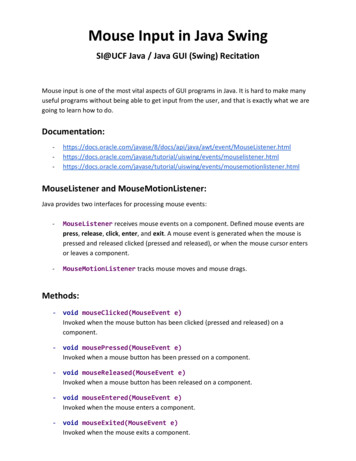
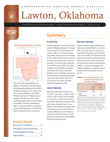
Wang et al. Biological Procedures Online(2019) 21:20expressed genes in female and male gonads at the samestage of development. Wayne analysis was then performedto identify the genes with higher expression in all fourstages of development, and these genes were defined assex-biased expressed genes that may participate in regulating gonadal sexual differentiation.One hundred and thirty-seven male-biased and 187female-biased expressed genes were obtained (Fig. 1 andAdditional file 3 Table. S1, Additional file 4 S2). Thesegenes were classified into 3 categories. The first categoryincluded genes functionally annotated and reported toplay an important role in sex determination and differentiation, such as Sox9 [22, 23], Xist [24], Amh [25],Insl3 [26], Emx2 [26], Dhh [26], Wnt4 [27, 28], Foxl2[29] and Fst [26]. The second category included geneswith functional annotations but no reported involvementin sex determination and differentiation, includingErbb3, Mt3, Gas6, Lzts1, Lrrn1, Olfm1 and Lgr6. Thethird category had genes with no functional annotations,such as Gm30587, GM6705, Gm11734 and Gm31399.Validation of Representative Sex-Biased Gene Expressionby qPCRTo validate the screened sex-biased expressed genes, twelvegenes (Cst9, Zbtb7c, Lgr6, Gas6, Erbb3, Erbb4, Zfp819, Rec8,Sox8, Sox9, Wnt4 and Foxl2) were randomly selected andtheir ovarian and testicular expression levels at different stagesof development were detected by qPCR. The results showedthat Cst9, Erbb3, Erbb4, Zfp819, Sox8 and Sox9 had biased expression in male gonads, whereas Lgr6, Zbtb7c, Gas6, Rec8,Page 4 of 13Wnt4 and Foxl2 had biased expression in female gonads(Fig. 2). The results of qPCR were consistent with RNA-seq.Protein–Protein Interaction Network of Mouse GonadalSex-Biased Gene ExpressionThe protein–protein interaction (PPI) network of mousegonadal sex-biased expressed genes was constructed byusing String and Cytoscape software. Twenty nodal geneswere connected via more than 10 edges in the network;Wnt4, Dhh, Amh, Bmp2, Fst, Sox9, Spp1, Cd44, Cyp11a1,Cyp17a1, Erbb3, Erbb4, Fshr, Lhcgr, Mapk4, Prkg2, Calb1,Nos1, Acta2 and Cacna1d (Additional file 1 Figure S1 andTab. 2). There were defined as hub genes in the network,suggesting they may play a key regulatory role in theprocess of gonadal sexual differentiation.Among these 20 hub genes, Sox9, Cyp17a1, Cyp11a1,Dhh, Amh, Fshr, Lhcgr, Wnt4, Fst and Bmp2 are wellknown sex differentiation genes. The function of the other10 hub genes with respect to sexual differentiation remains unknown. Interestingly, Erbb3, Erbb4, Prkg2 andMapk4 encode protein kinases. Protein kinases catalyzeprotein phosphorylation and transfer the γ-phosphategroups of adenosine triphosphate (usually ATP) to aminoacid residues of substrate proteins. The four protein kinase genes suggest that protein phosphorylation may playan important role in sexual differentiation.Time Series Cluster Analysis of Gonadal Gene Expressionduring DevelopmentTime series cluster analysis was performed to identifythe global trends and model profiles of gonadal geneFig. 1 Venn diagram of genes with higher expression in murine male (left) and female (right) gonads at different stages of development
Wang et al. Biological Procedures Online(2019) 21:20Page 5 of 13Fig. 2 Validation of the identified sex-biased gene expression by qPCR. log2FC: Male/FemaleTable 2 Hubs of the protein–protein interaction network constructed from the mouse gonadal sex-biased expressed genesGene SymbolDefinitionGene typeWnt4wingless-type MMTV integration site family, member 4protein codingDhhdesert hedgehogprotein codingAmhanti-Mullerian hormoneprotein codingBmp2bone morphogenetic protein 2protein codingFstfollistatinprotein codingSox9SRY (sex determining region Y)-box 9protein codingSpp1secreted phosphoprotein 1protein codingCd44CD44 antigenprotein codingCyp11a1cytochrome P450, family 11, subfamily a, polypeptide 1protein codingCyp17a1cytochrome P450, family 17, subfamily a, polypeptide 1protein codingErbb3erb-b2 receptor tyrosine kinase 3protein codingErbb4erb-b2 receptor tyrosine kinase 4protein codingFshrfollicle stimulating hormone receptorprotein codingLhcgrluteinizing hormone/choriogonadotropin receptorprotein codingMapk4mitogen-activated protein kinase 4protein codingPrkg2protein kinase, cGMP-dependent, type IIprotein codingCalb1calbindin 1protein codingNos1nitric oxide synthase 1, neuronalprotein codingActa2actin, alpha 2, smooth muscle, aortaprotein codingCacna1dcalcium channel, voltage-dependent, L type, alpha 1D subunitprotein coding
Wang et al. Biological Procedures Online(2019) 21:20expression according to signal densities in the 12.5, 13.5and 16.5 dpc, and 6 dpp temporal sequence. We identified 26 possible gene expression profile patterns (numbered 0–25), which represent the overall expressionpatterns (Fig. 3a, 4a). Eleven patterns (No. 0, 1, 9, 10, 12,13, 14, 15, 16, 17 and 25) showed significance (P 0.05)in female gonadal development, and 10 patterns (No. 0,3, 9, 12, 13, 14, 15, 16, 23 and 25) showed significance(P 0.05) in male gonadal development (Fig. 3a, 4a).For both female and male gonads, genes in pattern No. 25showed increased expression levels during gonadal development, suggesting these genes may be crucial for gonadal development. Significantly enriched GO terms from pattern No.25 are illustrated in Fig. 3b and 4b. For the female gonads,pattern No. 25 contained 102 genes and significantly enrichedGO terms closely correlated with mRNA splicing, positiveregulation of the MAPK cascade, N-glycan fucosylation, andnegative regulation of oocyte development (Fig. 3b). This listwas also significantly enriched for genes associated with thePage 6 of 13p53 signaling, N-Glycan biosynthesis signaling, and cGMPPKG signaling pathways (Fig. 3c). To further understand theimportance of pathway interactions, and to screen key pathways for significant roles in female gonadal development, webuilt a Pathway-Act-Network according to the direct or systemic interactions assigned between pathways in the KEGGdatabase (Fig. 3d). Key pathways were identified, includingapoptosis signaling and protein processing in endoplasmicreticulum signaling pathways, as shown in Fig. 3d. In addition,we found 9 unannotated ncRNAs in pattern No. 25,designated LOC102639808, LOC102640507, LOC102637464,LOC102637824, LOC102638234, LOC102634191, LOC102635948
first-strand cDNA was synthesized using SMARTScribe Reverse Transcriptase and then purified using SPRI Ampure Beads. After amplification using the Polymerase Mix, double-stranded complementary DNA was purified using SPRI Ampure Beads. High-throughput sequencing was performed by