Transcription
MEETING THECHALLENGES OFEM SAMPLE PREPARATIONTHE LEICA NANOTECHNOLOGYPRODUCT PORTFOLIOThe highly comprehensive product portfolio forpreparation of biological, medical, and industrial samples.
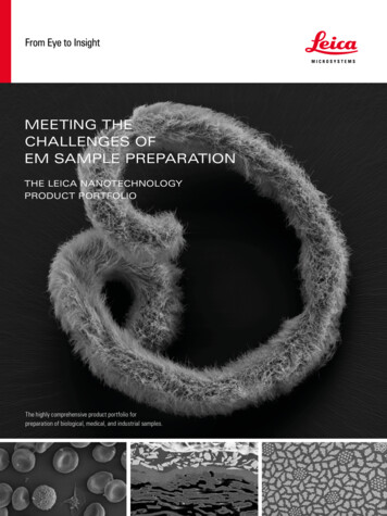
2.3SAMPLE PREPARATIONWITH LEICA MICROSYSTEMS –THE PORTFOLIO THAT GIVES YOUSUCCESS FOR YOUR APPLICATIONTRIMMING &MECHANICAL PREPARATIONEM TXP, EM RAPID, EM TRIM2ION BEAM MILLINGEM TIC 3X, EM RES102ULTRAMICROTOMY &CRYO-ULTRAMICROTOMYEM UC7, ARTOS 3D, EM FC7, EM KMR3SAMPLE TRANSFEREM VCT500, EM VCMCRYO CLEMTHUNDER Imager EM Cryo CLEMCRYO PREPARATIONEM ICE, EM GP2, EM AFS2, EM CTDCOATING &FREEZE FRACTURINGEM ACE200, EM ACE600, EM ACE900TISSUE PROCESSINGEM TPCONTRASTINGEM AC20CRITICAL POINT DRYINGEM CPD300CONCENTRATING ON WORKFLOW SOLUTIONS, WE PROVIDEA PRODUCT RANGE THAT IS ALIGNED TO YOUR NEEDS INTEM, SEM, LM, AND AFM INVESTIGATIONS.Cover images: top: Nematode Eubostrichus dianae with ectosymbiotic bacteria layer, critical point dried with the EM CPD300 (source: Mag. N. Leisch, Universityof Vienna, Austria); bottom left: Human Erythrocytes and Lymphocytes, critical point dried with the EM CPD300 (source: Dr. W. Müller, University of Utrecht,Netherlands); bottom middle: cross section of abrasive paper, prepared with the EM TIC 3X (source: Wolfgang Grünewald, TU Chemnitz, Germany); bottom right:cross-section of a Nb3Sn superconductor, prepared with the EM TXP and EM TIC 3X (source: Wolfang Grünewald, TU Chemnitz, Germany).
THE COMPLETE PORTFOLIO FOR EM SAMPLETRIMMING &MECHANICALPREPARATIONION BEAMMILLING Accurate location and preparation ofEM TXPmicrotargetsTarget preparation device for milling,sawing, drilling, grinding and polishing In-situ stereomicroscope observationsamples prior to examination by SEM, Automatic process control to produce a mirrorlike surface qualityTEM and LM techniques. A perfectsystem to pre-prepare the sample priorto the ion beam milling techniques.EM RAPIDAdvanced specimen trimming devicefor TEM, SEM, LM. 0.5, 1, 10, 100 µm step advance Adjustable cutting speed 300–20,000 rpm Advance indication on LCD displayEM TRIM2Specimen trimming device for TEM,SEM, LM. 1 µm step advance Perpendicular viewing of the sample LED illumination Cutting speed 20,000 rpmEM TIC 3XThe Triple Ion Beam Milling Systemallows production of cross sectionsand planed surfaces for SEMmicrostructure analysis (EDS, WDS,Auger, EBSD) and AFM investigations. Broad and deep cross sections as well asuniform large area milling Interchangeable stages – Standard stage,Multiple sample stage, Cooling stage, Rotarystage EM VCT500 option for environmentally sensitiveand / or cryogenic sample transferThe EM TIC3X outfitted with anEM VCT500 docking station is theideal solution for environmentallysensitive sample and / or cryogenicsample transfer.EM RES102Unique ion beam milling device withtwo modified saddlefield ion sourcesof variable ion energy for optimumresults. It combines the preparation ofTEM, SEM, and LM samples in a singlebenchtop unit. External control of the milling process via LAN Preparation of samples up to 25 mm diameter Fully computer-controlled milling para meters
PREPARATIONULTRAMICROTOMY&CRYO-ULTRAMICROTOMY4.5EM UC7Ultramicrotome for ultrathin sectioningof biological and industrial samples. Knife usage monitoring Feed range from 1 nm up to 15 µm Fully motorized knife stage and AutoTrimfunction Vibration decoupled gravity strokeEM FC7Low temperature ultrathincryosectioning of biological andindustrial samples. Can be mountedon the EM UC7 and the ARTOS 3D. Temperature range from 110 C to -185 C Individual temperature settings of specimen,knife, and gas Easy section collection using micromani pulatorand EM CRION ionizer EM VCT500 option for environmentallyprotected sample transferThe EM FC7 outfitted with anEM VCT500 transfer port is the idealsolution for environmentally sensitivesample and / or cryogenic sampletransfer.SAMPLETRANSFERCRYO CLEMARTOS 3DArray Tomography Solution forautomatic creation and collection ofhundreds of serial-section ribbonsready for array tomography with aSEM. Fast setup with programs pre-defined by theuser for different section carriers Wrinkle-free sorting and positioning of ribbonson the section carrier ready for SEM imaging Uses the same small section carrier through theentire workflow from sectioning to imaging Also and Ideal solution for CLEM as transparentsection carriers are availableEM KMR3Balanced-break glass knife maker forproducing 45 glass knives from6.4 mm, 8 mm, and 10 mm glass. Highly reproducible, outstanding knife quality Automatic reset of the breaking and scoringmechanism Ergonomic design for comfortable useEM VCT500Versatile vacuum cryo transfer systemfor contamination-free transferof specimens between differentpreparation and analysis instruments. Workflow specimen monitoring Links workflow from preparation to analysis Connects to more than one SEM Various specimen holders for SEM, FIB-SEM,freeze-fracture and moreEM VCMLN2 cooled workstation forcontamination-free specimenmanipulation. All sample transfers from loading under vacuum Improved connectivity given by a movableloading sphere, adaptors to the Cryo CLEM andCryo-TEM transfer holdersTHUNDER Imager EM Cryo CLEMThe system ensures contaminationfree sample transfer and loading fromcryo sample preparation instrumentsto Leica fix stage light microscope.Maintains sample vitrified during cryoimaging. Rapid screening of large areas and fastdetermination of regions of interest in theelectron microscope under controlled cryoconditions The cryo objective with low working distance(0.28 mm) and with NA 0.9 for high resolution(364 nm) ensures fast and specific localizationof target structures in EM
CRYOPREPARATIONEM ICEHigh pressure system for freezingaqueous samples delivers optimalsample preservation. Offers thehighest flexibility to meet multipleapplication demands. Programmable sequential freezing of nine(3 3) samples Automated LN2 re-filling of the sample storagedewar Recovery time between freezing cycles is oneminute Retrofitable light stimulation and/or electricalstimulation modeEM ICE Light Stimulation (LS)All the features of EM ICE standard,in addition offers fully integrated lightstimulation. Software integrated programming for LS Automatic recondition of the specific lightmodule Modules with different LEDs (wave lengths):UV, blue, red, green, amber Detailed log file of each experiment Light stimulation precision of 1 millisecondEM ICE Electrical Stimulation (ES)All the features of EM ICE standard,in addition offers fully integratedelectrical stimulation. Millisecond precision Complete coordination of electrical discharge atthe moment of freezing Capturing and imaging action potential andmembrane trafficking eventsEM GP2Automatic plunge freezer for EM grids. Automatic single sided and multiple sided Single sided sensor blotting Fast, easy, and safe filling of the secondarycryogen with the unique liquifying head Controllable secondary cryogen temperature Environmental chamber with adjustabletemperature and humidity Intuitive control via touch panelEM AFS2Freeze substitution and lowtemperature embedding for light andelectron microscopy. –140 C to 70 C working range Transfer function – LN2 gas regulation in thechamber to minimize contamination LED UV polymerization Stereomicroscope viewing AFS smart-remote observation of the processand delivery of critical information via SMSEM FSPAutomatic reagent handling /dispensing system for freezesubstitution and PLT. One step preparation Safer, convenient handling Flexible built-in UV light for polymerization Up to 20 samples per runEM CTDCryo tool dryer Combines heated air flow and heating plate forreliable de-icing Maximum temperature 50 C
6.7COATING &FREEZEFRACTURINGEM ACE200Desk-top coater for homogeneouscoatings of conductive metal or carbonfor EM. Fully automated instrument,options include: Carbon thread evaporation Sputtering Both methods with interchangeable heads Quartz crystal measurement Planetary rotation Glow dischargeEM ACE600Fully automated, versatile highvacuum coater produceing very thin,fine-grained, conductive metal andcarbon coatings. Up to two angledcoating sources configurable. For highresolution analysis, required for FESEM and TEM applications. Sputtering Carbon thread evaporation Carbon rod evaporation E-beam evaporation Glow discharge 104 mm automated rotating stage withplanetary option EM VCT500 option for cryo-coating, freezefracture, double-replica, and controlledenvironmental transfer with the VCT shuttleThe EM ACE600 outfitted with anEM VCT500 is the ideal solutionfor contamination-free cryo-SEMsample preparation with completeenvironmental control.EM ACE900High-end system for freeze fractureapplications. High vacuum, a 3-axismovable microtome, and low anglee-beam coating with rotation ensurethe best results for TEM replicasand together with the EM VCT500,contamination-free cryo-SEM blockface imaging. Large closed cryo-shield Rotating cryo stage High resolution low angle e-beam coating ofcarbon / metal Gate valves for e-beam sources and load lock(sample and knife exchange) EM VCT500 optionTISSUEPROCESSINGEM TPAutomated tissue processor for LMand EM sample preparation. Programming of all processing steps Integrated touch-screen-based software Consistent, reproducible performance Processing of multiple tissues in one run Maintaing of environmental conditions duringpreparationCONTRASTINGEM AC20Automatic contrasting of ultrathinsections for electron microscopy.CRITICALPOINTDRYINGEM CPD300Critical point dryer for biological(pollen, tissue, plants and insects) andindustrial (Micro Electro MechanicalSystems (MEMS), hydro or aerogels)samples. 60 runs per one set of Ultrostains Low reagent consumption High contrast Reduced process times by Leica filler / sampleholder concept Minimized CO2 consumption and minimal userinteraction time Integrated waste separator avoids directcontact with chemical waste
Leica Mikrosysteme GmbH · Vienna · AustriaT 43 1 486 8050-0 · F 43 1 486 8050-30www.leica-microsystems.com/emLeica LNT Product Portfolio · Engilsh 01/2020 · Copyright by Leica Mikrosysteme GmbH, Vienna, Austria, 2020. Subject to modifications. LEICA and the Leica Logo are registered trademarks of Leica Microsystems IR GmbH.CONNECTWITH US!
THE PORTFOLIO THAT GIVES YOU SUCCESS FOR YOUR APPLICATION Cover images: top: Nematode Eubostrichus dianae with ectosymbiotic bacteria layer, critical point dried with the EM CPD300 (source: Mag. N. Leisch, University . Ergonomic design for comfortable use ULTRA MICROTOMY & CRYO-ULTRA MICROTOMY The EM FC7 outfitted with an EM VCT500 transfer .