Transcription
European Review for Medical and Pharmacological Sciences2020; 24: 10155-10161Changes in intestinal flora in preeclampsiarats and effects of probiotics on theirinflammation and blood pressureB.-M. SUN1, L. MENG2, H. LIU3, D. BAO4Department of Obstetrics and Gynecology, Linyi Central Hospital, Linyi, ChinaDepartment of Disease Management Centre, Rizhao Hospital of Traditional Chinese Medicine,Rizhao, China3Department of Pharmacy Intravenous Admixture, Weifang People’s Hospital, Weifang, China4Department of Obstetrics and Gynecology, PLA Rocket Force Characteristic Medical Center,Beijing, China12Abstract. – OBJECTIVE: The aim of thisstudy was to investigate the changes in intestinal flora in preeclampsia rats and the effectsof probiotics on their inflammation and bloodpressure.MATERIALS AND METHODS: A total of 40Specific Pathogen Free (SPF) Wistar rats wererandomly selected in this study. Abdominal operation was performed to reduce uterine bloodperfusion, so as to establish the model of preeclampsia in rats. All rats were randomly divided into two groups, namely, observation group(treated with probiotics, n 20) and control group(not treated with probiotics, n 20). Subsequently, the changes in serum endotoxin level duringintervention, the 24-h urinary 99mTc-diethylene triamine pentaacetic acid (DTPA) excretion rate, and intestinal flora colonization ability after intervention were compared betweenthe two groups. The distribution of intestinalflora after intervention was recorded in the twogroups. Meanwhile, vascular endothelial function and blood pressure following interventionwere compared between the two groups as well.In addition, the changing trend of inflammatorycytokines during intervention in the two groups,and the correlation of colonization ability of intestinal flora with changes in systolic bloodpressure and high-sensitivity C-reactive protein(hs-CRP) level in patients were analyzed.RESULTS: At 3 days and 1 week after intervention, serum endotoxin level in the observation group was significantly lower than that inthe control group at the same period (p 0.05).Following intervention, observation group exhibited remarkably higher excretion rate of 24h urinary 99mTc-DTPA (p 0.05), stronger colonization ability of intestinal flora (p 0.05), higher levels of Bifidobacteria and Lactobacillus inintestinal flora (p 0.05), lower level of endothelin-1 (ET-1) (p 0.05) and higher level of nitric oxide (NO) (p 0.05) than the control group. In ad-dition, the systolic blood pressure and diastolic blood pressure in the observation group werebasically normal, which were both notably lower than those in the control group (p 0.05). At 3days and 1 week after intervention, the levels ofserum inflammatory cytokine hs-CRP in the observation group was markedly lower than thatin the control group (p 0.05) at the same period. Furthermore, the colonization ability of intestinal flora was negatively associated with thechanges in systolic blood pressure and hs-CRPlevel in patients (p 0.05).CONCLUSIONS: Treating preeclampsia ratswith probiotics can effectively reduce the levelof serum endotoxin, improve the body’s capacity to eliminate metabolites and the colonizationability of intestinal flora, maintain the stabilityof intestinal flora, enhance vascular endothelial function, and reduce blood pressure and thebody’s inflammatory responses.Key Words:Preeclampsia, Rats, Intestinal flora, Probiotics, Inflammatory response, Changes in blood pressure.IntroductionPreeclampsia is a special disease during pregnancy, which can affect all organs and systems ofthe whole body. It may lead to poor prognosis andeven clinical death of pregnant women and fetuses1. Recently, with the popularization of prenatalcare, as well as clinical diagnosis and treatmentmethods, the incidence rates of preeclampsia havebeen greatly reduced. However, preeclampsia remains a high-risk factor severely influencing theprognosis of pregnant women and fetuses2. Liet al3 have revealed that over 100,000 pregnantCorresponding Author: Hui Liu, BM; e-mail: 12124841@cumt.edu.cn10155
B.-M. Sun, L. Meng, H. Liu, D. Baowomen worldwide annually die of preeclampsiaor eclampsia. Therefore, much more attentionshould be paid to the diagnosis and treatment ofpreeclampsia.Due to diverse physiological changes of themother during pregnancy, great physiological andimmune functional changes will happen in theintestinal flora4. Intestinal flora may be dysfunctional, thus resulting in maternal maladjustment,fetal development restriction, spontaneous abortion, as well as pregnancy-induced hypertension5.Sun et al6 have indicated that the changes in intestinal flora are related to the occurrence and development of preeclampsia to some extent. In thisstudy, intervention with probiotics was carriedout for preeclampsia rats. Meanwhile, its effectson changes in the intestinal flora, inflammatorycytokines, and blood pressure were analyzed.Our findings aimed to better investigate the correlation between preeclampsia and changes in theintestinal flora of pregnant women.Materials and MethodsGeneral DataA total of 40 Specific Pathogen Free (SPF)Wistar rats (provided by the Laboratory AnimalCenter of our hospital, No.: SCXK2018-0011)with a body mass of (250 15) g were randomlyselected in this study. This research was approvedby the Ethics Committee of Linyi Central Hospital. All laboratory rats were adaptively fed in theSPF Laboratory Animal Center of our hospital for1 week. During the process, natural lighting andnormal circadian rhythm were guaranteed, andthe rats were given free access to food and water.Meanwhile, the temperature of the feeding centerwas kept at 24 C, with a humidity of 40-50%.Female rats and the sexually mature male ratswere cooped up at a ratio of 2:1-3:1 overnight, andthe vaginal secretions of female rats were takenregularly for examination. If sperms were found,the pregnancy would start and be observed. Usingthe random number table method, all rats wereassigned into two groups, namely, observationgroup (treated with probiotics, n 20) and controlgroup (not treated with probiotics, n 20).Preeclampsia Rat Modeling MethodThe rats were given abdominal operation to reduce uterine blood perfusion on the 14th day afterpregnancy. Specifically, the rats received generalanesthesia induction with isoflurane on the 14th10156day after pregnancy. 3% pentobarbital sodium (4mL/kg) was injected into the abdominal cavity.Next, the abdominal cavity was cut along themidline of the lower abdomen to fully expose thedistal abdominal aorta and uterine and ovarianarteries. Then, titanium clips were applied to thebifurcation of the proximal iliac artery at the distal abdominal aorta to reduce uterine blood supply. The resulting placental ischemia and anoxiachanges in rats might lead to pregnancy-inducedhypertension. Thereafter, the abdominal cavitywas closed, and invasive carotid catheterizationwas conducted at 9:00 a.m. On the 14th day afterpregnancy, continuous blood pressure was measured by connecting transducers. Blood pressurewas measured again on the 19th day after pregnancy. The elevation of arterial pressure by morethan 30% indicated successful modeling.Preparation of Gavage ProbioticSolutionProbiotic powder (provided by Guangdong HuanKai Microbial Co., Ltd., Batch No.:201802037, Guangzhou, China) freeze-dried in arefrigerator at -80 C was taken and activated forlater use. At 1 day before gavage, the activatedthird-generation probiotic culture solution wascentrifuged at 4500 r/min for 15 min. The solution was then taken out. 0.9% of normal salinewas utilized for cleaning and re-suspension twiceto form the probiotic suspension with the number of viable probiotics in paper cups 108 cfu/mL. Thereafter, the solution was stored in therefrigerator at 4 C for subsequent application. Inthe observation group, 1 mL/100 g probiotic suspension was given to rats according to their bodyweight, and blood pressure was measured on the14th day after pregnancy. Following continuousintervention for 1 week, the rats were killed byneck dissection.Observation IndicatorsThe changes in serum endotoxin level duringintervention, the 24-h urinary 99mTc-diethylenetriamine pentaacetic acid (DTPA) excretion rate,and intestinal flora colonization ability afterintervention were compared between the twogroups. Next, the distribution of intestinal floraafter intervention was recorded in the two groups.Vascular endothelial function and blood pressurefollowing intervention were compared betweenthe two groups as well. In addition, the changingtrend of inflammatory cytokines during intervention, and the correlation of colonization ability of
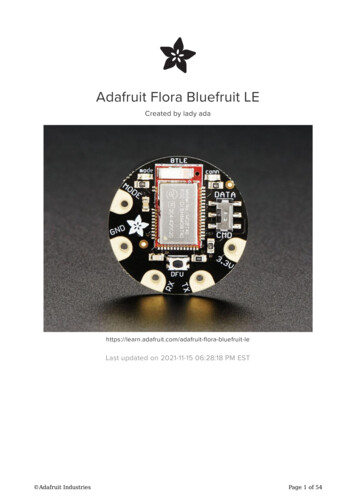
Intestinal flora in preeclampsia ratsintestinal flora with changes in the systolic bloodpressure and high-sensitivity C-reactive protein(hs-CRP) level in patients were analyzed.Evaluation StandardsEndotoxins in intestinal flora were examinedusing azo color development method, with reference value 0.1 EU/mL. Subsequently, the colonization ability of intestinal flora was evaluatedbased on the Bifidobacterium/Enterobacterium(B/E) level, with normal value 1. The distributionof intestinal flora was determined according to thedistribution of Bifidobacteria, Escherichia coli,Lactobacillus and Enterococcus. Later, intestinalmucosa permeability was detected by radiationchromatography, and 24-h urinary excretion ofmacromolecular substance 99mTc-DTPA was calculated. Examined indicators related to vascularendothelial function included endothelin-1 (ET-1,normal value 43.5 ng/L-58.4 ng/L, nitric oxide(NO, normal value 13.8 μmol/L-34.6 μmol/L),and the inflammatory cytokine hs-CRP (normalvalue 10 mg/L) were also detected.Statistical AnalysisStatistical Product and Service Solutions(SPSS) 20.0 (IBM Corp., Armonk, NY, USA)was adopted for statistical analysis. Measurementdata were expressed as mean standard deviation ( x s). The comparison of the mean and ratebetween two groups was conducted via t-test andχ2-test, respectively. p 0.05 was considered statistically significant.ResultsChanges in the Serum Endotoxin LevelDuring Intervention in the two GroupsBefore and at 3 days and 1 week after intervention, serum endotoxin level in the observation group was (1.31 0.30) EU/mL, (0.56 0.13)EU/mL, and (0.06 0.01) EU/mL, respectively.Figure 1. Changes in serum endotoxin level duringintervention in the two groups.Meanwhile, serum endotoxin level in the controlgroup was (1.30 0.30) EU/mL, (1.12 0.28) EU/mL, and (0.86 0.17) EU/mL, respectively. Theresults showed that at 3 days and 1 week afterintervention, the serum level of endotoxin inobservation group was notably lower than thatin control group at the same period (t 11.473 and29.711, p 0.000) (Figure 1).Comparison of 24-h Urinary99mTc-DTPA Excretion Rate andIntestinal Flora Colonization AbilityBetween the two GroupsFollowing intervention, the 24-h urinary99mTc-DTPA excretion rate (p 0.05) was remarkably higher and the intestinal flora colonization ability (p 0.05) was markedly stronger inthe observation group than those in the controlgroup (Table I).Distribution of Intestinal Flora AfterIntervention in the two GroupsObservation group exhibited significantlyhigher levels of Bifidobacteria and LactobacillusTable I. Comparison of the 24-h urinary 99mTc-DTPA excretion rate and the colonization ability of intestinal flora afterintervention between the two groups (x– s).24-h urinary 99mTc-DTPAexcretion rate (%)Colonization ability ofintestinal floraObservation group12.1 1.51.3 0.2Control group 9.1 0.70.8 0.1t11.46214.142p 0.000 0.00010157
B.-M. Sun, L. Meng, H. Liu, D. BaoTable II. Distribution of intestinal flora after intervention in the two groups (LgN/g, x– s).BifidobacteriaLactobacillusEscherichia coliEnterococcus8.1 0.86.9 0.67.5890.0009.1 0.89.0 0.80.5590.5787.3 0.67.3 0.50.0001.000Observation group9.3 0.9Control group7.6 0.5t10.443p 0.000in intestinal flora than the control group (p 0.05).However, no evident differences were observedin the levels of Escherichia coli and Enterococcus between the two groups (p 0.05) (Table II).Comparison of Vascular EndothelialFunction Between the two GroupsAfter intervention, ET-1 level in the observation group was remarkably lower than that in thecontrol group (p 0.05). However, NO level inobservation group was higher than control group(p 0.05) (Table III).Comparison of Blood Pressure AfterIntervention Between the two GroupsSystolic blood pressure and diastolic bloodpressure were basically normal after interventionin observation group, which were both markedlylower than those in the control group (p 0.05)(Table IV).Changing Trend of InflammatoryCytokines During Intervention inthe two GroupsBefore and at 3 days and 1 week after intervention, hs-CRP in observation group was(23.3 1.8) mg/L, (10.3 0.2) mg/L, and (6.8 0.2)mg/L, respectively. However, hs-CRP in the control group was (23.4 1.8) mg/L, (18.5 1.3) mg/L,and (13.3 0.8) mg/L, respectively. These resultssuggested that the level of serum inflammatory cytokine hs-CRP in observation group wasprominently lower than that in control group at 3days and 1 week after intervention (t 39.429 and49.853, p 0.000) (Figure 2).Table IV. Comparison of blood pressure after interventionbetween the two groups (mmHg, x– s).Systolic bloodpressureDiastolic bloodpressureObservation group 134.5 3.980.6 3.1Control group155.8 5.198.8 5.3t20.98218.747p 0.000 0.000Correlation Analyses of IntestinalFlora Colonization Ability withChanges in Systolic Blood Pressureand hs-CRP Level in PatientsIntestinal flora colonization ability was foundnegatively associated with changes in systolic blood pressure and hs-CRP level in patients(p 0.05) (Figure 3-4).DiscussionThe species and quantity of intestinal flora inmammals are generally in a relatively balancedstate. They may vary with changes in diet, en-Table III. Comparison of vascular endothelial function afterintervention between the two groups (x– s).ET-1(ng/L)NO(μmol/L)Observation group37.4 2.862.3 3.9Control group62.6 5.236.5 1.8t26.98637.988p 0.000 0.00010158Figure 2. Changing trend of inflammatory cytokinesduring intervention in the two groups.
Intestinal flora in preeclampsia ratsFigure 3. Correlation analysis of the colonization abilityof intestinal flora with systolic blood pressure in patients.docrine status, sanitary conditions, diseases, andage7. The composition of intestinal flora in pregnancy stage will change significantly. In particular, the diversity of maternal intestinal flora inthe third trimester of pregnancy will be evidentlyreduced. This may be accompanied by increasedactinomycetes and deformities, decreased levelsof Lactobacillus and Bifidobacteria, as well as theappearance of physiological enteritis and dyspepsia8. Safari and Girard9 have illustrated that thenumber of soft-walled bacteria increases significantly in the intestinal flora of the Asian population in the second and third trimester of pregnancy. However, pregnant women with preeclampsiahave enhanced vascular tension and permeability,weakened body immunity and increased asepticinflammatory responses due to changes in theneurotransmitter level in the autonomic nervoussystem. Ultimately, this may increase pathogenicFigure 4. Correlation analysis of the colonization ability ofintestinal flora with hs-CRP level.microorganisms, such as Clostridium perfringensand Brefeldin in the intestinal tract, and distinctively reduce probiotics, especially Coprococcus,Lactobacillus and Bifidobacteria10.To better maintain the balance of intestinal florain pregnant women with preeclampsia, probioticswere mainly used for intervention in this study.The changes in the serum endotoxin level betweenthe two groups during intervention were compared. The results revealed that serum endotoxinlevel in the observation group was remarkablylower than that in the control group at the same period (at 3 days and 1 week after intervention). Thissuggested that probiotic therapy for preeclampsiarats could efficaciously reduce the level of serumendotoxin in rats. The 24-h urinary 99mTc-DTPAexcretion rate and intestinal flora colonization ability were compared between the two groups as well.Our results discovered that following intervention,the 24-h urinary 99mTc-DTPA excretion rate wasevidently higher, and the intestinal flora colonization ability was notably stronger in the observationgroup than those in the control group. It can be inferred that probiotic therapy for preeclampsia ratscan more effectively facilitate kidney metabolismand enhance the colonization ability of intestinalflora. In the meantime, the distribution of intestinal flora after intervention was compared betweenthe two groups. The results demonstrated that theobservation group exhibited significantly higherlevels of Bifidobacteria and Lactobacillus in theintestinal flora than control group. The abovefindings indicated that treating preeclampsia ratswith probiotics successfully upregulated the levelof probiotics in the intestinal tract and reducedthe number of pathogenic bacteria. According tothe comparison of the changes in vascular endothelial function and blood pressure after intervention between the two groups, it was found thatET-1 level in observation group was remarkablylower than that in the control group. However,NO level in observation group was significantlyhigher. Besides, systolic blood pressure and diastolic blood pressure in the observation groupwere basically normal, which were both overtlylower than those in the control group. It could beinferred that probiotic treatment for preeclampsiarats could reduce vascular endothelial cell damageand vascular permeability to a certain extent. Thistreatment method was also valuable for stabilizingblood pressure of preeclampsia rats. At last, thechanges in the level of the inflammatory cytokinehs-CRP during intervention were analyzed in thetwo groups. The results revealed that the level of10159
B.-M. Sun, L. Meng, H. Liu, D. Baoserum inflammatory cytokine hs-CRP in the observation group was significantly lower than thatin the control group at the same period (at 3 daysand 1 week after intervention). Furthermore, thecolonization ability of intestinal flora was discovered through correlation analyses to be negatively correlated with the changes in systolic bloodpressure and hs-CRP level in patients. The abovefindings suggested that treating preeclampsia ratswith probiotics was able to effectively enhance thecolonization ability of intestinal flora, and reduceblood pressure and the body’s inflammatory responses in rats.In this study, preeclampsia rats in observation group were intervened by probiotics to efficaciously modulate the integrity of intestinalbarrier in rats11. This may also contribute to themaintenance of intestinal flora balance in preeclampsia rats, and effectively change the metabolic abnormalities caused by pregnancy andpreeclampsia-induced hypertension12, eventuallyslowing down the progression of preeclampsia13.The reason may be that the applied probioticspromote the proteolysis and fermentation capability of probiotic strains, such as Lactobacillusand Bifidobacteria14. Ultimately, this can activateangiotensin-converting enzyme II to inhibit plasma activity and effectively lower blood pressure15.The potential phosphatidylinositol-3-kinase/protein kinase B (PI3K/Akt) signal transductionpathway of Lactobacillus is enhanced as well,which regulates vascular permeability16 and improves vascular endothelial function17. Besides,utilized probiotics are also capable of decreasingthe levels of pathogenic Enterobacteriaceae andEnterococcus in the intestinal tract of rats. Thismay further improve intestinal biological barriercapability18, modulate Th1/Th2 cytokine level19,reduce the body’s inflammatory responses of preeclampsia, and enhance the immunity capabilityof the body20.ConclusionsTo sum up, the novelty of this study was thatthe application of probiotics for the treatment ofpreeclampsia rats can effectively reduce the levelof serum endotoxin, improve the body’s capacityto eliminate metabolites and the colonizationability of intestinal flora, maintain the stabilityof intestinal flora, enhance vascular endothelial function, and reduce blood pressure and thebody’s inflammatory responses.10160Conflict of InterestThe Authors declare that they have no conflict of interests.References1) Whittemore JC, Stokes JE, Price JM, Suchodolski JS.Effects of a synbiotic on the fecal microbiome andmetabolomic profiles of healthy research cats administered clindamycin: a randomized, controlledtrial. Gut Microbes 2019; 10: 521-539.2) Dalton A, Mermier C, Zuhl M. Exercise influenceon the microbiome-gut-brain axis. Gut Microbes2019; 10: 555-568.3) L i C, Zhang J, Xu H, Chen Y, G ao S, Guo SN, HuangB, G ao R, Long Z, Jiang M. Effect of Dahuang Danpi Decoction on Lactobacillus bulgaricus growthand metabolism: in vitro study. Medicine (Baltimore) 2019; 98: e13941.4) Mirsepasi -L auridsen HC, Vallance BA, K rogfelt KA,Petersen AM. Escherichia coli pathobionts associated with inflammatory bowel disease. Clin Microbiol Rev 2019; 32: e00060-18.5) A bbasi J. Are probiotics money down the toilet? Orworse? JAMA 2019; 321: 633-635.6) Sun Y, Geng W, Pan Y, Wang J, X iao P, Wang Y.Supplementation with Lactobacillus kefiranofaciens ZW3 from Tibetan Kefir improves depression-like behavior in stressed mice by modulating the gut microbiota. Food Funct 2019; 10:925-937.7) Stadlbauer V, Horvath A, Komarova I, Schmerboeck B,Feldbacher N, K lymiuk I, Durdevic M, R ainer F, BleslA, Stiegler P, L eber B. Dysbiosis in early sepsiscan be modulated by a multispecies probiotic: arandomised controlled pilot trial. Benef Microbes2019; 10: 265-278.8) Shirani V, Jazi V, Toghyani M, A shayerizadeh A, Shari fi F, Barekatain R. Pulicaria gnaphalodes powder inbroiler diets: consequences for performance, guthealth, antioxidant enzyme activity, and fatty acidprofile. Poult Sci 2019; 98: 2577-2587.9) Safari Z, Gerard P. The links between the gut microbiome and non-alcoholic fatty liver disease(NAFLD). Cell Mol Life Sci 2019; 76: 1541-1558.10) Baranowski JR, Claud EC. Necrotizing enterocolitisand the preterm infant microbiome. Adv Exp MedBiol 2019; 1125: 25-36.11) Selma-Royo M, Tarrazo M, G arcia-M antrana I, Go mez-G allego C, S alminen S, Collado MC. Shapingmicrobiota during the first 1000 days of life. AdvExp Med Biol 2019; 1125: 3-24.12) Paparo L, Nocerino R, Di Scala C, Della GG, Di Costanzo M, Buono A, Bruno C, Berni CR. Targetingfood allergy with probiotics. Adv Exp Med Biol2019; 1125: 57-68.13) M azloom K, Siddiqi I, Covasa M. Probiotics: how effective are they in the fight against obesity? Nutrients 2019; 11: 258.
Intestinal flora in preeclampsia rats14) Moller GB, Da CGM, Nicoletto BB, A lves FD,Schneider CD. Supplementation of probiotics andits effects on physically active individuals and athletes: systematic review. Int J Sport Nutr ExercMetab 2019; 29: 481-492.15) De A lmeida CV, de C amargo MR, Russo E, A medei A.Role of diet and gut microbiota on colorectal cancer immunomodulation. World J Gastroenterol2019; 25: 151-162.16) Rigo -A drover M, K nipping K, G arssen J, van L impt K,K nol J, Franch A, C astell M, Rodriguez-L agunas MJ,Perez-C ano FJ. Prevention of rotavirus diarrhea insuckling rats by a specific fermented milk concentrate with prebiotic mixture. Nutrients 2019; 11:189.17) De Sire R, Talocco C, Petito V, Lopetuso LR, GrazianiC, G asbarrini A, Scaldaferri F. [Microbiota and in-flammatory bowel disease: an update.]. RecentiProg Med 2018; 109: 570-573.18) Wang Y, Gong L, Wu YP, Cui ZW, Wang YQ, HuangY, Zhang XP, L i WF. Oral administration of Lactobacillus rhamnosus GG to newborn piglets augments gut barrier function in pre-weaning piglets.J Zhejiang Univ Sci B 2019; 20: 180-192.19) Tan H, Zhao J, Zhang H, Zhai Q, Chen W. Novel strains of Bacteroides fragilis and Bacteroidesovatus alleviate the LPS-induced inflammation inmice. Appl Microbiol Biotechnol 2019; 103: 23532365.20) Yilmaz I, Dolar ME, Ozpinar H. Effect of administering kefir on the changes in fecal microbiotaand symptoms of inflammatory bowel disease: arandomized controlled trial. Turk J Gastroenterol2019; 30: 242-253.10161
intestinal flora4. Intestinal flora may be dysfunc-tional, thus resulting in maternal maladjustment, fetal development restriction, spontaneous abor-tion, as well as pregnancy-induced hypertension 5. Sun et al6 have indicated that the changes in in-testinal flora are related to the occurrence and de-velopment of preeclampsia to some extent. In this