Transcription
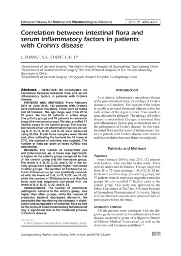
European Review for Medical and Pharmacological Sciences2017; 21: 4913-4917Correlation between intestinal flora andserum inflammatory factors in patientswith Crohn’s diseaseJ. ZHANG1, S.-L. CHEN2, L.-B. LI3Department of General Surgery, The Eighth People’s Hospital of Guangzhou, Guangdong ChinaDepartment of Gastrointestinal Surgery The First Affiliated Hospital of Sun Yat-sen University,Guangdong China3Department of General Surgery, Dongguan People’s Hospital, Guangdong China12Abstract. – OBJECTIVE: We investigated thecorrelation between intestinal flora and seruminflammatory factors in patients with Crohn’sdisease.PATIENTS AND METHODS: From February2014 to June 2016, 132 patients with Crohn’swere enrolled in this study. There were 84 malesand 48 females. The age range was from 28 to72 years. We had 62 patients in active stage(the activity group) and 70 patients in remissionstage (the remission group). We also enrolled 71healthy cases in the control group. The expression levels of serum inflammatory factors including IL-6, IL-17, IL-22, and IL-33 were measuredusing ELISA. Fresh feces samples were dilutedand, after cultivating the bacteria for 48 hours at37 C, the number of colonies was counted. Thenumber of flora per gram of feces (CFU/g) wasdetermined.RESULTS: The number of Escherichia coliand Enterococcus sp. in feces was significantly higher in the activity group compared to thatof the control group and the remission group.The levels IL-1, IL-17, L-22, and IL-33 in the activity group were significantly higher than thoseof other groups. The number of Escherichia coli and Enterococcus sp. was positively correlated with the levels of IL-6, IL-17, IL-22, and IL-33,while the number of Bifidobacteria and Bacilluslactic acid was negatively correlated with thelevels of IL-6, IL-17, IL-22, and IL-33.CONCLUSIONS: The number of conditionalpathogenic bacteria in the activity group, washigher than other groups, while the number ofprobiotics bacteria decreased distinctly. Weconcluded that monitoring the changes in distribution and composition of intestinal flora as wellas the levels of blood inflammatory factors couldplay a significant role in the treatment processof Crohn’s disease.Key Words:Crohn’s disease, Intestinal flora, Inflammatory factor.IntroductionAs a chronic inflammatory granuloma diseaseof the gastrointestinal tract, the etiology of Crohn’sdisease is still unclear1. The location of the lesionsis usually at terminal ileum and adjacent colon, buteach section of the digestive tract from mouth toanus, all could be affected2. The etiology of Crohn’sdisease is unidentified. Changes in intestinal floraand inflammatory factors play an important role inthe pathogenesis of Crohn’s disease3. In this work,intestinal flora and the levels of inflammatory factors in patients with Crohn’s disease were studied,and the correlation between them was analyzed.Patients and MethodsPatientsFrom February 2014 to June 2016, 132 patientswith Crohn’s, were enrolled in this study. Therewere 84 males and 48 females. The age range wasfrom 28 to 72 years (average 38.5 12.3). 62 patients were in active stage (the activity group), and70 patients were in remission stage (the remissiongroup). We also enrolled 71 healthy cases in thecontrol group. This study was approved by theEthics Committee of The First Affiliated Hospitalof Guangdong Pharmaceutical University. Signedwritten informed consents were obtained from allparticipants before the study.Inclusion CriteriaAll the patients were consistent with the diagnosis guideline made by the inflammatory boweldisease cooperative group of a Digestive Branchof Chinese Medical Association2, as well as theAmerican and European guidelines4,5.Corresponding Author: Jian Zhang, MM; e-mail: ze0d97@163.com4913
J. Zhang, S.-L. Chen, L.-B. LiTable I. General clinical condition.GroupsActivity groupRemission groupHealth controlT/X2 valuep-valueCasesAgeGender ratio (Male/Female)BMI627071-38.8 23.439.4 21.838.3 29.20.430.6845/1739/3134/370.580.5721.3 2.222.4 3.220.6 3.30.330.69Table II. Number of intestinal flora (CFU/g, –x s).GroupsActivity groupRemission groupHealth controlT/X2 valuep-valueCasesAgeGender ratio (Male/Female)BMI627071-38.8 23.439.4 21.838.3 29.20.430.6845/1739/3134/370.580.5721.3 2.222.4 3.220.6 3.30.330.69Exclusion Criteria(1) Patients with cardiovascular and cerebrovascular disease; (2) Patients with tumors of eachsystem, (3) Patients with autoimmune and inflammatory diseases, and (4) Patients who took probiotic preparations, antibiotics, and other drugswithin four weeks before the examination date.Serum Inflammatory FactorsThe expression levels of serum inflammatoryfactors including IL-6, IL-17, IL-22, and IL-33were measured using ELISA. All reagents used inthis study were purchased from Wuhan BoshideBiological Engineering Co., Ltd. and we followedthe instructions provided by the manufacturer.Intestinal FloraFresh feces samples (0.5 g) were collected anddiluted (10-8). 10 μl of diluted sample was takenfor inoculation. For Escherichia coli and Enterococcus sp. cultures we used aerobic medium,and for Bacillus lactic acid and Bifidobacteria weused the anaerobic medium. After cultivating thebacteria for 48 hours in 37 C, the number of colonies was counted. We then stained (gram stain)the bacteria and conducted biochemical identification. The number of bacteria per gram of feces(CFU/g) was determined.Statistical AnalysisWe used SPSS17.0 software (SPSS Inc., Chicago, IL, USA) for our statistical analysis. All measurement data were expressed as mean standard4914deviation. The variance analysis of repeated measurement data was used for statistical treatment.For comparison between groups, we used thet-test. The paired t-test was used for comparisonin each group. The chi-square test was used forenumeration data. p 0.05 meant the differencewas statistically significant.ResultsThe age, gender ratio, BMI and other indexesof subjects were compared, with no statistically significant difference among the groups( p 0.05) (Table I). The number of Escherichiacoli and Enterococcus sp. in feces was significantly higher in the activity group comparedto that of the control group and the remissiongroup. The difference was statistically significant ( p 0.05). No significant difference wasdetected between the control group and the remission group (Table II).The levels IL-1, IL-17, L-22, and IL-33 in theactivity group were significantly higher than thoseof the control group and the remission group. Thedifference was statistically significant (p 0.05).No significant difference was detected between the control group and the remission group(p 0.05) (Table III).The number of Escherichia coli and Enterococcus sp. was positively correlated with the levels of IL-6, IL-17, IL-22, and IL-33, while thenumber of Bifidobacteria and Bacillus lactic acid
Correlation between intestinal flora and serum inflammatory factors in Crohn’s diseaseTable IV. Correlation between intestinal flora and inflammatory factors.GroupsIL-1IL-17IL-22IL-33Escherichia coliEnterococcusBifidobacteriaBacillus lactic .06-0.490.170.55-1.27-0.33was negatively correlated with the levels of IL-6,IL-17, IL-22, and IL-33 (p 0.05) (Table IV).DiscussionThe bacterial flora in human digestive tractcan reach more than 500 species. They are mostly concentrated in the colon and small intestine. Under physiological conditions, these bacteria are divided into three categories, includingphysiological bacteria, conditional pathogenicbacteria and pathogenic bacteria6-8. However,due to the effect of maintaining normal functionof host gastrointestinal tract, the physiologicalbacteria and conditional pathogenic bacteriabelong to the intestinal autochthonous flora9. Insome pathological conditions, such as Crohn’sdisease, the abnormal distribution of intestinalautochthonous flora could trigger the release ofinflammatory factors10.Prior studies showed that in patients with inflammatory bowel disease in the active stage,the balance of Bifidobacteria and Bacillus lacticacid was broken. Other reports suggested thatin patients with inflammatory bowel disease,the number of Clostridium sp. declined significantly in the intestinal flora while the number ofEscherichia coli increased distinctly11. Patientswith Crohn’s disease often suffer from mucosalfissuring ulcers, which affects all layers of intestinal wall, stiffen the intestinal tube, and eventually cause intestinal obstruction12. Damage tothe intestinal tract, triggers the release of a largenumber of various inflammatory factors, whichchanges local micro-environment and affectsintestinal pH. These changes alter the intestinal micro-environment and put the pathogenicbacteria in an advantageous position13-16. Amongthe inflammatory factors, IL-1, IL-17, IL-22, andIL-33 are the most common factors17. IL-1 playsan important role in the inflammatory processof Crohn’s disease by inducing the expression ofcycloxygenase-2, phospholipase A2, nitric oxidesynthase, interferonγ and adhesion molecule18.IL-17 is largely secreted by CD4 T cells, andpromotes the generation of epithelial cells, endothelial cells, and fibroblasts. The release IL17 may accelerate the formation of the fibroticscar at the damaged part of intestinal tissues anddestroy the structure of intestinal canal19. IL-33can activate NF-κB and MAPK, promote theproduction of Th2 cytokines, participate in theallergic reaction, and also can interact with IL-6to promote the micro-thrombus in intestinalmicrocirculation 20. The joint action of the above-mentioned inflammatory factors can disturbthe intestinal environment and further affectsthe distribution of intestinal flora21. In this study,we discovered that the levels of inflammatoryfactors including IL-1, IL-17, L-22, and IL-33in the activity group were significantly higherthan those in the remission group and the healthcontrol group. Prior studies showed 22,23 that thelevel of IL-22 in patients with Crohn’s disease inactive stage increased significantly. These results suggested that there was a mutual adjustmentamongst various inflammatory factors in the pathogenesis of Crohn’s disease.Our results showed that the average number ofEscherichia coli in feces samples taken from patients with Crohn’s disease in the active stage was(8.2 2.1) CFU/g, and the average number of Enterococcus sp. was (7.9 1.7) CFU/g, which weresignificantly higher than those in other groups. Wediscovered that the Crohn’s disease in active stagehad clinical manifestation such as systemic aggravating, and was accompanied by some changes ofintestinal flora distribution and composition. Ourfindings were consistent with results reported inthe previous studies. Results obtained from previous studies24, revealed that an imbalance in intestinal flora was frequently occurred in patientswith inflammatory bowel disease. Additionally,the number of conditional pathogenic bacteria suchas Escherichia coli and Enterococcus sp. in theactivity group, was higher than other groups, whilethe number of probiotics bacteria represented byBifidobacteria and Bacillus lactic acid decreaseddistinctly. This may lead to intestinal mucosal bar4915
J. Zhang, S.-L. Chen, L.-B. Lirier dysfunction, decline in immunity, and deterioration of patient’s condition.The number of Escherichia coli and Enterococcus sp. was positively correlated with the levels of IL-6, IL-17, IL-22, and IL-33, while thenumber of Bifidobacteria and Bacillus lactic acidwas negatively correlated with the levels of IL-6,IL-17, IL-22, and IL-33. These results suggestedthat the evaluation of inflammatory factor levelscould primarily prompt the distribution of intestinal flora and also could comprehensively assessthe progression of Crohn’s disease.ConclusionsMonitoring the changes in distribution andcomposition of intestinal flora as well as the levels of blood inflammatory factors could play a significant role in the treatment process of Crohn’sdisease.Conflict of interestThe authors declare no conflicts of interest.References1) Bruè C, M ariotti C, Rossiello I, Saitta A, GiovanniniA. Demyelinizing neurological disease after treatment with tumor necrosis factor-α antagonists.Case Rep Ophthalmol 2016; 7: 345-353.2) The inflammatory bowel disease cooperative groupof Digestive branch of Chinese M edical A ssociation.Consensus on the standard of diagnosis and treatment of inflammatory bowel disease in China.Modern Digest Intervent 2008; 13: 139-145.3) A lhagamhmad MH, Day AS, L emberg DA, L each ST.An overview of the bacterial contribution to Crohndisease pathogenesis. J Med Microbiol 2016; 65:1049-1059.4) Nguyen GC, Loftus EV Jr, Hirano I, Falck-Ytter Y, Sin gh S, Sultan S, AGA Institute Clinical Guidelines Com mittee. American Gastroenterological AssociationInstitute Guideline on the Management of Crohn’sDisease After Surgical Resection. Gastroenterology 2017; 152: 271-275.5) Peyrin -Biroulet L, Bouhnik Y, Roblin X, Bonnaud G,Hagège H, Hébuterne X; gastroenterologist nominalgroup. French national consensus clinical guidelines for the management of Crohn’s disease. DigLiver Dis 2016; 48: 726-733.6) Zhu HX, Zhao J, Chen LP, Pang SJ. The diagnosticvalue of three detection techniques of ultrasound inthe active stage of Crohn’s disease. Eur Rev MedPharmacol Sci 2017; 21: 57-61.49167) Lopez J, Grinspan A. Fecal microbiota transplantation for inflammatory bowel disease. Gastroenterol Hepatol (NY) 2016; 12: 374-379.8) Huang Y, Chen Z. Inflammatory bowel disease related innate immunity and adaptive immunity. AmJ Transl Res 2016; 8: 2490-2497.9) Forbes JD, Van Domselaar G, Bernstein CN. Microbiome survey of the inflamed and noninflamed gutat different compartments within the gastrointestinal tract of inflammatory bowel disease patients.Inflamm Bowel Dis 2016; 22: 817-825.10) Wang W, Chen L, Zhou R. Increased proportionsof Bifidobacterium and the Lactobacillus groupand loss of butyrate producing bacteria in inflammatory bowel disease. J Clin Microbiol 2014; 52:398-406.11) Verbeke KA, Boesmans L, Boets E. Modulating themicrobiota in inflammatory bowel diseases: prebiotics, probiotics or faecal transplantation. ProcNutr Soc 2014; 73: 490-497.12) Paroni M, M agarotto A, Tartari S, Nizzoli G, L arghiP, Ercoli G, Gianelli U, C aprioli F. Uncontrolled IL17 Production by intraepithelial lymphocytes in acase of non-IPEX autoimmune enteropathy. ClinTransl Gastroenterol 2016; 7: e182.13) van Onkelen RS, Gosselink MP, van Meurs M, MeliefMJ, Schouten WR, L aman JD. Pro-inflammatorycytokines in cryptoglandular anal fistulas. TechColoproctol 2016; 29: 243-248.14) DeFilippis EM, Longman R, H arbus M, Dannenberg K,Scherl EJ. Crohn’s disease: evolution, epigenetics, and the emerging role of microbiome. Targeted Therapies Curr Gastroenterol Rep 2016; 18:13.15) Xiao Y, Wang XQ, Yu Y, Guo Y, Xu X, Gong L, ZhouT, L i XQ, Xu CD. Comprehensive mutation screening for 10 genes in Chinese patients sufferingvery early onset inflammatory bowel disease.World J Gastroenterol 2016; 22: 5578-5588.16) Wang Z, Wang Z, Wang J, Diao Y, Qian X, Zhu N.T-bet-expressing B cells are positively associatedwith Crohn’s disease activity and support Th1 inflammation. DNA Cell Biol 2016; 35: 628-635.17) Paroni M, M agarotto A, Tartari S, Nizzoli G, L arghiP, Ercoli G, Gianelli U, Pagani M, Elli L, A brignaniS, Conte D, Geginat J, C aprioli F. Uncontrolled IL17 production by intraepithelial lymphocytes in acase of non-IPEX autoimmune enteropathy. ClinTransl Gastroenterol 2016; 7: e182.18) Edison N, Belhanes-Peled H, Eitan Y, Guthmann Y,Yeremenko Y, R affeld M, Elmalah I, Trougouboff P.Indolent T-cell lymphoproliferative disease of thegastro-intestinal tract following treatment withAdalimumab in resistant Crohn’s colitis. Hum Pathol 2016; 8177: 30141.19) X iao Y, Wang XQ, Yu Y, G uo Y, Xu X, G ong L,Z hou T, L i XQ, Xu CD. Comprehensive mutation screening for 10 genes in Chinese patientssuffering very early onset inflammatory boweldisease. World J Gastroenterol 2016; 22: 55785588.
Correlation between intestinal flora and serum inflammatory factors in Crohn’s disease20) L ee HW, Chung SH, Moon CM, Che X, K im SW, ParkSJ, Hong SP, K im TI, K im WH, Cheon JH. The correlation of serum IL-12B expression with diseaseactivity in patients with inflammatory bowel disease. Medicine (Baltimore) 2016; 95: e3772.21) G ren ST, Janciauskiene S, S andeep S, Jonigk D, KvistPH, G erwien JG, H åk ansson K, G rip O. The protease inhibitor cystatin C down-regulates therelease of IL-β and TNF-α in lipopolysaccharideactivated monocytes. J Leukoc Biol 2016; 100:811-822.22) Faubion WA Jr, Fletcher JG, O’Byrne S. EmergingBiomARKers in Inflammatory Bowel Disease(EMBARK) study identifies fecal calprotectin, serum MMP9, and serum IL-22 as a novel combination of biomarkers for Crohn’s disease activity:role of cross-sectional imaging. Am J Gastroenterol 2013; 108: 1891-1900.23) Monteleone I, Franchi L, Biancone L, Pallone F,Monteleone G. Enhanced expression of the Faspathway inhibitor, Flip, in the mucosa of patients with Crohn’s disease. Eur Rev Med PharmacolSci 2004; 8: 192-192.24) Nemoto H, K ataoka K, Ishikawa H. Reduced diversityand imbalance of fecal microbiota in patients withulcerative colitis. Dig Dis Sci 2012; 57: 2955-29644917
bution and composition of intestinal flora as well as the levels of blood inflammatory factors could play a significant role in the treatment process of Crohn's disease. Key Words: Crohn's disease, Intestinal flora, Inflammatory factor. European Review for Medical and Pharmacological Sciences 2017; 21: 4913-4917 J. ZHANG1, S.-L. CHEN2, L.-B .