Transcription
656PRACTICE GUIDELINESnature publishing groupCMEACG Clinical Guidelines: Diagnosis and Management ofCeliac DiseaseAlberto Rubio-Tapia, MD1, Ivor D. Hill, MD2, Ciarán P. Kelly, MD3, Audrey H. Calderwood, MD4 and Joseph A. Murray, MD1Downloaded from https://journals.lww.com/ajg by BhDMf5ePHKav1zEoum1tQfN4a kJLhEZgbsIHo4XMi0hCywCX1AWnYQp/IlQrHD3 uzoGmB NjVk3YO3KzuaG30yoF7itqYn/bBlbXeAKq4 on 05/27/2019This guideline presents recommendations for the diagnosis and management of patients with celiac disease.Celiac disease is an immune-based reaction to dietary gluten (storage protein for wheat, barley, and rye) thatprimarily affects the small intestine in those with a genetic predisposition and resolves with exclusion of glutenfrom the diet. There has been a substantial increase in the prevalence of celiac disease over the last 50 yearsand an increase in the rate of diagnosis in the last 10 years. Celiac disease can present with many symptoms,including typical gastrointestinal symptoms (e.g., diarrhea, steatorrhea, weight loss, bloating, flatulence, abdominalpain) and also non-gastrointestinal abnormalities (e.g., abnormal liver function tests, iron deficiency anemia, bonedisease, skin disorders, and many other protean manifestations). Indeed, many individuals with celiac disease mayhave no symptoms at all. Celiac disease is usually detected by serologic testing of celiac-specific antibodies. Thediagnosis is confirmed by duodenal mucosal biopsies. Both serology and biopsy should be performed on a glutencontaining diet. The treatment for celiac disease is primarily a gluten-free diet (GFD), which requires significantpatient education, motivation, and follow-up. Non-responsive celiac disease occurs frequently, particularly in thosediagnosed in adulthood. Persistent or recurring symptoms should lead to a review of the patient’s original diagnosisto exclude alternative diagnoses, a review of the GFD to ensure there is no obvious gluten contamination, andserologic testing to confirm adherence with the GFD. In addition, evaluation for disorders associated with celiacdisease that could cause persistent symptoms, such as microscopic colitis, pancreatic exocrine dysfunction, andcomplications of celiac disease, such as enteropathy-associated lymphoma or refractory celiac disease, should beentertained. Newer therapeutic modalities are being studied in clinical trials, but are not yet approved for use inpractice. Given the incomplete response of many patients to a GFD-free diet as well as the difficulty of adherenceto the GFD over the long term, development of new effective therapies for symptom control and reversal ofinflammation and organ damage are needed. The prevalence of celiac disease is increasing worldwide and manypatients with celiac disease remain undiagnosed, highlighting the need for improved strategies in the future forthe optimal detection of patients.Am J Gastroenterol 2013; 108:656–676; doi:10.1038/ajg.2013.79; published online 16 April 2013INTRODUCTIONThis clinical guideline addresses the diagnosis, treatment, andoverall management of patients with celiac disease (CD), including an approach to the evaluation of non-responsive CD. Whileit is primarily directed at the care of adult patients, variationspertinent to the pediatric population have been included.Each section will provide specific recommendations basedon the current literature and a summary of the evidence supporting those recommendations. The GRADE system was used toevaluate the quality of supporting evidence (1) (Table 1). A “strong”recommendation is made when the benefits clearly outweighthe negatives and the result of no action. “Conditional” is usedwhen some uncertainty remains about the balance of benefit/potential harm. The quality of the evidence is graded fromhigh to low. “High”-quality evidence indicates that furtherresearch is unlikely to change the authors’ confidence in theestimate of effect. “Moderate”-quality evidence indicates thatfurther research would be likely to have an impact on the confidence of the estimate, whereas “Low”-quality evidence indicatesthat further study would likely have an important impact on theconfidence in the estimate of the effect and would likely changethe estimate.1Division of Gastroenterology and Hepatology, Mayo Clinic, Rochester, Minnesota, USA; 2Department of Pediatrics, Wake Forest University School of Medicine,Winston-Salem, North Carolina, USA; 3Celiac Center, Division of Gastroenterology, Beth Israel Deaconess Medical Center & Harvard Medical School,Boston, Massachusetts, USA; 4Gastroenterology, Department of Medicine, Boston University School of Medicine, Boston, Massachusetts, USA.Correspondence: Joseph A. Murray, MD, Division of Gastroenterology and Hepatology, Mayo Clinic, 200 1st Street SW, Rochester, Minnesota 55905, USA.E-mail: murray.joseph@mayo.eduReceived 8 January 2013; accepted 26 February 2013The American Journal of GASTROENTEROLOGYVOLUME 108 MAY 2013 www.amjgastro.com
Celiac Disease GuidelinesTable 1. Criteria for assigning grade of evidenceType of evidenceRandomized trial highObservational study lowAny other evidence very lowDecrease grade if Serious ( 1) or very serious ( 2) limitation to study quality Important inconsistency ( 1) Some ( 1) or major ( 2) uncertainty about directness Imprecise or sparse data ( 1) High probability of reporting bias ( 1)Increase grade if Strong evidence of association—significant relative risk of 2 ( 0.5)based on consistent evidence from two or more observationalstudies, with no plausible confounders ( 1) Very strong evidence of association—significant relative risk of 5 ( 0.2) based on direct evidence with no major threats tovalidity ( 2) Evidence of a dose–response gradient ( 1) All plausible confounders would have reduced the effect ( 1)Definition of grades of evidence High Further research is unlikely to change our confidence in theestimate of effect Moderate Further research is likely to have an important impact onour confidence in the estimate of effect and may change the estimate Low Further research is very likely to have an important impact onour confidence in the estimate of effect and is likely to change theestimate Very low Any estimate of effect is very uncertainReprinted with permission from Camilleri et al. (264).WHEN TO TEST FOR CDRecommendations(1) Patients with symptoms, signs, or laboratory evidencesuggestive of malabsorption, such as chronic diarrheawith weight loss, steatorrhea, postprandial abdominalpain, and bloating, should be tested for CD. (Strongrecommendation, high level of evidence)(2) Patients with symptoms, signs, or laboratory evidencefor which CD is a treatable cause should be consideredfor testing for CD. (Strong recommendation, moderatelevel of evidence)(3) Patients with a first-degree family member who has aconfirmed diagnosis of CD should be tested if they showpossible signs or symptoms or laboratory evidence of CD.(Strong recommendation, high level of evidence)(4) Consider testing of asymptomatic relatives with a firstdegree family member who has a confirmed diagnosisof CD. (Conditional recommendation, high level ofevidence) 2013 by the American College of Gastroenterology(5) CD should be sought among the explanations for elevatedserum aminotransferase levels when no other etiology isfound. (Strong recommendation, high level of evidence)(6) Patients with Type I diabetes mellitus (DM) should betested for CD if there are any digestive symptoms, orsigns, or laboratory evidence suggestive of CD. (Strongrecommendation, high level of evidence)Summary of the evidence. CD is one of the most common causes of chronic malabsorption (2). This results from injury to thesmall intestine with loss of absorptive surface area, reduction ofdigestive enzymes, and consequential impaired absorption ofmicronutrients such as fat-soluble vitamins, iron, and potentiallyB12 and folic acid (3). In addition, the inflammation exacerbatessymptoms of malabsorption by causing net secretion of fluidthat can result in diarrhea. The failure of absorption of adequatecalories leads to weight loss, and the malabsorption results inabdominal pain and bloating (3). These are common symptomsassociated with CD (4,5).CD remains underdiagnosed in the United States (6). CD maypresent in many ways (7). Currently, active case-finding (serologictesting for CD in patients with symptoms or conditions closelyassociated with CD) is the favored strategy to increase detection of CD (8). Active case-finding may increase detection of CDamong patients with symptoms attending a primary-care office,although this strategy is insufficient to detect most patients withCD (7). There is no consensus regarding which symptoms, laboratory abnormalities, and/or associated diseases require evaluationfor CD. The frequency of CD in common clinical scenarios variesfrom modestly elevated, such as irritable bowel syndrome, to substantially elevated, such as unexplained iron-deficiency anemia(Table 2) (9–11).The complexity of deciding who to test is exemplified bypatients with dyspepsia. The prevalence of biopsy-proven CDin patients with dyspepsia is 1%, similar to that of the generalpopulation (12), and therefore systematic screening for CD isnot recommended based on disease prevalence alone. However, treatment for dyspepsia can be a clinical challenge (13) anddyspepsia as a symptom of CD will readily respond to thegluten-free diet (GFD) (4,14). Thus, mucosal biopsies of theduodenum should be considered in patients with dyspepsia whoundergo investigation with upper endoscopy because of persistentsymptoms despite initial therapy, are aged 55 years old, and/orpresent alarm symptoms (e.g., weight loss or clinical evidence ofanemia) (15).The frequency of CD is substantially increased in patients whohave a first-degree family member affected with CD (16,17). Theprecise risk is highest in monozygous twins, next in human leukocyte antigen (HLA)-matched siblings, siblings, and finally parentsand children of patients with CD (16). A lower rate probably applies to second-degree relatives (18). Members of families whohave more than one individual already identified with CD areat higher risk of CD and recommendations for screening shouldextend to all other family members, including second-degree relatives (19). The estimates of prevalence of CD in family membersThe American Journal of GASTROENTEROLOGY657
658Rubio-Tapia et al.Table 2. Conditions in which CD occurs more frequently thanin the general population and/or for whom a GFD may bebeneficialCD common ( 2 times prevalenceof general population)CD less common but treatableSymptomatic malabsorptionPulmonary hemosiderosisDiarrhea with weight lossUnexplained male or femaleinfertilityChronic diarrhea with or withoutabdominal painDyspepsiaChronic iron deficiency and anemiaAmenorrheaMetabolic bone disease andpremature osteoporosisChronic fatiguePostprandial bloating andgaseousnessApparent malabsorption of thyroidreplacement medicationUnexplained weight lossEpilepsy or ataxiaAbnormal elevated liver enzymesConstipationIncidental discovery of villous atrophy endoscopically or histologicallyRecurrent abdominal painDermatitis herpetiformisPeripheral neuropathyOral aphthous ulcersGrowth failureDiscolored teeth or developmentallysynchronous enamel lossThyroid diseaseIrritable bowel syndromeDown’s and Turner’s syndromesvary substantially, with one large multicenter study in the UnitedStates showing a rate as low as 5% in both first- and seconddegree relatives (18). Other studies, especially those that are community-based, show a rate that is substantially higher, affectingup to 20% in siblings and 10% in other first-degree relatives (16).The clinical implications are that newly diagnosed patients withCD should inform their first-degree family members of the potential increased risk for CD and the recommendation for testing. Inaddition, health-care providers should determine whether there isa family history of CD in patients with symptoms or signs suggestive of CD and if so consider screening the patient.Testing of truly symptomless first-degree relatives is reasonablebut controversial. Even those patients who initially thought themselves to be without symptoms on direct questioning at the time ofdetection often report improved health after adapting to the GFDbecause of disappearance of symptoms that may not have beenpreviously explained (20). Others may have symptoms that theydid not consider abnormal until after they initiated a GFD andthese symptoms resolve (21). Asymptomatic patients detected byscreening do not experience new symptoms after onset of a GFD(22). The majority of patients with CD identified on the basis ofscreening reported dietary adherence and improvements in qualityThe American Journal of GASTROENTEROLOGYof life on the GFD (20). A small proportion of patients, however,reported increased health-related anxiety after diagnosis (23).Overall satisfaction with the diagnosis was high (93%) (20).Abnormal liver blood tests, in particular elevations of alanineaminotransferase and aspartate aminotransferase, are commonlyseen in clinical care, although the prevalence of clinically significant liver disease is low (24). In CD, hypertransaminasemia isoften a subclinical finding that is gluten dependent (25). Patientswith unexplained elevation of liver enzymes should be assessedfor CD (26). There are reasonable data to show that glutendependent hypertransaminasemia will normalize in most patients( 95%) on a GFD (27). Rarely, CD can be associated with severeliver disease (28,29).There is evidence that CD is substantially more common inpatients with Type I DM than in the general Caucasian population. The estimates vary between 3 and 10% (30–32). In children,it has been suggested that yearly or every-other-year screeningfor CD be undertaken utilizing serology. Patients with Type I DMwho are undergoing upper endoscopy should undergo duodenalbiopsies to rule out CD if they have never been tested previously.After gastrointestinal symptoms, the second most commonmanifestation of CD in patients with Type I DM is diminished orimpaired bone mineralization. There is some evidence suggestingthat there is added disease burden to patients already strugglingwith the management of Type I DM. In addition, there is goodevidence that gastrointestinal symptoms present at diagnosis willrespond to a GFD with overall improvement in quality of liferelated to GI symptoms. The impact of the treatment of CD onthe management of Type I DM is mixed (33). Some data suggestan increase in absorption, leading to the need for increased insulin doses. Other data suggest improvement of DM controlled byreduction of hypoglycemic events, especially postprandial.Testing for CD in asymptomatic patients with Type I DM iscontroversial. No significant adverse outcomes were identifiedin children with Type 1 DM identified by screening who delaytherapy with a GFD for up to 2 years (34). However, it is necessaryto look at the potential long-term impact of CD in Type I DM aswell (35). A large study from Sweden showed an increased risk ofdiabetic retinopathy in patients with coexistent Type I DM andCD (36). Patients with undiagnosed CD and Type 1 DM had ahigher prevalence of retinopathy (58% vs. 25%) and nephropathy (42% vs. 4%) (37). Treatment with a GFD for 1 year was safein patients with coexistent Type I DM and CD (37). The effect(if any) of a GFD on DM-related complications requires furtherinvestigation.Parents of children with Type 1 DM or the children of parents with Type 1 DM are at increased risk of CD, estimated to be 4% (38–40). Because many patients with unrecognized CD mayactually have symptoms that improve on a GFD, informing suchparents of the risk of CD is suggested. Also, a family history ofeither CD or Type 1 DM indicates an increased risk of CD in thepatient and CD should be considered. There are no data to support a recommendation about when to stop screening for CD inchildren with Type 1 DM, but screening is not necessary in theabsence of HLA-DQ2 and -DQ8.VOLUME 108 MAY 2013 www.amjgastro.com
Celiac Disease GuidelinesDIAGNOSIS OF CDRecommendations(1) Immunoglobulin A (IgA) anti-tissue transglutaminase(TTG) antibody is the preferred single test for detectionof CD in individuals over the age of 2 years. (Strongrecommendation, high level of evidence)(2) When there exists a high probability of CD wherein thepossibility of IgA deficiency is considered, total IgA shouldbe measured. An alternative approach is to include bothIgA and IgG-based testing, such as IgG-deamidated gliadin peptides (DGPs), in these high-probability patients.(Strong recommendation, moderate level of evidence)(3) In patients in whom low IgA or selective IgA deficiencyis identified, IgG-based testing (IgG DGPs and IgG TTG)should be performed. (Strong recommendation, moderatelevel of evidence)(4) If the suspicion of CD is high, intestinal biopsy shouldbe pursued even if serologies are negative. (Strongrecommendation, moderate level of evidence)(5) All diagnostic serologic testing should be donewith patients on a gluten-containing diet. (Strongrecommendation, high level of evidence)(6) Antibodies directed against native gliadin are notrecommended for the primary detection of CD. (Strongrecommendation, high level of evidence)(7) Combining several tests for CD in lieu of TTG IgA alonemay marginally increase the sensitivity for CD but reducesspecificity and therefore are not recommended in low-riskpopulations. (Conditional recommendation, moderatelevel of evidence)(8) When screening children younger than 2 years of age forCD, the IgA TTG test should be combined with DGP(IgA and IgG). (Strong recommendation, moderate levelof evidence)Summary of the evidence. The use of TTG-IgA testing and itsaccuracy in the primary-care setting and referral cohorts hasbeen extensively studied (9). The sensitivity of the TTG-IgA foruntreated CD is about 95% (41). The specificity is also 95% orgreater. The higher the titer of the test, the greater the likelihood ofa true positive result (9). The test is most commonly based on anenzyme-linked immunosorbent assay test and less commonly onradioimmunoassay (42). There are recognized differences in testperformance between the various commercially available test kits,but overall there is consistency in the sensitivity and specificity ofthe test (42–44).In the past, several antibody tests have been developed to detectCD (45). Antibodies may be directed against native or alteredcereal derived peptides. Anti-gliadin antibodies (AGA) have beenused for decades and are reasonably accurate when there is a highpretest prevalence of CD (46). However, it was with the adventof auto-antibodies, first directed against reticulin, then endomysium antibodies (EMA), and finally TTG antibodies, that the trulyceliac-specific testing was developed (47). The identification of 2013 by the American College of GastroenterologyTTG IgA antibody as the target antigen for IgA EMA antibodies was a major advance (48). This antigen was initially producedby extraction from the liver or purification from human red cellsand, most recently, by recombinant protein production. TTGbased assays have brought accurate serology for CD into the reachof most doctors and hospitals. The College of American Pathology laboratory proficiency survey has included TTG antibodytesting for several years and most laboratories in the United Statesthat provide TTG testing participate. Other similar systems are inplace outside the United States.IgA deficiency is more common in CD than in the general population. It affects anywhere between 1 in 400 to 1 in 800 membersof the general population, but occurs in 2–3% of patients with CDand 1% of those getting tested for CD (49,50). In patients in whomthere is a high pre-test prevalence of CD, the measurement of IgAlevels should be considered, especially if IgA-based celiac serologytest is negative. One approach is to measure total IgA at the beginning of testing to determine whether IgA levels are sufficient and,if not, to incorporate IgG-based testing into the serology testingcascade. DGPs IgG and/or TTG IgG would then be the preferredtest in this circumstance (51,52). EMA IgG is not widely available.It has been suggested that IgA deficiency should be consideredif the TTG-IgA levels are undetectable (53,54). However, not allassays can detect this with any accuracy or the result is merelyreported as negative. While there are limited data on the sensitivity of each of these tests for CD in an IgA-deficient person, thismay be about 80–90% individually and higher if the tests are combined. If the suspicion for CD is high, intestinal biopsy should bepursued even when serologies are negative. Finding IgA deficiency should prompt evaluation for other diseases that may cause villous atrophy, such as giardiasis, small-bowel bacterial overgrowth,or common variable immunodeficiency (55).The antibodies directed against gliadin or its deamidated products as well as the self-antigen TTG are dependent on the ingestion of gluten. The reduction or cessation of dietary gluten leadsto a decrease in the levels of all these celiac-associated antibodiesto normal concentrations. While little is known about the precisedynamics of the reduction, a weakly positive individual may become negative within weeks of strict adherence to GFD (56). After6–12 months of adhering to a GFD, 80% of subjects will test negative by serology (57). By 5 years, more than 90% of those adheringto the GFD will have negative serologies (58).While antibodies directed against native gliadin (AGA) havebeen in use for several decades, there is a wide variability in theirdiagnostic accuracy (43). Both IgA and IgG AGA have sensitivities and specificities inferior to those of the TTG-IgA and DGPIgA assays (57) and should no longer be included in the routinetesting strategy for CD.No one test for CD has a perfect sensitivity or specificity. Thus,individual tests may be combined in commercially availablepanels. This strategy may increase the sensitivity if any positivetest is regarded as an overall positive result; however, the increasedsensitivity comes at the expense of a reduction of specificity (59).Unless all patients who test positive in the panel undergo histological confirmation of CD, this practice may lead to incorrectThe American Journal of GASTROENTEROLOGY659
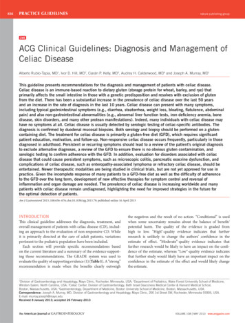
660Rubio-Tapia et al.Low probability( 5%)High probability( 5%)TTGA IgA IgAlevelPositive TTGANegative TTGALow IgADuodenal biopsyTTGA IgG DGP IgGDuodenal biopsyTTGA IgAAny positiveBoth negativeBoth positiveCD unlikelyCDNegative TTGANormal IgACD unlikelyAll negativeBiopsy/serologydisagreement HLA DQ2 and DQ8 genotyping Measure IgA level TTGA/DGPIgG Work-up for other causes ofvillous atrophy (see text)Figure 1. Celiac disease (CD) diagnostic testing algorithm. DGP, deamidated gliadin peptide; HLA, human leukocyte antigen; Ig, immunoglobulin; TTGA,tissue transglutaminase antibody.and over diagnosis followed by unnecessary treatment with GFD.Conversely, if the threshold is set that all tests within the panelmust be positive for a “positive” panel test, then the specificity andhence positive predictive value (PPV) for CD will be increased,but at the expense of sensitivity (9). One diagnostic approach isshown in Figure 1.There is some evidence that both TTG and EMA are less sensitive in young children (less than 2 years of age) (60,61). In this agegroup the sensitivity of AGA and DGP antibodies is higher thanboth the TTG and EMA (61–63). In general, AGA have a low sensitivity and specificity and are not recommended as a screeningtest for CD (64,65). Although DGP tests perform less well thanTTG and EMA tests, they are superior to the AGA (66). For thisreason it is preferable to combine the TTG with DGP tests whenscreening young children.CONFIRMATORY TESTING IN CDRecommendations(1) The confirmation of a diagnosis of CD should be basedon a combination of findings from the medical history,physical examination, serology, and upper endoscopy withhistological analysis of multiple biopsies of the duodenum.(Strong recommendation, high level of evidence)(2) Upper endoscopy with small-bowel biopsy is a criticalcomponent of the diagnostic evaluation for persons withsuspected CD and is recommended to confirm the diagnosis. (Strong recommendation, high level of evidence)(3) Multiple biopsies of the duodenum (one or two biopsiesof the bulb and at least four biopsies of the distal duodenum) are recommended to confirm the diagnosis of CD.(Strong recommendation, high level of evidence)The American Journal of GASTROENTEROLOGY(4) Lymphocytic infiltration of the intestinal epithelium in theabsence of villous atrophy is not specific for CD and othercauses should also be considered. (Strong recommendation, high level of evidence)Summary of the evidence. Gastrointestinal symptoms alonecannot accurately differentiate CD from other common gastrointestinal disorders (e.g., 20–50% of patients with CD fulfilledthe Rome criteria for irritable bowel syndrome) (4,67). A metaanalysis showed a pooled prevalence of irritable bowel syndrometype symptoms of 38% (95% confidence interval (CI), 27–50%)in patients with CD (68). Improvement of gastrointestinal symptoms or clinical exacerbation after re-introduction of gluten has avery low PPV for CD (36% and 28%, respectively) and should notbe used for diagnosis in the absence of other supportive evidence(69). Moreover, ingestion of gluten can cause gastrointestinalsymptoms including abdominal pain and bloating in the absenceof CD (70). A GFD improved gastrointestinal symptoms in about60% of patients with diarrhea-predominant irritable bowelsyndrome, especially those with HLA-DQ2 (71).A positive CD-specific serology (TTG, DGP, and EMA) inpatients with villous atrophy confirms the diagnosis of CD (43).TTG-IgA may be negative in 5–16% of patients with biopsy-confirmed CD tested when following a gluten-containing diet (41,57).IgA EMA-negative CD has been described in patients with normal IgA (72). Thus, a negative CD-specific serology in patientswith villous atrophy does not completely exclude the diagnosis ofCD though it does make it much less likely. Other causes of villousatrophy are summarized in Table 3.Histological response to GFD in patients with villous atrophystrongly supports a diagnosis of CD. HLA typing and histologicalresponse may help to rule out or confirm the diagnosis of CD inpatients with sero-negative CD (73,74).VOLUME 108 MAY 2013 www.amjgastro.com
Celiac Disease GuidelinesSmall-intestinal biopsy has been central to the confirmation of the diagnosis of CD since the late 50s (75). Traditionally,the diagnosis of CD required three intestinal biopsies: a biopsyon a gluten-containing diet (diagnosis), a biopsy after a periodon GFD, and a biopsy after a gluten challenge (76). Subsequentstudies demonstrated that a biopsy at the time of diagnosis inchildren without subsequent intestinal biopsies was able to correctly diagnose 95% of cases (77). The availability of CD-specificserological tests facilitated the recognition of many CD patientsand the wide spectrum of clinical manifestations (6,18). A positive serological test is supportive of the diagnosis but no single testis 100% specific for CD and the diagnostic accuracy varies dramatically between laboratories (43). Indeed, a large internationalstudy found that laboratory sensitivity ranged from 63 to 93% andspecificity ranged from 96 to 100% when comparing TTG assaysamong various research and clinical laboratories (42). Serologicaltests may perform less well in the clinical setting than research (apositive result of both TTG and EMA had a sensitivity of 81%) (78).Table 3. Other causes of villous atrophy in duodenumTropical sprueSmall-bowel bacterial overgrowthAutoimmune enteropathyHypogammaglobulinemic sprueDrug-associated enteropathy (e.g., olmesartan)Whipple diseaseCollagenous sprueCrohn’s diseaseEosinophilic enteritisIntestinal lymphomaIntestinal tuberculosisInfectious enteritis (e.g., giardiasis)Graft versus host diseaseMalnutritionAcquired immune deficiency syndrome enteropathyA diagnosis of CD requires the demonstration of histologicalchanges associated with the disease, which can be classified according to Marsh, Marsh modified (Oberhuber), or the more recent,simplified Corazza classification (79–81) (Table 4). Small-bowelbiopsy is also useful for the differential diagnosis of malabsorptivedisorders (82,83).A recent guideline promulgated by the European Society ofPediatric Gastroenterology, Hepatology, and Nutrition (ESPGHAN)proposed that it may be possible to avoid any intestinal biopsy inchildren who meet the following criteria: characteristic symptomsof CD, TTG IgA levels 10 upper limit of normal (confirmedwith a positive EMA in a different blood sample), and positiveHLA-DQ2 (84). A TTG antibody IgA 5 upper limit of normal was observed in 9% of 236 adult patients with suspected CDand had a PPV for CD of 86.4% (85). PPV was 97.4% among 150symptomatic children who met the “triple test” ESPGHAN criteria (86). Among 3,031 family members (25% younger than 18years old) of patients with CD, TTG antibody IgA was abnormalin 336 (11%); of these, 88 (26%) had TTG antibody IgA 100 U(87). Population-based data are not available to know how frequent the “triple test” criteria are met by unselected populations.In the absence of standardization of TTG assays, use of a predefined threshold to select a population to avoid intestinal biopsymay not be the optimal strategy (88). Prospective data to validateESPGHAN recommendation in children or adults are lacking.Histological abnormalities associated with CD can be patchy(89–93). Multiple biopsies of duodenum should be performed ifthe diagnosis of CD is considered. Among 132,352 patients without known CD who underwent duodenal biopsy in
2000 - 2009: Th e Mayo Clinic Experience. J Pediatr Gastroenterol Nutr 2013 ; 56 : 51 - 5 . 106 . Kuppa r K , Ahos nr M , nen at Il S et al. C liac e disasee wihout t villoo-urs t a phy in hcildren: a ve ospudi str pt ce . J yPdiaer 2010 ; 157 : 373 - 80 , t 380 e1 .