Transcription
CHAPTER 10ANATOMYOF THEMUSCULARSYSTEMCHAPTER OUTLINEKEY TERMSSkeletal Muscle Structure, 281Connective Tissue Components, 281Size, Shape, and Fiber Arrangement, 282Attachment of Muscles, 282Muscle Actions, 283Lever Systems, 283First-class levers, 284Second-class levers, 284Third-class levers, 284How Muscles Are Named, 285Hints on How To Deduce Muscle Actions, 286Important Skeletal Muscles, 286Muscles of Facial Expression, 287Muscles of Mastication, 288Muscles That Move the Head, 288Trunk Muscles, 289Muscles of the Thorax, 289Muscles of the Abdominal Wall, 289Muscles of the Back, 290Muscles of the Pelvic Floor, 290Upper Limb Muscles, 293Muscles Acting on the Shoulder Girdle, 293Muscles That Move the Upper Arm, 294Muscles That Move the Forearm, 295Muscles That Move the Wrist, Hand, and Fingers, 295Lower Limb Muscles, 300Muscles That Move the Thigh and Lower Leg, 301Muscles That Move the Ankle and Foot, 304Posture, 306How Posture Is Maintained, 307Cycle of Life, 308The Big Picture, 308Case Study, eprime moversynergisturvival depends on the ability to maintain a relativelyconstant internal environment. Such stability often requires movement of the body. For example, we mustgather and eat food, defend ourselves, seek shelter, and maketools, clothing, or other objects. Whereas many different systems of the body have some role in accomplishing movement, it is the skeletal and muscular systems acting togetherthat actually produce most body movements. We have investigated the architectural plan of the skeleton and have seenhow its firm supports and joint structures make movementpossible. However, bone and joints cannot move themselves.They must be moved by something. Our subject for now,then, is the large mass of skeletal muscle that moves theframework of the body: the muscular system (Figures 10-1and 10-2).Movement is one of the most characteristic and easily observed “characteristics of life.” When we walk, talk, run,breathe, or engage in a multitude of other physical activitiesthat are under the “willed” control of the individual, we doso by the contraction of skeletal muscle.There are more than 600 skeletal muscles in the body.Collectively, they constitute 40% to 50% of our body weight.And, together with the scaffolding provided by the skeleton,muscles also determine the form and contours of our body.Contraction of individual muscle cells is ultimately responsible for purposeful movement. In Chapter 11 the physiology of muscular contraction is discussed. In this preliminary chapter, however, we will learn how contractile unitsare grouped into unique functioning organs—or muscles.
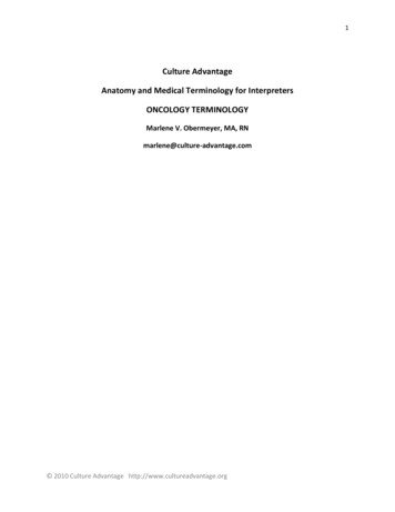
Anatomy of the Muscular System Chapter 10Facial is majorSerratus anteriorBiceps brachiiRectus abdominisLinea albaFlexors of wristand fingersExtensors of wristand fingersExternal abdominal obliqueAdductorsof thighTensor fasciae lataeRetinaculumVastus lateralisSartoriusRectus femorisVastus medialisPatellaPatellar tendonGastrocnemiusTibialis anteriorExtensor digitorumlongusPeroneus longusSoleusPeroneus brevisLSuperior extensorretinaculumSRIFigure 10-1 General overview of the body musculature. Anterior view.279
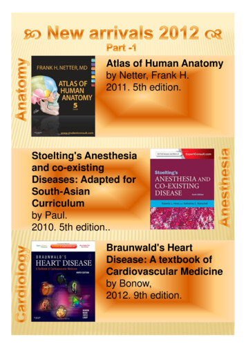
280Unit 2Support and MovementSternocleidomastoidSeventh cervical vertebraSplenius capitisTrapeziusDeltoidInfraspinatusTeres minorTeres majorTriceps brachiiLatissimus dorsiExternal abdominalobliqueExtensorsof the wristand fingersGluteus maximusSemitendinosusHamstringgroupAdductor magnusBiceps femorisSemimembranosusGracilisIliotibial tractGastrocnemiusCalcaneal tendon(Achilles tendon)Peroneus longusSoleusPeroneus brevisSLRIFigure 10-2 General overview of the body musculature. Posterior view.
Anatomy of the Muscular System Chapter 10The manner in which muscles are grouped, the relationshipof muscles to joints, and how muscles attach to the skeletondetermine purposeful body movement. A discussion of muscle shape and how muscles attach to and move bones is followed by information on specific muscles and muscle groups.The chapter will end with a review of the concept of posture.SKELETAL MUSCLE STRUCTURECONNECTIVE TISSUE COMPONENTSThe highly specialized skeletal muscle cells, or muscle fibers,are covered by a delicate connective tissue membrane calledthe endomysium (Figure 10-3). Groups of skeletal musclefibers, called fascicles, are then bound together by a tougherconnective tissue envelope called the perimysium. The muscle as a whole is covered by a coarse sheath called the epimysium. Because all three of these structures are continuouswith the fibrous structures that attach muscles to bones orother structures, muscles are firmly harnessed to the structures they pull on during contraction. The epimysium, perimysium, and endomysium of a muscle, for example, may becontinuous with fibrous tissue that extends from the muscle281as a tendon, a strong tough cord continuous at its other endwith the fibrous periosteum covering a bone. Or the fibrouswrapping of a muscle may extend as a broad, flat sheet ofconnective tissue called an aponeurosis, which usuallymerges with the fibrous wrappings of another muscle. Sotough and strong are tendons and aponeuroses that they arenot often torn, even by injuries forceful enough to breakbones or tear muscles. They are, however, occasionallypulled away from bones. Fibrous connective tissue surrounding the muscle organ and outside the epimysium andtendon is called fascia. Fascia is a general term for the fibrousconnective tissue found under the skin and surroundingmany deeper organs, including skeletal muscles and bones.Fascia just under the skin (the hypodermis) is sometimescalled superficial fascia, and the fascia around muscles andbones is sometimes called deep fascia.Tube-shaped structures of fibrous connective tissuecalled tendon sheaths enclose certain tendons, notably thoseof the wrist and ankle. Like the bursae, tendon sheaths havea lining of synovial membrane. Its moist, smooth surface enables the tendon to move easily, almost without friction, inthe tendon sheath.Figure 10-3 Structure of a muscle organ. Note that the connective tissue coverings, the epimysium, perimysium, and endomysium, are continuous with each other and with the tendon. Note also that muscle fibers areheld together by the perimysium in groups called fascicles.
282Unit 2Support and MovementABCDEFigure 10-4 Muscle shape and fiber arrangement. A, Parallel. B, Convergent. C, Pennate. D, Bipennate.E, Sphincter.fibers composing a muscle is significant because of its relationship to function. For instance, a muscle with the bipennate fiber arrangement can produce a stronger contractionthan a muscle having a parallel fiber arrangement.1. Identify the connective tissue membrane that: (a) coversindividual muscle fibers, (b) surrounds groups of skeletalmuscle fibers (fascicles), and (c) covers the muscle as awhole.2. Name the tough connective tissue cord that serves to attach amuscle to a bone.3. Name three types of fiber arrangements seen in skeletalmuscle.Figure 10-5 Attachments of a skeletal muscle. A muscle originates at a relatively stable part of the skeleton (origin) and inserts atthe skeletal part that is moved when the muscle contracts (insertion).SIZE, SHAPE, AND FIBER ARRANGEMENTThe structures called skeletal muscles are organs. They consist mainly of skeletal muscle tissue plus important connective and nervous tissue components. Skeletal muscles varyconsiderably in size, shape, and arrangement of fibers. Theyrange from extremely small strands, such as the stapediusmuscle of the middle ear, to large masses, such as the muscles of the thigh. Some skeletal muscles are broad in shapeand some are narrow. Some are long and tapering and someare short and blunt. Some are triangular, some quadrilateral,and some irregular. Some form flat sheets and others formbulky masses.Arrangement of fibers varies in different muscles. Insome muscles the fibers are parallel to the long axis of themuscle (Figure 10-4, A). In some they converge to a narrowattachment (Figure 10-4, B), and in some they are obliqueand pennate (Figure 10-4, C) like the feathers in an oldfashioned plume pen or bipennate (double-feathered) (Figure 10-4, D). Fibers may even be curved, as in the sphinctersof the face, for example (Figure 10-4, E). The direction of theATTACHMENT OF MUSCLESMost of our muscles span at least one joint and attach toboth articulating bones. When contraction occurs, one boneusually remains fixed and the other moves. The points of attachment are called the origin and insertion. The origin isthat point of attachment that does not move when the muscle contracts. Therefore the origin bone is the more stationary of the two bones at a joint when contraction occurs. Theinsertion is the point of attachment that moves when themuscle contracts (Figure 10-5). The insertion bone thereforemoves toward the origin bone when the muscle shortens. Incase you are wondering why both bones do not move, because both are pulled on by the contracting muscle, one ofthem is normally stabilized by isometric contractions ofother muscles or by certain features of its own that make itless mobile.The terms origin and insertion provide us with usefulpoints of reference. Many muscles have multiple points oforigin or insertion. Understanding the functional relationship of these attachment points during muscle contractionhelps in deducing muscle actions. Attachment points of thebiceps brachii shown in Figure 10-5 help provide functionalinformation. Distal insertion on the radius of the lower armcauses flexion to occur at the elbow when contraction occurs. It should be realized, however, that origin and insertion
Anatomy of the Muscular System Chapter 10are points that may change under certain circumstances. Forexample, not only can you grasp an object above your headand pull it down, you can also pull yourself up to the object.Although origin and insertion are convenient terms, they donot always provide the necessary information to understandthe full functional potential of muscle action.MUSCLE ACTIONSSkeletal muscles almost always act in groups rather thansingly. As a result, most movements are produced by the coordinated action of several muscles. Some of the muscles inthe group contract while others relax. The result is a movement pattern that allows for the functional classification ofmuscles or muscle groups. Several terms are used to describemuscle action during any specialized movement pattern. Theterms prime mover (agonist), antagonist, synergist, and fixatorare especially important and are discussed in the followingparagraphs. Each term suggests an important concept that isessential to an understanding of such functional muscle patterns as flexion, extension, abduction, adduction, and othermovements discussed in Chapter 9. The term prime moveror agonist is used to describe a muscle or group of musclesthat directly performs a specific movement. The movementproduced by a muscle acting as a prime mover is described asthe “action” or “function” of that muscle. For example, the biceps brachii shown in Figure 10-5 is acting as a prime moverduring flexion of the forearm.Antagonists are muscles that, when contracting, directlyoppose prime movers (agonists). They are relaxed while theprime mover is contracting to produce movement. Simultaneous contraction of a prime mover and its antagonist muscle results in rigidity and lack of motion. The term antagonist is perhaps unfortunate, because muscles cooperate,rather than oppose, in normal movement patterns. Antagonists are important in providing precision and control during contraction of prime movers.Synergists are muscles that contract at the same time asthe prime mover. They facilitate or complement primemover actions so that the prime mover produces a more effective movement.Fixator muscles generally function as joint stabilizers. Theyfrequently serve to maintain posture or balance during contraction of prime movers acting on joints in the arms and legs.Movement patterns are complex, and most muscles function not only as prime movers but also as antagonists, synergists, or fixators. A prime mover in a particular movementpattern, such as flexion, may be an antagonist during extension or a synergist or fixator in other types of movement.1. Identify the point of attachment of a muscle to a bonethat: (a) does not move when the muscle contracts; (b)moves when the muscle contracts.2. What name is used to describe a muscle that directly performsa specific movement?3. What type of muscles helps maintain posture or balance duringcontraction of muscles acting on joints in the arms and legs?4. Name the type of muscles that generally function as jointstabilizers.283LEVER SYSTEMSWhen a muscle shortens, the central body portion, called thebelly, contracts. The type and extent of movement is determined by the load or resistance that is moved, the attachment of the tendinous extremities of the muscle to bone(origin and insertion), and by the particular type of joint involved. In almost every instance, muscles that move a part donot lie over that part. Instead, the muscle belly lies proximalto the part moved. Thus muscles that move the lower arm lieproximal to it, that is, in the upper arm.Knowledge of lever systems is important in understanding muscle action. By definition, a lever is any rigid bar freeto turn about a fixed point called its fulcrum. Bones serve aslevers, and joints serve as fulcrums of these levers. A contracting muscle applies a pulling force on a bone lever at thepoint of the muscle’s attachment to the bone. This causes theinsertion bone to move about its joint fulcrum.A lever system is a simple mechanical device that makesthe work of moving a weight or other load easier. Levers arecomposed of four component parts: (1) a rigid rod or bar(bone), (2) a fixed pivot, or fulcrum (F), around which therod moves (joint), (3) a load (L) or resistan
Anatomy of the Muscular System Chapter 10 281 Figure 10-3 Structure of a muscle organ. Note that the connective tissue coverings, the epimysium, perimy-sium, and endomysium, are continuous with each other and with the tendon. Note also that muscle fibers are held together by the perimysium in groups called fascicles. SIZE, SHAPE, AND FIBER ARRANGEMENT The structures called skeletal
