Transcription
Introduction to Dental AnatomyVickie P. Overman, RDH, MEdContinuing Education Units: N/AThis continuing education course is intended for dental students and dental hygiene students. Maintainingthe teeth in a state of health is of utmost importance for complete digestion and nutrition. Not only do theteeth serve several functions in the chewing process, but they also affect our speech and appearance.Conflict of Interest Disclosure StatementThe author reports no conflicts of interest associated with this work.ADA CERPThe Procter & Gamble Company is an ADA CERP Recognized Provider.ADA CERP is a service of the American Dental Association to assist dental professionals in identifyingquality providers of continuing dental education. ADA CERP does not approve or endorse individual coursesor instructors, nor does it imply acceptance of credit hours by boards of dentistry.Concerns or complaints about a CE provider may bedirected to the provider or to ADA CERP rviewThe oral cavity is the first area in which digestion of the food we eat begins. All of surrounding andsupporting structures in the mouth contribute to the digestive process in one way or another. The majorstructures that are found in and around the oral cavity include the lips, cheeks, tongue, hard palate, softpalate, teeth, gums, salivary glands and the upper and lower jaws. Maintaining the teeth in a state of healthis of utmost importance for complete digestion and nutrition. Not only do the teeth serve several functionsin the chewing process, but they also affect our speech and appearance.Learning ObjectivesUpon the completion of this course, the dental professional will be able to: List by name and number the different teeth located in the human dentition. Define the function of each type of tooth. Identify where each type of tooth is located in the mouth. Identify the four parts of a tooth.1 Crest Oral-B at dentalcare.com Continuing Education Course, Revised May 20, 2001
Course Contents Distal – Away from the midline of the mouthBasic TerminologyThe Four Parts of A ToothTooth IdentificationTooth Numbering SystemsConclusionCourse TestReferencesAbout the AuthorBuccal – Any area on the cheek side of the teethLingual – Any area on the tongue side of theteethFacial – Any area on the cheek or lip side of theteeth. Is often used interchangeably with buccalbut mostly in the anterior portion of the mouth.Basic TerminologyBefore beginning the study of the teeth themselvesit is necessary to define some terms that are basicto learning about dental anatomy.Palatal – Any area on the tongue side of themaxillary teethOcclusal – Any area on the chewing surfaces ofback teeth.Human Dentition – The teeth that are located inthe upper and lower jaws are collectively referredto as the human dentition.Incisal – Any area on the biting surfaces of thefront teeth.Maxillae – The upper jaw is known as themaxillae.The Four Parts of A ToothEach tooth in the mouth contains four differenttissues that serve different functions. The teethare made up of two major parts: the crown andthe root. The crown of the tooth is what is visiblein the mouth (Figure 1). The root of the toothis the portion which normally not visible in themouth and is anchored within the bone (Figure 2).Within each tooth, the four different tissues thatare present are the enamel, the dentin, the pulpand the cementum.Maxillary Teeth – The teeth located in themaxillae form an arch and are referred to asmaxillary teeth.Mandible – The lower jaw is called the mandible.Mandibular Teeth – The teeth located in themandible are referred to as mandibular teeth.As humans, we have two sets of teeth during ourlifetime.1. Enamel – Makes up the protective outersurface of the crown of the tooth.2. Dentin – Makes up the majority of the innersurface of the tooth. It cannot normally beseen except on x-rays.3. Pulp – This is the area inside the tooth thatholds the nerves and blood vessels of thetooth. It is in the center of the tooth and is inboth the crown and the root of the tooth.4. Cementum – Makes up the outer surface ofthe root of the tooth. It is much softer thanenamel.Primary Dentition – The first set of teeth we get.These are often referred to as baby teeth. Thereare 20 teeth in the primary dentition.Permanent Dentition – The second set of teethwe get. These are often referred to as adult teeth.There are 32 teeth in the permanent dentition.There are several terms that help to definelocations on and around the teeth. These termsare used often to refer to specific areas of themouth when describing conditions there.Tooth IdentificationIn both the maxillary and mandibular arch thereare similar teeth. There are four types of teethin both arches. These include the incisors, thecanines, the premolars and the molars. Each ofthese teeth are located in a different area of themouth and serve different functions (Figure 3).Posterior – Towards the back of the mouth.Anterior – Towards the front of the mouthMesial – Towards the midline of the mouth.2 Crest Oral-B at dentalcare.com Continuing Education Course, Revised May 20, 2001
Figure 1. Crown of the tooth.Figure 3. Four Types of Teeth.4 premolars in each arch and two are locatedbehind each canine in the arch. These teeth aresmaller than the molars and are responsible forcrushing food in the chewing process. Theseteeth are also only present in the permanentdentition. The primary dentition only consists ofincisors, canines and molars.Figure 2. Root of the tooth.Incisors – The four front teeth in the mouth areknown as incisors. They are located in boththe maxillary and mandibular arches. The twocenter teeth are known as central incisors and theteeth on either side of them are known as lateralincisors. All of these teeth are responsible forcutting or biting food. They act like scissors.Molars – There are normally 6 molars in eacharch; three on the left and three on the right side.They are referred to as first, second and thirdmolars. Some people never develop third molarsand often these are the molars that are so farback in the mouth that they have difficulty comingin and may have to be taken out. The role of themolars in chewing is to grind the food.Canines – The teeth located distal to the lateralincisors are known as canines. These teethform the corners of the mouth. There are 2canines in the maxillary arch and 2 canines in themandibular arch. These teeth are responsible fortearing food particles when chewing.Tooth Numbering SystemsIn order to effectively and efficiently refer toteeth we often use numbering or letteringsystems. There are several systems that areused throughout the world. These includePremolars – The teeth located distal to thecanines are known as premolars. There are3 Crest Oral-B at dentalcare.com Continuing Education Course, Revised May 20, 2001
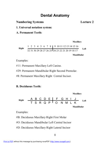
Figure 5. Universal Numbering Systemfor Primary Dentition Phase.Figure 4. Universal Numbering Systemfor Permanent Dentition Phase.ConclusionThe teeth have two major parts, the crown andthe root. When looking at a cross section of thetooth the four main tissues that make up the toothare the enamel, the dentin, the cementum and thepulp. All of these parts play important roles in theproper functioning of the dentition.the Universal Numbering System, the PalmerNotation System and the International NumberingSystem. The most widely used system in U.S.dental schools is the Universal NumberingSystem. This consists of assigning numbers tothe teeth in the permanent dentition from 1 to32 starting with the upper right third molar andcontinuing over to the upper left third molar andthen down to the lower left third molar and ontoto the lower right third molar. For example: Themandibular right canine tooth would be tooth #27(Figure 4).The primary dentition is made up of 20 teeth, whilethe permanent or adult dentition contains 32 teeth.Most dental professionals refer to a numbering orlettering system when identifying the teeth. Thereare four main types of teeth, the incisors, thecanines, the premolars and the molars. Each typeof tooth serves a different function in eating.Using the Universal Numbering System theprimary dentition is identified using letters.Beginning at the second molar on the upper right,the teeth in the maxillary arch are assigned lettersA – J. Then continuing with the mandibular leftsecond molar and around to the mandibular rightsecond molar, the teeth are assigned letters K – T(Figure 5).Understanding dental anatomy is essential inorder to begin to develop an appreciation for therole of teeth in digestion, appearance, speechand sensory input. Without the proper function ofthe teeth, usually due to disease such as decayor cavities, a person's health, appearance andnutrition can be affected.4 Crest Oral-B at dentalcare.com Continuing Education Course, Revised May 20, 2001
To receive Continuing Education credit for this course, you must complete the online test. Pleasego to www.dentalcare.com and find this course in the Continuing Education section.Course Test Preview1.Which teeth are responsible for tearing food?a. Caninesb. Molarsc. Central incisorsd. Premolars2.How many teeth are in the primary dentition?a. 32b. 10c. 20d. 153.Which tooth tissue contains the blood vessels and nerves of the tooth?a. Enamelb. Pulpc. Cementumd. Dentin4.Where are the mandibular teeth located?a. Upper jawb. Lower jawc. Posteriord. Anterior5.What is the area that is referred to when talking about the surface of the tooth that is on thecheek side?a. Buccalb. Lingualc. Palatald. Occlusal6.What number refers to the lower right canine tooth?a. 32b. 22c. 27d. 287.What tissue covers the outer surface of the crown of the tooth?a. Enamelb. Dentinc. Pulpd. Cementum8.How many molars are located in each arch?a. 6b. 3c. 2d. 125 Crest Oral-B at dentalcare.com Continuing Education Course, Revised May 20, 2001
9.The premolars are located in what relationship to the canines?a. Mesial to themb. Distal to themc. Lingual to themd. Facial to them10.Tooth #b refers to which tooth?a. The permanent maxillary right first molarb. The primary mandibular right second molarc. The permanent mandibular left second molard. The primary maxillary right first molar6 Crest Oral-B at dentalcare.com Continuing Education Course, Revised May 20, 2001
References1. Woelfel, JB, RC Scheid. Dental Anatomy, Its Relevance to Dentistry. Fifth ed. Williams and Wilkins,Baltimore. 1997, pp. 1-118.About the AuthorVickie P. Overman, RDH, MEdClinical Associate ProfessorUniversity of North Carolina School of DentistryCB# 7450Chapel Hill, NC 27599-7450Telephone: 919-966-2800Fax: 919-966-6761E-mail: vickie overman@dentistry.unc.edu7 Crest Oral-B at dentalcare.com Continuing Education Course, Revised May 20, 2001
Dental Anatomy, Its Relevance to Dentistry. Fifth ed. Williams and Wilkins, Baltimore. 1997, pp. 1-118. About the Author Vickie P. Overman, RDH, MEd Clinical A ssociate Professor University of North Carolina School of Dentistry CB# 7450 Chapel Hill, NC 27599-7450 Telephone: 919-966-2800 Fax: 919-966-6761 E-mail: vickie_overman@dentistry.unc.edu. Title : ce104 Author: P&G Crest Oral-B Subject .File Size: 482KBPage Count: 7