Transcription
Therapeutics and Clinical Risk ManagementDovepressopen access to scientific and medical researchReviewTherapeutics and Clinical Risk Management downloaded from https://www.dovepress.com/ by 54.70.40.11 on 20-Aug-2018For personal use only.Open Access Full Text ArticleFat embolism after fractures in Duchennemuscular dystrophy: an underdiagnosedcomplication? A systematic reviewThis article was published in the following Dove Press journal:Therapeutics and Clinical Risk Management10 October 2017Number of times this article has been viewedDavid Feder 1Miriam Eva Koch 1Beniamino Palmieri 2Fernando Luiz AffonsoFonseca 1Alzira Alves de SiqueiraCarvalho 3Pharmacology Department, Faculdadede Medicina do ABC, Santo André,São Paulo, Brazil; 2Departmentof General Surgery and SurgicalSpecialties, University of Modenaand Reggio Emilia Medical School,Surgical Clinic, Modena, Italy;3Neuroscience Department, Faculdadede Medicina do ABC, Santo André,São Paulo, Brazil1IntroductionCorrespondence: David FederPharmacology Department, Faculdadede Medicina do ABC, Av. Lauro Gomes,2000 Santo Andre, São Paulo 09060-870,BrazilEmail feder2005@gmail.comDuchenne muscular dystrophy (DMD) is the most frequent infantile hereditarymyopathy, an X-linked disorder, caused by a mutation in the dystrophin gene. Theproduct of this gene, dystrophin, is a protein localized in the subsarcolemmal area ofmuscle fiber and is responsible for membrane stability preventing cell damage inducedby contraction.1,2Several clinical trials have established both the beneficial effect of steroids in DMDand the well-known risk of side effects associated with their daily use. Steroids arerecommended in the international standards of care guidelines for DMD because oftheir ability to slow the progression of weakness, reduce the development of scoliosis(curvature of the spine) and delay breathing and heart problems.1,2For many years, it has been known that steroids associated with ambulation loss leadto obesity and also damage the bone structure resulting in the bone density reductionand increased incidence of bone fractures which vary between 25% cases in longbones to 19% in vertebras.1,21357submit your manuscript www.dovepress.comTherapeutics and Clinical Risk Management 2017:13 1357–1361Dovepress 2017 Feder et al. This work is published and licensed by Dove Medical Press Limited. The full terms of this license are available at https://www.dovepress.com/terms.phpand incorporate the Creative Commons Attribution – Non Commercial (unported, v3.0) License (http://creativecommons.org/licenses/by-nc/3.0/). By accessing the work youhereby accept the Terms. Non-commercial uses of the work are permitted without any further permission from Dove Medical Press Limited, provided the work is properly attributed. For permissionfor commercial use of this work, please see paragraphs 4.2 and 5 of our Terms i.org/10.2147/TCRM.S143317Powered by TCPDF (www.tcpdf.org)Abstract: Duchenne muscular dystrophy is the most frequent lethal genetic disease. Severalclinical trials have established both the beneficial effect of steroids in Duchenne muscular dystrophy and the well-known risk of side effects associated with their daily use. For many years ithas been known that steroids associated with ambulation loss lead to obesity and also damage thebone structure resulting in the bone density reduction and increased incidence of bone fracturesand fat embolism syndrome, an underdiagnosed complication after fractures. Fat embolismsyndrome is characterized by consciousness disturbance, respiratory failure and skin rashes.The use of steroids in Duchenne muscular dystrophy may result in vertebral fractures, evenwithout previous trauma. Approximately 25% of patients with Duchenne muscular dystrophyhave a long bone fracture, and 1% to 22% of fractures have a chance to develop fat embolismsyndrome. As the patients with Duchenne muscular dystrophy have progressive cardiac andrespiratory muscle dysfunction, the fat embolism may be unnoticed clinically and may result inincreased risk of death and major complications. Different treatments and prevention measuresof fat embolism have been proposed; however, so far, there is no efficient therapy. The prevention, early diagnosis and adequate symptomatic treatment are of paramount importance.The fat embolism syndrome should always be considered in patients with Duchenne musculardystrophy presenting with fractures, or an unexplained and sudden worsening of respiratoryand cardiac symptoms.Keywords: Duchenne muscular dystrophy, fat embolism syndrome, fractures
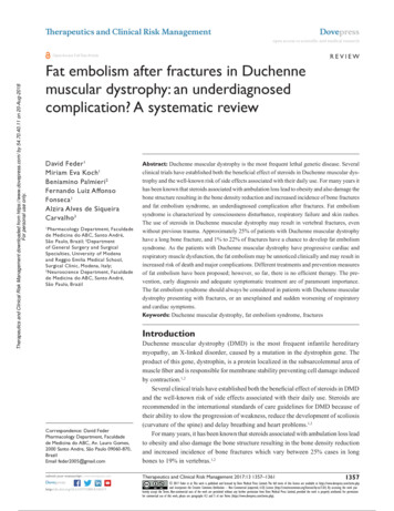
DovepressTherapeutics and Clinical Risk Management downloaded from https://www.dovepress.com/ by 54.70.40.11 on 20-Aug-2018For personal use only.Feder et alTherefore, the factors described earlier are the causeof DMD patient’s predisposition to develop fat embolismsyndrome (FES), an underdiagnosed complication after fractures, characterized by consciousness disturbance, respiratory failure and skin rashes. As the patients with DMD haveprogressive cardiac and respiratory muscle dysfunction, theFES may be unnoticed clinically and may result in increasedrisk of death and major complications. In addition, the minorcases are hardly diagnosed.3–5The presence of fat droplets in the microcirculation hasbeen found in about 95% of bone fractures and may cause afat embolism. The FES is present in about 19% of cases oftrauma being characterized by skin rash, respiratory failureand consciousness disturbance.3 However, the condition isusually asymptomatic as the embolus is destroyed beforecausing any injury.The diagnosis of FES is clinical. According to Gurd andWilson’s5 criteria, the cardinal symptoms constitute the syndrome triad: skin rash and respiratory and neurologic alterations. The minor symptoms include tachycardia, disturbanceof retina, fever and diuresis reduction.4 It also could provokethrombocytopenia over 50%, hemoglobin decrease over 20%and fatty macroglobulinemia.The clinical manifestations do not help in the early diagnosis as the symptoms may develop 24–72 h after trauma(and especially after fractures) when lipid microparticlesbecome impacted in the pulmonary microvasculature andother microvascular beds such as in the brain. The onset ofsymptoms may coincide with the agglutination and degradation of fat emboli.4,5Some hypotheses have been proposed as possible mechanisms of FES are important to highlight them. 1) The mechanictheory shows that trauma increases the intramedullary pressure, releasing lipid particles from the sinusoids, obstructingthe microcirculation.3 2) The lipase theory suggests that theincreased plasmatic level of lipase after lesion promotessaponification and causes mobilization of fat particles.3) According to the free fatty acid theory, trauma stimulatesthe inflammatory process, releasing free fatty acids, whichcause vasculitis. 4) In relation to the shock and coagulationhypothesis, trauma would lead to hypovolemia, reducing theblood flow velocity and activating the coagulation.4Different treatments and prevention measures of FEShave been proposed; however, so far, there is no efficienttherapy.6Materials and methodsThis is a systematic review of the literature. The databasesearch was finished in May 28, 2016. The content was1358Powered by TCPDF (www.tcpdf.org)submit your manuscript www.dovepress.comDovepress5HFRUGV LGHQWLILHG WKURXJK VHDUFKLQJ GDWDEDVHVQ 'XSOLFDWHG UHFRUGV UHPRYHGQ 5HFRUGV H[FOXGHG DIWHU DEVWUDFW OHFWXUHQ (OLJLEOH DUWLFOHVQ ([FOXGHG DUWLFOHV DIWHU LQFOXVLRQ FULWHULDQ 6WXGLHV LQFOXGHG LQ TXDOLWDWLYH V\QWKHVLVQ Figure 1 Literature flowchart.researched in the databases: PubMed, Lilacs, Science Direct,Wiley Online Library and HighWire. The keywords usedin the advanced search were the following: “Duchennedystrophy and fat embolism,” “Duchenne dystrophy andfracture” and “fracture and fat embolism epidemiology”(Figure 1; Table 1).ResultsAfter extensive review of the literature about FES andDMD, we found just five articles reporting patients with thisassociation. A total of 16 cases were described, and they aresummarized in Table 2.In 1992, the first case of DMD related to FES was published by Pender et al,7 describing a 12-year-old boy whosuffered a fracture followed by consciousness disturbance,respiratory failure and skin rash with a recovery.Amador et al8 reported a case of a 14-year-old boy, whosuffered a fracture after falling from the wheelchair 48 hbefore hospitalization. While the trauma was being treated,there was a neurological decay. The treatment was performedsuccessfully.McAdam et al9 presented the first article about fat embolism as a result of minor trauma without fracture in five boysTable 1 Databases and different keywordsKeywordsPubMed Lilacs Science Wiley HighWireDirect OnlineDuchenne muscular 4dystrophy fat embolismDuchenne2dystrophy fractureFracture 1fat embolism31143131122192000Therapeutics and Clinical Risk Management 2017:13
DovepressFat embolism after fractures in Duchenne muscular dystrophyTherapeutics and Clinical Risk Management downloaded from https://www.dovepress.com/ by 54.70.40.11 on 20-Aug-2018For personal use only.Table 2 Clinical data from five different reports: FES DMDStudyNAge(years)Use ofsteroidsNon-ambulantFES, majorcriteria 2FES, minorcriteria 1EvolutiondeathPender et al7Amador et al8Medeiros et al10McAdam et al1,9Stein et al1111851121414–1314–2315NoY, 1Y, 7Y, 4Y, 1YY, 1Y, 7Y, 5Y, 1Y, 1Y, 1Y, 8Y, 5Y, 1Y, 1Y, 1Y, 8Y, 5Y, 100340Note: Y, present; No, absent.Abbreviations: DMD, Duchenne muscular dystrophy; FES, fat embolism syndrome.with DMD. Four patients were taking corticosteroids dailyand had a fatal outcome. All patients fulfilled the clinicalcriteria for FES.Medeiros et al10 reported eight cases of fat embolism inpatients with DMD who had sustained low-energy trauma.Recently, Stein et al11 described a case report about apatient with DMD exhibiting cardiopulmonary, neurologicand visual signs consistent with FES after minor trauma.DiscussionIn this review, we discuss the causes of FES and fractures inpatients with DMD as well as the correlation among themand the difficulty of an accurate diagnosis.Even though FES has been known for decades, manyfactors remain undiscovered in the scientific community:could be the multifactorial pathophysiology of FES?What about the effective therapeuticapproaches and the real epidemiology ofits clinical manifestations?When examining the literature of FES, the occurrence can varygreatly among the studies reaching values of 22%, althoughthere is a great difficultly in diagnosing minor cases.12Although there is a difficulty to obtain an early diagnosis,a prompt treatment would be vital to have a better prognosis. Differential diagnosis includes aspiration pneumonia/pneumonitis, pulmonary embolism, traumatic brain injury,stroke and seizures, which could result in unhelpful diagnostics and procedures. In relation to thrombosis, conditions suchas cerebral infarction and pulmonary embolism are seriouslife-threatening complications of DMD, as activated in thecoagulation system as well.13–18Thus, it is important to keep in mind the possibility ofFES during postoperative situations or after fractures aspatients with DMD also have cardiac and respiratory muscledysfunctions from the disease itself, which could mask theright diagnosis of FES.We found only a few reports about fractures followed byFES in patients with DMD.Therapeutics and Clinical Risk Management 2017:13The occurrence of FES has also been described in5–14-year-old children experiencing long bone and pelvicfractures. It is estimated that FES incidence in children is100 times lower than in adults, and this could be due to thelower fat marrow content and small amounts of liquid triolein(glyceryl trioleate) in children’s bone marrow.19 On the otherhand, osteoporosis, obesity, immobility and steroid therapycontribute to the increased fat on the bone marrow.9Although the fat embolism is common after long bonefractures, the FES is not a well-recognized complication inpatients with DMD.The few remote cases reported could be explained by theheterogeneous presentation and problems with diagnostictesting. Except for the skin rush, there are no pathognomonicsigns. In addition, an asymptomatic latent period of about12–48 h usually precedes the symptoms.7The cases reported in the literature showed that the ageonset of FES in DMD started between 12 and 23 years old.Almost all patients were non-ambulant and were corticosteroid users suggesting that immobility and steroid use couldtrigger the FES (Table 2).The review articles stress the fragile bone formation andmuscle atrophy in patients with DMD which could predisposeto the occurrence of falls, resulting in ambulant loss. Theconsequent immobilization would predispose to obesity overloading the bone structure. Conversely, steroids increase theincidence of obesity and decrease bone density causing vertebralfractures and facilitating clinical evolution to fat embolism.Different treatments and prevention of FES have beenpublished; however, there is no efficient treatment until now.6The standard procedure consists of supporting measuresbeyond the early fixation of the broken bone8 but above all,early diagnoses should for instance be considered as part ofthe initial treatment. One of the most important measures inFES consists of ensuring good arterial oxygenation. Highflow rate oxygen is given to maintain the arterial oxygentension in the normal range.20 But in DMD, the increasedsupply of oxygen can aggravate hypoventilation and therefore lead to death.21 In patients with DMD, the oxygensubmit your manuscript www.dovepress.comDovepressPowered by TCPDF (www.tcpdf.org)1359
DovepressTherapeutics and Clinical Risk Management downloaded from https://www.dovepress.com/ by 54.70.40.11 on 20-Aug-2018For personal use only.Feder et alsupply must be made with assisted ventilation (invasive ornot).21 In addition, maintenance of intravascular volume isimportant, because shock can exacerbate the lung injurycaused by FES.20 In DMD, due to the possibility of cardiacdysfunction, the supply of fluids should be taken carefullyand use of vasoactive drugs should be important.Aiming to improve life quality, this review highlyadvocates the adoption of the DMD Care ConsiderationsWorking Group for the medical staff in charge of DMDcases.21 According to the guideline, the patients’ nutritionand fracture history needs to be detailed. In addition, thefollowing examinations should be performed to preventcomplications: calcium, phosphorus, alkaline phosphatase,25 OH vitamin D level, magnesium and parathyroid hormonelevels, urine calcium, sodium and creatinine, and annually,bone densitometry for patients at risk (history of fractures,glucocorticoid use or Z-score , 2). Back X-rays should betaken in patients with back pain or with kyphoscoliosis torule out vertebral compression fractures. Possible interventions suggested are as follows: Vitamin D supplementation incase of deficiency, calcium, intravenous bisphosphonates forvertebral compression fractures and a diet rich in vitamin Dand calcium. For patients taking steroids, it is also importantto encourage weight-bearing activities, multivitamin supplements with vitamin D3 and to consider bisphosphonatesuse.22 It would be safer that patients carry an emergency cardwarning of the risk of FES after fractures.Ness and Apkon22 advocate the identification of fractureson admission and an implementation of a mitigation planthat focuses on ensuring no child acquires a fracture whilehospitalized, especially in high-risk patients such as neuromuscular ones.In additional, the study by James et al23 revealed that themain risk factor for fractures in patients with DMD/Beckermuscular dystrophy consisted of full-time wheelchair use.Consequently, it is suggested to pay special attention to assurethe use of seatbelts, instruct the families how to perform safetransfers and explain the risks. As for fractures in ambulatoryboys, the use of internal fixation and weight-bearing measures are recommended. In addition, before any surgery, it isvital to discuss the case with the child’s neuromuscular teammembers, including cardiologist and pulmonologist.These measures can improve life quality and preventevents such as fractures and FES.ConclusionA high index of suspicion is needed to make the diagnosisof the fatal FES.1360Powered by TCPDF (www.tcpdf.org)submit your manuscript www.dovepress.comDovepressAs the patients with DMD have progressive cardiac andrespiratory muscle dysfunction, the FES may be unnoticedclinically and may result in increased risk of death and majorcomplications. So, we do recommend to always keep in mindthe diagnoses of FES.Early detection and treatment is the key to a successfuloutcome and also to reduce the progression and severity ofclinical picture.DisclosureThe authors report no conflicts of interest in this work.References1. McAdam LC, Mayo AL, Alman BA, Biggar WD. The Canadianexperience with long term deflazacort treatment in Duchenne musculardystrophy. Acta Myol. 2012;31(1):16–20.2. Morgenroth VH, Hache LP, Clemens PR. Insights into bone health inDuchenne muscular dystrophy. Bonekey Rep. 2012;1:9.3. Kwiatt ME, Seamon MJ. Fat embolism syndrome. Int J Crit Illn InjSci. 2013;3(1):64–68.4. Mellor A, Soni N. Fat embolism. Anaesthesia. 2001;56(2):145–154.5. Gurd AR, Wilson RI. The fat embolism syndrome. J Bone Joint Surg Br.1974;56B:408–416.6. Bulger EM, Smith DG, Maier RV, Jurkovich GJ. Fat embolism syndrome. A 10-year review. Arch Surg. 1997;132(4):435–439.7. Pender ES, Pollack CV Jr, Evans OB. Fat embolism syndrome in achild with muscular dystrophy. J Emerg Med. 1992;10:705–711.8. Amador EV, Villamarín FG, Quintero MP. Embolismo graso en unniño con distrofia muscular de Duchenne y fractura bilateral de fémur.Una rara asociación. Rev Fac Med. 2007;55(1):58–62.9. McAdam LC, Rastogi A, Macleod K, Douglas Biggar W. Fat embolismsyndrome following minor trauma in Duchenne muscular dystrophy.Neuromuscul Disord. 2012;22(12):1035–1039.10. Medeiros MO, Behrend C, King W, Sanders J, Kissel J, Ciafaloni E.Fat embolism syndrome in patients with Duchenne muscular dystrophy.Neurology. 2013;80(14):1350–1352.11. Stein L, Herold R, Austin A, Beer W. Fat embolism syndrome in a childwith Duchenne muscular dystrophy after minor trauma. J Emerg Med.2016;50(5):e223–e226.12. Talbot M, Schemitsch EH. Fat embolism syndrome: history, definition,epidemiology. Injury. 2006;37(4 suppl):S3–S7.13. Matsuishi T, Yano E, Terasawa K, et al. Basilar artery occlusion in a caseof Duchenne muscular dystrophy. Brain Dev. 1982;4(5):379–384.14. Biller J, Ionasescu V, Zellweger H, Adams HP, Schultz DT. Frequencyof cerebral infarction in patients with inherited neuromuscular diseases.Stroke. 1987;18(4):805–807.15. Gaffney JF, Kingston WJ, Metlay LA, Gramiak R. Left ventricular thrombus and systemic emboli complicating the cardiomyopathy of Duchenne’s muscular dystrophy. Arch Neurol. 1989;46(11):1249–1252.16. Riggs T. Cardiomyopathy and pulmonary emboli in terminal Duchenne’smuscular dystrophy. Am Heart J. 1990;119(3):690–693.17. Hanajima R, Kawai M. Incidence of cerebral infarction in Duchennemuscular dystrophy. Muscle Nerve. 1996;19(7):928.18. Saito T, Yamamoto Y, Matsumura T, Nozaki S, Fujimura H, Shinno S.Coagulation system activated in Duchenne muscular dystrophy patientswith cardiac dysfunction. Brain Dev. 2005;27(6):415–418.19. Gossling HR, Pellegrini VD Jr. Fat embolism syndrome: a review ofthe pathophysiology and physiological basis of treatment. Clin Orthop.1982;165:68–82.20. Shaikh N. Emergency management of fat embolism syndrome. J EmergTrauma Shock. 2009;2(1):29–33.Therapeutics and Clinical Risk Management 2017:13
Dovepress23. James KA, Cunniff C, Apkon SD, et al. Risk factors for first fracturesamong males with Duchenne or Becker Muscular Dystro
Keywords: Duchenne muscular dystrophy, fat embolism syndrome, fractures Introduction Duchenne muscular dystrophy (DMD) is the most frequent infantile hereditary myopathy, an X-linked disorder, caused by a mutation in the dystrophin gene. The product of this gene, dystrop