Transcription
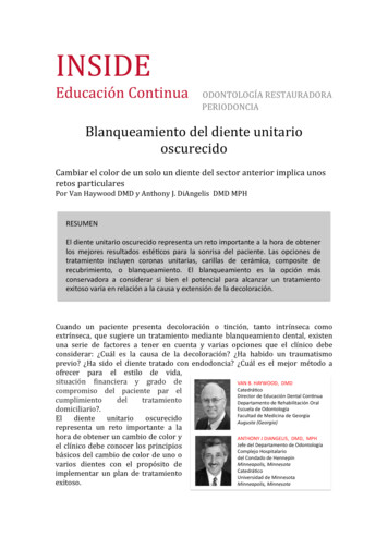
nuaBleaching the SingleODONTOLOGÍAESTAURADORAChangingthe color ofRjustone anterior tooth presents uniqByPVanERIODONCIAB. Haywood, DMD Anthony J. DiAngelis, DMD, MPChanging the color of just one anterior tooth presents uniquVanHaywood, Anthony J. DiAngelis, DMD, ingle dark teeth represent a major challenge to obtain best esthetic outcomoscurecidopatient’s smile. Treatment options may include single crowns, veneers, bondiABSTRACTbleaching. Bleaching is the most conservative option to consider, but the potforoutcomevariesbasedon theand extentof the discoloraSingledark teetharepresenta majorchallengeto obtainbest successfulectornteriorimplicaucausenospatient’s smile. Treatment options may include single crowns, veneers, bondinretosparticularesbleaching. Bleaching is the most conservative option to consider, but the Dsuccessfuloutcome varies based on the cause and extent of the discoloratmust be aware of the basic princhen a patientchanging the color of one or mopresents witheitherintrinin order to implement a successRESUMEN'aware of the basic princhena patientsic orextrin- mustmentbeplan.presentswithsic stainingor changing the color of one or o'importante'a'la'hora'de'obtener'eitherintrin- inorderto implementa msextrinlos' mejores' resultados' esté:cos' para' la' sonrisa' del' paciente.'Las'opciones'de'plan.andto mentThefirst and most importasic stainingortratamiento' incluyen' coronas' unitarias,'becarillas'de'composite'de'a candidateforcerámica,'tooth bleaching,theresiderationis to determine thdiscolorationTheInitialExaminationa variety of factors andes'optionstheof thetooth discoloration.Arecubrimiento,' o' blanqueamiento.' El' isblanqueamiento'la' hecauseconservadora' a' considerar' si' bien' el' potencial' para' alcanzar' un' tratamiento'befor tooth bleaching,is to determinethecludes evaluationof the colorfora candidatethe discoloration?Is there theretooth 'extensión'de'la'decoloración.'istraumaa varietyinvolved,of factorsorandoptionsfor theofthe andtoothAchasthe affectedteeththediscoloration.adjacent r.Whatisthecausetooth been endodontically treated?1). Additionally, loration?IstheretoothWhat is the best deliverymethod fordiographs, and pulp testing matraumainvolved,or financialhas the affectedand theadjacent gingivathe patient’slifestyle,situation, ointrínsecacomoWhatisdarkthe teethbest deliverymethodfordiographs,andpulpalpulp nancialRadiographslengefor colorchangeand thesituation,clinician propriate.any other entayvariasopcionesqueelclínicodebeand commitment level to home care?betaken2).of aFromsinglethisdarktooth, oloración?¿HahabidountraumatismoSingle dark wdetermination is made of ¿Cuálestheelclinicianmejormétodoalenge for color changeandanyothersymptomthanbecomiHAYWOOD,DMDtooth is vital or not. A vital tooProfessor(Figure2). dueFromexaminatiofrecerparaelestilodevida,be darkertothistraumaand reDirector of Dentaldeterminationismadeof g into the dentalVAN B. HAYWOOD, DMD Continuing tedrá5co out loss of vitality. Vital teethcompromisodelpacienteparelDepartment of OralProfessorDirector de Educación Dental Con5nua bedarkerduetotraumaandrediscolor from internal or orof DentalDepartamento de Rehabilitación Oral bleedingthemetamorphosisdental tubuleSchool of scuela de Odontología domiciliario?.Medical College of Georgiaoutlossofvitality.Vitalteeth ad de Medicina de Georgia EldienteunitariooscurecidoAugusta, Georgiadiscolorinternalor r ool of Dentistrysorption,calcificmetamorphosistooth mayhave becomedarker yor leakingANTHONYJ. of GeorgiaANTHONY J DIANGELIS, DMD, MPH samereasonsas a restorationsvital tooth, bAugusta,GeorgiaDMD, MPHDIANGELIS,Jefe del Departamento de Odontología proximalorlingualsurfaces.A noelclínicodebeconocerlosprincipioshave experienced pulpal death.ChiefDepartmentComplejo Hospitalario toothmayhavebecomedarkerfrthat has received endodontic trebásicosdelcambiodecolordeunooofDentistrydel Condado de Hennepin ANTHONYJ.samereasonsasavitaltooth,bmay also later darken, iosdientesconelpropósitodeDIANGELIS,DMD, MPHhaveexperiencedpulpaldeath.Catedrá5co MedicalCenteris a poor seal of the endodonticAChief d de Minnesota thathas dontic3).of erdarken,especiallyHennepin CountyEven if a tooth tests as nonProfessorMedical Centerismaya poorof theendodonticnot sealrequireendodontictheUniversity of MinnesotaMinneapolis, denMinneapolis, MinnesotaEven ifanda toothtests symptomas non-vthologyno ity of Minnesotathere is no radiographic evidenc September42 INSIDE DENTISTRY2010 insidedentistry.netMinneapolis,Minnesotathology and no clinical symptom
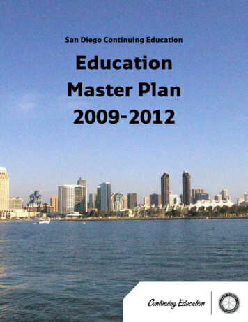
ments is indicative of connective tissueMost traumas to primary teeth areThe use of the “X” on the teeth to berepair of the fracture.2,38luxation injuries that frequently result inbleached was also helpful when the paPulp necrosis as evidenced by periapi- radiographic evidence of PCO. Althoughtient already had crowns on some teeth,cal radiolucency is an infrequent sequelathis may or may not result in crown dis- and placing bleaching material on themto PCO occurring in approximately 7%coloration, it ceases to be a concern forwas a waste of material. While this trayto 16% of cases; consequently, prophy- the patient, parent, or clinician as thesystem was simple and effective, it didnot always result in a perfect match oflactic endodontic therapy is not recom- tooth is eventually exfoliated. The onlymended by most authors.28,39-41 Teethindication for bleaching primary teeth,the teeth. All the teeth would lighten,with PCO likely have diminished heal- which are generally very light, is traumabut often the darker tooth was not ableElExamenInicialing capacity,and it is not well establishedthat caused the tooth to become dark andto lighten as much as the normal teeth,INSIDECONTINUINGand theoutcomewas lighterwhether EDUCATIONaysecondaryor addi- consideraciónthe patient is being eterminarlaresultantcausadelational dental treatment causes necro- cally by the darker teeth. There is no in- teeth, but still with one tooth hevaloraciónsis. In someinstances,suchas lprepardarker thanothers. Some youngerpatientswhohaverecommendeda a1).Adicionalmente,pueden44to the increased deposition of under- restorative procedure. A recent articlethe form of a discolored incisor presentsbleaching efficacy. It isusingnot eticwherechallenge.developmentof the toonthebleach”darkera toothtooth,either,but theuse of reslying dentin. Additionally,necesariasthere maymay ráficasytestspulpares.toothis incomplete,PCO in theervoirstherapyendodonticbefore thetherapydefinitivehas notbeen shownto increasebe a gradual diminution in responseendodonticimplementingprior permanentconservativeapproachto managingbleachingmaterialgoes throughAn improvethe use of thtray when onother teeth a(Figure 6). Intional non-scis fabricated.either side of(Figure 7 andgiven one syrial and applitooth mold aTeeth will bleto different cotermine howwill bleach firtesting. to development of a periapical radiolu- PCO-induced discoloration is bleach- the enamel and dentin to the pulp in 5to electrical and thermal pulpto 15 minutes, and bleaches under resPCO occurs more frequentlyin teeth cency in a tooth with PCO, based on two ing without endodontic therapy.torations and from one surface to thewith open apices and in more severemajor considerations: (1) the technicaldisplace- difficulty and complications that may Tray Bleachingluxation injuries involvingother (facial to lingual). It has also beenment.2,34 Extrusive and lateralluxation occur in treating these teeth; and (2) There are a number of types of bleach- shown to bleach beyond the bordersinjuries in immature permanent teeththeir review of a study that demonstrat- ing techniques to consider for both vitalof the tray, generally to the cementoeand non-vital teeth, but these types mayhave demonstrated high rates of PCO.35ed a 97.9% success rate for teeth treatednamel junction (CEJ), even if the toothbe divided mainly into those performedA recent study by Netto andcolleagueswithout periapical radiolucencies vs ais only partially erupted.reported the chances of PCO in in- 62.5% success rate for teeth treated within-office or those continued at home.The ideal bleaching tray is fabricatedtruded permanent teeth to be six timesperiapical radiolucencies.42 SpecificWith the advent of nightguard vitalon a horseshoe-shaped cast with nogreater than in mature teeth,open vs clinical situations will dictate clinical bleaching involving tray application vestibule to provide good adaptationclosed apex, and that PCO occurred indecisions; however, given the relativelyof 10% carbamide peroxide, a methodof the bleaching tray material. Also, the26.7% of such injuries.36 PCO can oc- low incidence of pulp necrosis in teethfor bleaching single dark teeth becamecast should be trimmed such that thecur in subluxated and crown-fracturedwith PCO, endodontic treatment usuallymore readily available, and did not in- central incisors are vertical to avoidteeth, although with less frequency.37is not recommended in the absence ofvolve the use of highly caustic chemi- folds on the facial. One challenge inFIG. 1FIG. 2FIG. 3FIGAs mentioned previously, PCO is aa periapical radiolucency or symptoms.cals.43 The original recommendationfabrication of the single-tooth or reguroot frac- CLINICALlar y%bleachingtray is trimming the castcommon occurrence afterNonetheless,if a periapicallesionde- un%fora singledark toothwasto makeaFig.%1 % %lateral%muy%oscurecido%un% incisivo%CLINICAL EXAMPLES (3.) A radiogra(1.) A clinical examination demonstrates a single, very dark lateral incisortures. The location of PCO is thought no-reservoirtray,andwithoutabradingteethor colorthe is related to %el%adjacentincisivo%centralcentral%adyacente%y% varias%eithererthethedarka moderatelydarkcentralincisorwitha crownonen%theincisorand severalchallengingandfraughtwithcomplicato be indicative of the type of healing. darkbleachall theteeth.Thetooththat wasgingiva.Thisoutcome isaccomplishedchamber,leaking restorations, caries,gingivalareas.(2.) Aradiographfindsno pulpchamberin theslightlydark centralincisoráreas%gingivales%oscuras.%Fig.% 2 % La%radiogra a%no%detecta%cámara%pulpar%en% el%incisivo%failedtherapy. (4.) Enda silverpointon thelateral incisor.titrated tookapproachneeded usPCO in the apical segmentonly is sug- andtions(Figure4). Theusedarkestof operatorydarkerAgenerallylonger,tosobleachingan “X” wasby trimmingthe l%más%oscurecido.%Se%attemptedindividual toothgestive of hard-tissue callus formation, ingmicroscopesthe treatments.hands of a skilledmade on that tooth mold of the trayrather than the sides (Figure5). on a tooth with calcific menecesita%una%intentaAva%evaluaAva% de%wasblanqueamiento%uAlizando% tratamientos%de% dientes%subsequent perforation and file fractuwhereas PCO in the coronal segment orclinician is warranted and improves theso the patient could continue to bleachindividualizados.%in both coronal and apical fractureseg- chances of a successful outcome.that tooth longer than the other teeth.Single-Tooth Bleaching TraySeptemberteeth2010 areinsidedentistry.netments is indicative of connective tissue44 INSIDEMostDENTISTRYtraumas to primaryThe use of the “X” on the teeth to beAn improvement on this concept isrepair of the fracture.2,38luxation injuries that frequently result inbleached was also helpful when the pa- the use of the “single-tooth” nteoscurecidoporquepuedehaberPulp necrosis as evidencedby periapi- radiographic evidence of PCO. Althoughtient already had crowns on some teeth,tray when one tooth is darker, but thecal radiolucency is an infrequentsequelathis mayor may notresultin crowndis- andmaterial on acinguelableachingdecoloración(Figura).are reasonably acceptableto PCO occurring in approximately 7%coloration, it ceases to be a concern forwas a waste of material. While this tray(Figure 6). In this tray design, a evitalpuedetrayto 16% of cases; consequently,prophypatient, parent,or clinician as thesielsystemwas simpleandvitaleffective,oit no.did Untionalnon-scalloped,no-reservoirnot always result in a perfect match oflactic endodontic therapy esultantedentrodelosnot recom- tooth is eventually exfoliated. The onlyis fabricated. Then the teeth molds onmended by most authors.28,39-41 Teethindication for bleaching primary teeth,the teeth. All the teeth would lighten,either side of the dark tooth are removedtúbulossinsuvitalidad.Eldientevitalwith PCO likely have diminishedheal- dentinarioswhich are generallyvery perderlight, is traumabut often the darkertooth was notable puede(Figure 7decolorarseand Figure 8). The porpatient ising capacity, and it is not ficante,caries,ofiltracionesqueestablishedthat caused the tooth to become dark andto lighten as much as the normal teeth,given one syringe of bleaching matewhether a secondary trauma or addi- the patient is being affected psychologi- and the resultant outcome was lighterrial and applies it only to the single aleslingualesdeltional dental treatment causesnecro- acallyby the darker teeth. Thereis no in- teeth,tooth moldoandsleeps in the appliance.stillsuperficieswith one tooth slightlydicationforendodontictherapy.sis. In some instances, ehaberseoscurecidoporlasmismasrazonesperoanding a tooth with PCO for an abutment, itIn contrast, younger patients whohave recommended using a reservoirto different color levels. The goal is to entehatherecibidomay be prudent to perform prophylacticsustain TDIswhere developmentof thethe darker tooth,but the useElof resterminequehow lightsingle dark st.If the color of thesingleendodontic therapy before G. 1FIG. 2endodóncicobasándoseúnicamenteenlosCLINICAL EXAMPLES (1.)A clinicalexaminationa single,very dark ienteunitarioand a moderately dark central incisor with a crown on the adjacent central incisor and severaldark gingival areas. (2.) decoloradoA radiograph finds no ltadodeunand a silver point on the darkest lateral incisor. A titrated approach to bleaching was needed using individual tooth clínicapudiendohaberocurrido44 INSIDE DENTISTRY September 2010 rollecualquierproblemapulpar. FIG. 3FIG. 4Fig.% 3 % La% radiogra-a% indicará% si% el% color% oscuro% está%CLINICAL EXAMPLES (3.) A radiograph will indicate whethrelacionado%con%is materiales%depositados%en% la%incámara%erthe dark colorrelated to materialsremainingthe pulppulpar,% on,reabsorción%chamber,restorations,caries, internalorfailed endodontic therapy. (4.) Endodontic therapy ttempted on a tooth with calcific metamorphosis, withFig.%4 %subsequentperforation and file fracture in the PDL.con% metamorfosis% calcificante,% con% subsecuente%perforación%y%fractura%en%el%PDL.%
exo.1- escolarhaexperimentadounaLDT.7- onesdentarias.9,14- nadasconeldeporte.15- gravante.24- ioralaslesionesporluxación(3,8- s(69- ‐73%).2,28- ricadelostestspulpares.
nasecuelainfrecuentedelaOCPqueocurresolamenteen7- oresnorecomiendalaendodonciaprofiláctica.28,39- ientesconsideracionesprincipales:1. ocurrirduranteeltratamientodeesosdientes.2. menoscabo
ntehastalauniónamelo- ienteunitario.Enestediseño, atratar(Figuras7y8).
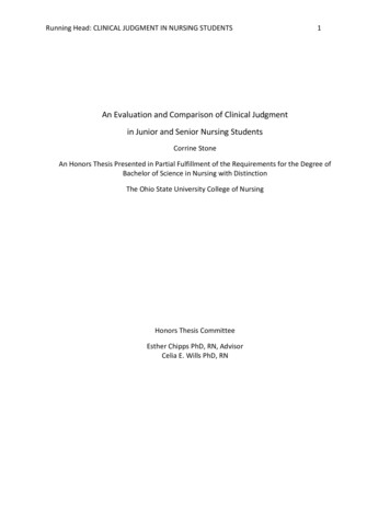
UCATIONquently restored with an acgual composite that matchedlor. However, in subsequentooth may have discoloredIn this situation, the deciching favors external bleache going inside the tooth tocomposite will weaken there 11). However, the choiceside the endodontic toothwhether the treating dentisthe extent to which the pulps debrided during endodonas well as the height in thethe cement and filler.Bleachingeaching is the oldest formg. Attempts to bleach singledate back to the 1800s, andsingle dark tooth was onebleaching research areas.45f materials have been used,en peroxide has been theorite. The high concentrarogen peroxide could beernally or internally, andved heat and light. The-vital in-office bleachingnvolved the placement ofgen peroxide into the pulpnd increasing the chemiby the use of heat or light.his technique lacks preciseo the amount of lightening.ally, when cases of externalresorption were evaluated,our common concerns listhad received trauma; 2) highons of peroxide were used;t was used to enhance thend 4) there was no seal overercha. Although the dentistrol the trauma, eliminationr three areas under dentaluld be done to lessen theresorption and loss of theer possibilities for resorpe the fact that 10% of teetha connection between thecementum, with possibleof hydrogen peroxide intoding areas, lowering the pH.ching product with a highervary catalase are attemptsesorption issues.Bleach Techniquein in-office bleaching ledstep of “walking bleaching.”nique, the gutta-percha wasmm below the CEJ and aFIG. 5FIG. 6FIG. 7FIG. 8FIG. 9CASO%EJEMPLO%1%%Fig.% 5 % Recortando% el% molde% solamente% desde% )%hasta%que% se% extrae% todo% el% vesGbulo% y% se% produce% un%hueco% en% el% paladar% evitará% el% peligro% de% dañar% los%dientes% respecto% al% recortado% tradicional% así% e%vacío.%%FigN% 6 % Los% dientes% unitarios% que% ha% dadosamente% mediante% radiograQas.% La% prácPca%más% segura% es% blanquear% este% diente% en% solitario%hasta% que% la% respuesta% del% diente% y% el% máximo%aclaramiento% pueda% ser% determinado% Fig.% 7 % La%cubeta% de% blanqueamiento% de% diente% unitario% no%Pene% reservorio% ni% espaciadores,% y% se% exPende%sobre% la% encía% 1% o% 2% mm% pero% evitando% los%movimientos% del% frenillo.% Los% dientes% que% no% van% a%blanquearse% se% recortan% de% la% cubeta% manteniendo%el% resto% de% la% cubeta% intacta.% Fig.% 8% % La% cubeta% �s%sobre% el% paladar% que% la% cubeta% tradicional% para%preservar% la% integridad% de% la% cubeta% % cuando% se%recortan% los% dientes% adyacentes.% Los% límites% de% la%cubeta% se% esconden% bajo% las% rugosidades% y% van%dispuestos% sobre% todas% las% áreas% de% tejido.% Fig.% 9% s%aproximadamente% 8% semanas% de% blanqueamiento%de% diente% unitario.% A% menudo,% los% pacientes%%interrumpen% el% tratamiento% antes% de% que% el% proporcionaalpacienteunajeringademate
had experienced a TDI.6 Approximately 30% of children have sustained a TDI to their primary dentition, and 25% of all school-aged children have experienced a TDI.7-9 Other reports document that luxations represent the majority of primary teeth injuries, whereas crown fractures constitute the most commonly