Transcription
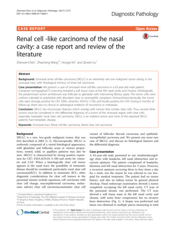
Zhenwei-Chen et al. Diagnostic Pathology (2017) 12:75DOI 10.1186/s13000-017-0660-1CASE REPORTOpen AccessRenal cell -like carcinoma of the nasalcavity: a case report and review of theliteratureZhenwei-Chen1, Zhaoming-Wang2*, Hongqi-Shi1 and Qinwei-Liu1AbstractBackground: Sinonasal renal cell-like carcinoma (SRCLC) is an extremely rare low malignant tumor arising in thesinonasal tract, with histological mimicry of renal cell carcinoma.Case presentation: We present a case of sinonasal renal cell-like carcinoma in a 63-year-old male patient.Computer tomography(CT) scanning revealed a soft tissue mass at the left nasal cavity and choana. Histologically,the predominant tumor architecture was follicular to glandular with intervening fibrous septa. The tumor cells wereuniform cuboidal to polyhedral with abundant clear or eosinophilic cytoplasm. Immunohistochemically, the tumorcells were strongly positive for CK7, EMA, vimentin, SOX10, S-100, and focally positive for CA9. During 6 months offollow-up, there was no clinical or radiological evidence of recurrence or metastasis.Conclusion: SRCLC has microscopic features which overlap with tumors that contain clear cells. Thus, several othertumors must be considered in the differential diagnosis of a tumor of the sinonasal region with clear cells,especially metastatic renal clear cell carcinoma. SRCLC is an indolent tumor and none of the reported SRCLCpatients had metastatic disease.Keywords: Sinonasal tract, Renal cell-like carcinoma, Renal clear cell carcinomaBackgroundSRCLC is a rare, low-grade malignant tumor, that wasfirst described in 2002 [1, 2]. Microscopically, SRCLC isuniformly composed of a varied histological appearance,with glandular and follicular areas in various proportions, nested, solid, or papillary patterns may also beseen. SRCLC is characterized by strong positive expression for CK7, EMA,SOX10, S-100 and rarely for vimentin and CA9. When a histologically clear cell tumorappears in the nasal tract, the possibility of metastaticdisease should be considered, particularly renal clear cellcarcinoma(RCC). In addition to metastatic RCC, otherdiagnostic considerations for clear cell tumors in theparanasal sinuses include squamous cell carcinoma withclear cell change, mucoepidermoid carcinoma, melanoma, salivary clear cell carcinoma,metastatic clear cell* Correspondence: wangzhaoming@sina.com2Department of Pathology, The First Affiliated Hospital of Zhejiang University,79 Qingchun Road, Hangzhou 310000, Zhejiang Province, People’s Republicof ChinaFull list of author information is available at the end of the articlevariant of follicular thyroid carcinoma and epithelialmyoepithelial carcinoma and. We present one more rarecase of SRCLC and discuss its histological features andthe differential diagnosis.Case presentationA 63-year-old male presented to our otorhinolaryngology clinic with headache, left nasal obstruction and recurrent epistaxis. The patient complained of headache,dizziness and left nasal obstruction for 3 years. However,a recurrent epistaxis occurring three to four times a dayfor a week, was the reason he was referred to our hospital for medical treatment. The patient had no tumorhistory, and also no kidney tumor by general physicalcheckup. Nasal endoscopy examination showed a tumorcompletely occupying the left nasal cavity. CT scan ofthe paranasal sinuses was performed. The CT scanshowed a soft tissue mass in the left nasal cavity andchoana, with some bone compression but no invasivebone destruction (Fig. 1). A biopsy was performed andtissue was obtained in multiple pieces measuring in total The Author(s). 2017 Open Access This article is distributed under the terms of the Creative Commons Attribution 4.0International License (http://creativecommons.org/licenses/by/4.0/), which permits unrestricted use, distribution, andreproduction in any medium, provided you give appropriate credit to the original author(s) and the source, provide a link tothe Creative Commons license, and indicate if changes were made. The Creative Commons Public Domain Dedication o/1.0/) applies to the data made available in this article, unless otherwise stated.
Zhenwei-Chen et al. Diagnostic Pathology (2017) 12:75Page 2 of 5Fig. 1 a A coronal CT image showing a soft tissue mass filling the left nasal cavity, causing obstruction of the left frontal and ethmoid sinuses.b Axial enhanced CT showed a soft tissue mass in the left nasal cavity featuring mild to moderate heterogeneous enhancement. The tumorextends posteriorly to the choana and nasopharyngeal region, with an indistinct border from the soft tissue of the lateral nasopharyngeal wall1.5X1X0.6 cm. Microscopically, the tumor consisted ofglands and follicles, with focal micropapillary pattern,lined by cells with abundant clear or eosinophilic cytoplasm (Fig. 2a, b). The cells were uniform, cuboidal topolyhedral and the nuclei were slightly irregular toshrunken with coarse chromatin. Several intranuclearinclusions could be seen [Fig. 2c]. Some nuclei were in alinear arrangement away from the basal aspect of thecells, usually in the center of cytoplasm or more apical,with a classic feature of clear cell papillary renal cellcarcinoma (Fig. 2d). No necrosis, mitotic activity, orperineural invasion was identified in the tumor. Immunohistochemically, the tumor cells showed strongcytoplasmic expression for CK7 (Fig. 3a), S-100 (Fig. 3b),vimentin (Fig. 3c), EMA (Fig. 3d), nuclear expression forSOX10 (Fig. 3e), focal membranous for CA9 (Fig. 3f ),and were negative for thyroglobulin, actin, calponin,PAX8, CD10, CK20, CDX2, CD117, DOG-1 and SMA.Based on the histological and immunophenotypical findings, a diagnosis of sinonasal renal cell-like carcinomawas rendered. Later, surgery was performed under general anesthesia, followed by adjuvant radiation therapy.The patient is well with no disease progression at6 months of follow up. The patient had a renal CT scanboth at the time or following the initial diagnosis. Noevidence of renal carcinoma was found.Fig. 2 a The tumor consists of nests and follicles of polyhedral cells with abundant optically clear cytoplasm. Nuclear pleomorphism and mitoticactivity are minimal. The follicles are filled with basophilic secretions(magnification 100). b There is also micropapillary configuration that lacksfibrovascular cores(magnification 100). c There are several intranuclear inclusions, which are quite unique to this tumor type (magnification 400).d Some nuclei are in linear arrangement away from the basal aspects of the cells, which is a consistent feature in clear cell papillary renal cellcarcinoma (magnification 200)
Zhenwei-Chen et al. Diagnostic Pathology (2017) 12:75Page 3 of 5Fig. 3 a Immunohistochemical examination showed positive expression of CK7(magnification 100). b Immunohistochemical examination showedpositive expression S-100(magnification 100). c Immunohistochemical examination showed positive expression of Vimentin (magnification 200).d Immunohistochemical examination showed positive expression of EMA(magnification 100). e Immunohistochemical examination showed nuclearpositive expression of SOX10(magnification 200). f Immunohistochemical examination showed focally positive expression of CA9(magnification 400)DiscussionZur et al. first described a rare sinonasal tract carcinoma in2002 as a histologic mimicker of renal clear cell carcinoma,which was named “sinonasal renal cell-like carcinoma” [1].Additional reports have been identified, and there are currently 14 patients described in the English literature [1–9].According to the review by BISHOP JA [6], the patientshad a broad age distribution range from 22 to 75 years(mean 54). There was a female predominance of 9:4. Theclinical manifestations, which are relatively nonspecific, include nasal obstruction, epistaxis, headache, epiphora andolfactory impairment. The most common site of involvement is the nasal cavity, followed by paranasal sinuses andnasopharynx [1, 3, 7–10].Microscopically, SRCLC is composed of mixtures offollicular to glandular structures with intervening fibroussepta. The neoplastic cells are uniform, cuboidal to polyhedral with abundant clear to eosinophilic cytoplasm.There are slightly irregular to shrunken nuclei with coarsechromatin and a low nuclear: cytoplasmic ratio. The mitoses are inconspicuous and tumor necrosis, perineuralinvasion are absent [1–3, 6–9]. Some neoplastic cells contain intranuclear inclusions, which are a quite distinctivefeature of this tumor [3, 7]. SRCLC is less vascular andpleomorphic than renal clear cell carcinoma. All the histological features described above are illustrated in our case.Because of the presence of clear microscopic appearance, SRCLC have to be differentiated from many neoplasms. The top differential diagnosis is metastatic renalclear cell carcinoma(RCC), which not infrequently metastasizes to the nasal cavity [11, 12]. Strong staining forCK7, S-100 and SOX10 supports SRCLC and conversely,strong staining for PAX8 and CD10 supports metastaticRCC. In our case, the tumor cells were positive for CK7,S-100, SOX10 and negative for PAX-8 and CD10, arguing against a metastatic clear cell RCC. However, sinceCA9, EMA and vimentin are also expressed in bothRCC and SRCLC [7, 13], they are of limited value indifferentiating the two tumors. Although there aredifferences in the immunophenotype, clinical historyand radiologic examinations are necessary to a definiteseparation.
Zhenwei-Chen et al. Diagnostic Pathology (2017) 12:75SRCLC also may be confused with a wide variety of tumors, including squamous cell carcinoma with clear cellchange, clear cells mucoepidermoid carcinoma, ballooncell melanoma, salivary clear cell carcinoma, metastaticclear cell variant of follicular thyroid carcinoma andepithelial-myoepithelial carcinoma.The clear-cell variant of squamous cell carcinoma alsooccurs in the sinonasal tract. When the tumor is composed entirely of clear cells, its separation from SRCLCbased on hematoxylin and eosin stained sections alone isproblematic. The presence of high molecular weightcytokeratin, such as CK5/6, P63, and 34βE12, in theclear cells is the best way to establish the squamous nature of the tumor. Mucoepidermoid carcinoma(MEC) isthe most common malignant salivary gland neoplasm,but it also arises in other rare locations, such as thesinonasal tract [14, 15]. Occasionally, clear cells predominate over other cell types, rendering distinction fromSRCLC challenging. However, the SRCLC lack the multiple, distinct cell types, such as mucinous cells, squamous cells, and intermediate cells, the latter two ofwhich are more diffusely p63 and CK5/6 positive, thattypify EMC. Salivary clear cell carcinoma (CCC), mostfrequently occurs in intraoral salivary gland sites, e.g.,the palate and the base of the tongue but it is rarelyfound in the nasal cavity [16–18]. CCC is mainly composed of polygonal epithelioid tumor cells arranged intosheets and separated by dense hyalinized stroma,whereas SRCLC can form true glandular, follicular structures, and some neoplastic cells have intranuclear inclusions. In addition, CCC has been shown to harborWESR1-ATF1 fusion gene [19], which it is easier for distinction between the two tumors. Pure clear-cell variantsof melanoma composed predominantly or entirely ofcells containing large amounts of clear, vacuolated cytoplasm, due to intracytoplasmic glycogen may be confused with SRCLC. In such cases, immunohistochemicalstaining is a valuable diagnostic adjunct, as SRCLC isuniformly positive for S100, SOX10 but negative forHMB-45 and Melan-A. Clear cell change may be presentin follicular thyroid carcinoma and appears quite vascular, a feature often seen in SRCLC. In such difficultcases, immunostains is helpful in the distinction as thyroid folliculogenic neoplasms are positive for TTF1 andthyroglobulin, both being negative in SRCLC.Epithelial-myoepithelial carcinoma (EMC) consists of abiphasic arrangement of inner luminal ductal cells andouter myoepithelial cells. Some EMC shows a classicaltubular growth, with clear, polygonal, abluminal myoepithelial cells and small eosinophilic luminal ducts, theepithelial cells are positive for cytokeratin and EMA.The clear cells stain positive for myoepithelial markers,such as calponin, S-100, and SMA, whereas SRCLC isnegative for calponin and SMA.Page 4 of 5At present, the histogenesis of SRCLC remains uncertain. New markers for seromucinous glands, includingsinonasal glands, namely DOG1, SOX10 and S100 havebeen defined [20, 21]. DOG1, a calcium-activated chloridechannel, initially described in gastrointestinal stromal tumors, but now known to be expressed in normal sinonasalseromucinous glands, it shows moderate apical luminalstaining of serous acini with weaker staining in mucousacini. SOX10, an important marker for melanocytic,schwannian and myoepithelial differentiation, is alsoexpressed in normal sinonasal seromucinous glands. S100is strongly positive in serous acini and weakly positive tonegative in the mucous acini. Our study shows that tumorcells in SRCLC are positive for two of these three seromucinous markers, suggesting possible seromucinous derivation of SRCLC. It has also been reported that markers ofseromucinous differentiation are present to varying degrees in the majority of low-grade non-intestinal-typeadenocarcinomas [21]; a term which has been used synonymously with renal cell -like carcinoma (SRCLC) in thenew edition of WHO classification of head and neck tumors [22]. With a common seromucinous phenotype, weconclude that SRCLC is likely a clear cell variant of sinonasal non-intestinal type adenocarcinoma.SRCLC has a very indolent clinical course. To date,none of the patients with SRCLC have experienced metastatic disease in the neck and distant sites [1, 3, 7, 9, 10],including our case, although the follow up is relativelyshort. However, owing to its rarity, more cases should becollected to better define short-and long-term clinical behavior, immunohistochemical features, pathogenesis andother relevant information.ConclusionsSRCLC is a very rare, low-malignant sinonasal neoplasm.The most striking feature of this tumor is its resemblance to the clear-cell type of renal cell carcinoma. Inaddition, a panel of sensitive and specific antibodieswould also be crucial in making differential diagnosis.Complete resection of this tumor and careful histologicalexamination is necessary to make a correct diagnosis.Early reports of the clinical behavior and natural historysuggest that this tumor appears to show very indolentbiological behavior with a favorable prognosis.AbbreviationsCCC: Salivary clear cell carcinoma; CT: Computer tomography;EMC: Epithelial-myoepithelial carcinoma; RCC: Renal clear cell carcinoma;SRCLC: Sinonasal renal cell-like carcinomaAcknowledgementsNot applicable.FundingNil.
Zhenwei-Chen et al. Diagnostic Pathology (2017) 12:75Availability of data and materialsThe dataset supporting the conclusions of this article is included within thearticle.Authors’ contributionsZWC and HQS made the pathological diagnosis and carried out histopathologicaland drafted manuscript. QWL performed the immunohistochemistry. ZMWparticipated in manuscript revision. All authors read and approved the finalmanuscript.Ethics approval and consent to participateNot applicable.Consent for publicationWritten informed consent was obtained from the patient for the publicationof this case report and any accompanying images.Competing interestsThe authors declare that they have no competing interests.Publisher’s NotePage 5 of 515.16.17.18.19.20.21.Springer Nature remains neutral with regard to jurisdictional claims inpublished maps and institutional affiliations.Author details1Department of Pathology, Jinhua Municipal Central Hospital, 351 MingyueRoad, Jinhua 321000, Zhejiang Province, People’s Republic of China.2Department of Pathology, The First Affiliated Hospital of Zhejiang University,79 Qingchun Road, Hangzhou 310000, Zhejiang Province, People’s Republicof China.22.case showing a large clear cell component and lacking typical epidermoidcells and intermediate cells. Med Mol Morphol. 2017;50:117–21.Peison B, Benisch B, Schwartz IS, Gordon RE. Clear-cell mucoepidermoidcarcinoma arising in the nasal cavity: case report with ultrastructuralobservations. Mt Sinai J Med. 1988;55:417–20.Suzuki K, Wanibuchi M, Akiyama Y, Ikeda J, Minamida Y, Hasegawa T, et al.Primary clear cell carcinoma of the skull base and paranasal cavity: a casereport. No Shinkei Geka. 2012;40:617–21.Imai T, Satoh I, Matsumoto K, Ito S, Asada Y, Kato K. et al.Clear cell carcinoma ofthe nasal cavity: A case report from histopathological viewpoint. Auris NasusLarynx. 2016;43:108–11.Zhao W, Yang L, Wang L, Zuo W, Yuan S, Yu J. et al.Primary clear cellcarcinoma of nasal cavity: report of six cases and review of literature. Int JClin Exp Med. 2014;7:5469–76.Antonescu CR, Katabi N, Zhang L, Sung YS, Seethala RR, Jordan RC, et al.EWSR1-ATF1 fusion is a novel and consistent finding in hyalinizing clear-cellcarcinoma of salivary gland. Genes Chromosomes Cancer. 2011;50:559–70.Chênevert J, Duvvuri U, Chiosea S, Dacic S, Cieply K, Kim J, Shiwarski D, et al.DOG1: a novel marker of salivary acinar and intercalated duct differentiation.Mod Pathol. 2012;25:919–29.Purgina B, Bastaki JM, Duvvuri U, Seethala RR. A Subset of Sinonasal NonIntestinal Type Adenocarcinomas are Truly Seromucinous Adenocarcinomas:A Morphologic and Immunophenotypic Assessment and Description of aNovel Pitfall. Head Neck Pathol. 2015;9:436–46.EI-Naggar AK, Chan JKC, Grandis JR, Takata T, et al. WHO Classification ofHead and Neck Tumours. Lyon: IAPR press; 2017. p. 24–6.Received: 11 April 2017 Accepted: 25 September 2017References1. Zur KB, Brandwein M, Wang B, Som P, Gordon R, Urken ML. Primary descriptionof a new entity, renal cell-like carcinoma of the nasal cavity: van Meegeren in thehouse of Vermeer. Arch Otolaryngol Head Neck Surg. 2002;128:441–7.2. Moh'd Hadi U, Kahwaji GJ, Mufarrij AA, Tawil A, Noureddine B. Low gradeprimary clear cell carcinoma of the sinonasal tract. Rhinology. 2002;40:44–7.3. Storck K, Hadi UM, Simpson R, Ramer M, Brandwein-Gensler M. Sinonasalrenal cell–like adenocarcinoma: a report on four patients. Head Neck Pathol.2008;2:75–80.4. Huang XJ, Chen JD, Shi QF. One case with primary clear-cell carcinoma ofnasal cavity. Zhonghua Er Bi Yan Hou Tou Jing Wai Ke Za Zhi. 2011;46:245–6.5. Negahban S, Daneshbod Y, Khademi B, Rasekhi AR, Soleimanpour H. Sinonasalclear cell adenocarcinoma: a case report. Acta Cytol. 2009;53:597–600.6. Bishop JA. Newly Described Tumor Entities in Sinonasal Tract Pathology.Head Neck Pathol. 2016;10:23–31.7. Shen T, Shi Q, Velosa C, Bai S, Thompson L, Simpson R, et al. Sinonasal renalcell-like adenocarcinomas: robust carbonic anhydrase expression. HumPathol. 2015;46:1598–606.8. Brandwein-Gensler M, Wei S. Envisioning the next WHO head and neckclassification. Head Neck Pathol. 2014;8:1–15.9. Kubik M, Barasch N, Choby G, Seethala R, Snyderman C. Sinonasal RenalCell-Like Carcinoma: Case Report and Review of the Literature. Head NeckPathol. 2016;10:1–5.10. Li P, Yin WH, Yao XJ, Wan L, Chen GR. Primary clear cell carcinoma of nasalcavity: report of a case. Zhonghua Bing Li Xue Za Zhi. 2011;40:52–3.11. Kokenek-Unal TD, Gumuskaya B, Ocal B, Alper M. A Rare Cause of NasalObstruction: Metastatic Renal Cell Carcinoma. Case Rep Pathol. 2016;2016:2560749.12. Mahajan R, Mayappa N, Prashanth V. Metastatic Renal Cell CarcinomaPresenting as Nasal Mass: Case Report and Review of Literature. Indian JOtolaryngol Head Neck Surg. 2016;68:374–6.13. Al-Ahmadie HA, Alden D, Qin LX, Olgac S, Fine SW, Gopalan A, et al.Carbonic anhydrase IX expression in clear cell renal cell carcinoma: animmunohistochemical study comparing 2 antibodies. Am J Surg Pathol.2008;32:377–82.14. Tajima S, Namiki I, Koda K. A clear cell variant of mucoepidermoid carcinomaharboring CRTC1-MAML2 fusion gene found in buccal mucosa: report of aSubmit your next manuscript to BioMed Centraland we will help you at every step: We accept pre-submission inquiries Our selector tool helps you to find the most relevant journal We provide round the clock customer support Convenient online submission Thorough peer review Inclusion in PubMed and all major indexing services Maximum visibility for your researchSubmit your manuscript atwww.biomedcentral.com/submit
Nasal endoscopy examination showed a tumor completely occupying the left nasal cavity. CT scan of the paranasal sinuses was performed. The CT scan . HMB-45 and Melan-A. Clear cell change may be present in follicular thyroid carcinoma and appears quite vascu-lar, a feature often seen in SRCLC. In such difficult