Transcription
Acute Kidney Injury (AKI)Undergraduate nurse educationYear TwoDeveloped Summer 2017
Aims and ObjectivesTo review physiology of the kidneyIdentify illness and disease that can affect renal functionTo be able to identify causes of Acute kidney injuryTo identify evidence based nursing interventions and management of patientswith Acute Kidney InjuryAcute Kidney Injury Year Two
Media AwarenessThousands dying of thirst on NHS: Watchdog forced to issue guidelines on giving patients waterTens of thousands dying in hospital from kidney failure linked to dehydrationNHS officials calculate up to 42,000 deaths a year could be avoidedNICE issuing guidelines to help staff prevent deaths from conditionEstimated a fifth of patients attending A&E every year suffer from the illnessAcute Kidney Injury Year Two
What is the problem?AKI seen in 13-18% of all people admitted to hospital. Older adults most affectedAKI in increasingly being seen in primary care without any acute illness.If AKI is highlighted early (community) this may prevent hospital admission orreduced length of stay.Associated mortality from 10-80%.Financial burden to the NHS estimated between 420 million and 600 million peryear – equivalent of 20,000 District Nurses!Acute Kidney Injury Year Two
Findings from Ipsos MORI poll June 2014Acute Kidney Injury Year Two
Findings from Ipsos MORI poll June 2014Acute Kidney Injury Year Two
Findings from Ipsos MORI poll June 2014I have definitely heard of itI think I have heard of itNo, I have not heard of itDon’t knowAcute Kidney Injury Year Two
Findings from Ipsos MORI poll June 2014Acute Kidney Injury Year Two
What does this mean?‘It is clear from analysis of the survey results that public knowledge levels aboutthe normal functioning of the human kidney are low. Kidneys do not appear to beconsidered by the public as vital organs that need to be considered and kepthealthy.’ThinkKidneys/Ipsos MORI 2014Almost Everything You Need to Know About Your KidneysAcute Kidney Injury Year Two
Recap:Physiology and functions ofthe kidneyAcute Kidney Injury Year Two
Acute Kidney Injury Year Two
Nephron
Functions of the Kidney1. Excretion of nitrogenous wastes2. Fluid homeostasis,3. Electrolyte homeostasis,4. Control of Blood pressure,5. Acid base balance,6. Erythropoisis,7. Vitamin D conversion,8. Calcium and Phosphate homeostasis,9. Excretion of drugs and toxins.Acute Kidney Injury Year Two
Definition and risk factors ofAKIAcute Kidney Injury Year Two
Definition and stagingThe renal association defines AKI as.“AKI is characterised by a rapid reduction in kidney function resulting in a failure tomaintain fluid, electrolyte and acid base balance homeostasis” (2011, page 3).AKI can be staged using a variety of methods from RIFLE, AKIN and KDIGO theserange from 1-3.UK Renal Association Acute Kidney Injury Clinical Practice Guidelines – 5th Edition,2011 (New version expected to be published Autumn dition.pdfAcute Kidney Injury Year Two
Risk Factors for AKIAge (above 75 years)Chronic Kidney DiseaseCardiac FailureAtherosclerotic Peripheral Vascular DiseaseLiver DiseaseDiabetesNephrotoxic medicationsSevere BurnsProlonged bouts of Diarrhoea and vomitingProfuse sweatingDiuretic AbuseAcute Kidney Injury Year Two
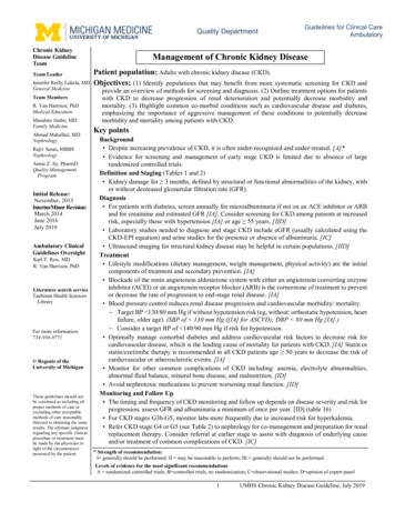
Who is at risk of developing acute kidney injury?The prevalence of CKD and AKIincreases with age. Between onequarter and one-third of all adultsaged over 64 years have CKD.The incidence of severe AKI is morethan fifty times higher in people agedover 80 years than in people agedunder 50 years.
AKI Classification Pre-renal: Failure to receive anadequate blood supply (40-80%)Renal: Intrinsic damage to the kidneytissue. This is actual damage of renalcells, commonly referred to as ATN (Acute Tubular Necrosis) (20-40%)Post-renal: Impaired renal drainage(obstruction of the renal tract) (210%)Sepsis, hypovolaemia, heart disease.Low cardiac outputInfection, diabetes, hypertension,disease.(Ischaemia)Calculi (stones) prostatic hyperplasia.Inability to pass urine ?
Causes of AKI: Pre-renalMost common cause of AKI Flow disruption to the kidneyDrugs - ACE inhibitorsFor example:Hypovolaemia - vomiting & diarrhoea,blood loss/ surgery, sepsisLow blood pressure – for a variety ofreasons (as above) fluid loss, bleeding,sepsis, heart failure.Blockage to flow – thrombosis,atherosclerosis.Decrease in effective circulatory volume low cardiac output - septic shock cirrhosis
Pre-renal AKIContinuumHypoperfusionPre-renal failureIntrinsic renal failurea)Tubular Necrosisb)Cortical NecrosisAcute Kidney Injury Year TwoCorrection of hypoperfusionrestores normal renal functionCorrection of hypoperfusion doesnot restore renal function
Causes of AKI: IntrinsicDamage to the kidney itselfFor example:Glomerulonephritis, infection.The main cause of intrinsic damage is from Acute Tubular Necrosis (ATN) caused by: Ischaemic injury: (can occur if blood flow to the kidneys is not improved followingone of the pre-renal insults) nephrotoxic injury: exposure to many drugs, poisons & endogenous compoundsin high concentrationsOccurs when there is damage to the structures of the nephron, such as the glomeruli,tubules, vessels or interstitium
Acute Tubular NecrosisPre-renal and intrinsic renal. Lack of blood flow andoxygen can result in cell injury and acute tubularnecrosis (ATN).Recovery of AKI is dependant upon the severity andextent of ATN.Diagnosing ATN is oneof exclusion.
Causes of AKIPost RenalProstatic hypertrophyA consequence of urinary tractobstruction.Cervical carcinomaFor example:Blocked catheterRenal calculiBladder tumoursRetroperitoneal fibrosisAcute Kidney Injury Year TwoUrethral strictureIntra-abdominal hypertension
Identification of AKIAcute Kidney Injury Year Two
Identification of AKIMeasuring changes in serum creatinine as well aschanges in urine volume in patients who haverisk factors can help identify AKI.The markers used are;Urine outputSerum creatinineeGFR (estimated glomerular filtration rate)Some hospitals have AKI alerts (whencreatinine is elevated by 26μmol/L)Acute Kidney Injury Year Two
KDIGO Staging of AKIStageSerum Creatinine121.5-1.9 times baselineOR26.5 µmol increase frombaseline2.0 -2.9 times baseline33 times baselineORIncrease in SrCr353.6µmolORInitiation of RRTAcute Kidney Injury Year TwoUrine Output 0.5 ml/kg/h for 6-12 hrs 0.5 ml/kg/h for 12 h 0.3 ml/kg/h for 24 hORAnuria for 12 h
Diagnosis: UrineMinimum expected urine output per day 0.5mls/kg/hr.How could this be measured?Consider acute hospitalsConsider communityAcute fluid balance charts/ urine colour charts and fluid estimations could bedocumented in patients’ assessment sheets and alert nursing staff to a problem.Patients who are incontinent – easy. Weigh pads 1ml / 1gPatients who are continent – education regarding importance of urine output andhow to recognise dehydration.
Urine as a marker of AKIUrine output is used as a decreasein output is often seen beforechanges in creatinine (Levin et al.,2007).Urine output is however, lessspecific than creatinineUrine output can remain normal inthe present of severe AKI especiallyin the presence of diuretics.Acute Kidney Injury Year Two
UrinalysisAssessment of urine can provide vital information as to whether the damage is withinthe kidneys or as a result of pre renal cause.Blood and protein enter the urine if the filtration system within the kidneys is notworking effectively.2 of blood or 2 protein is a significant finding and should be documented andappropriate individual informed.
Complications andManagementAcute Kidney Injury Year Two
Treatment of AKIAcute kidney injury is frequently reversible.Rapid recognition and treatment may preventirreversible nephron loss.Patients with underlying renal insufficiency may beprevented from reaching end- stage renal failure.
Specific complications of AKI:Fluid overloadHyperkalaemiaAcidosis,Acute Kidney Injury Year Two
Emergency treatment of AKI: FluidFluid management is extremely important as hypovolaemia exacerbates AKI.However rapid fluid infusion can result in overload which is also lifethreatening.Fluid challenge: What does this mean?Improving urine output will help resolve issues with hyperkalaemia/ fluidacidosisConsider the role of DiureticsAcute Kidney Injury Year Two
Fluid TherapyAssess; A-E assessment heart rate, blood pressure, capillary refill (should be 3secs), conscious level.Fluid status:If hypovolaemic a bolus of fluid (e.g. 250-500mls) should be considered, regularreview and repeat if required.If the patient is euvolaemic (normal fluid status) - maintenance fluids may beprescribed. This is based on estimated output plus 500mls.If the patient is overloaded and there is evidence of oedema- diuretics orHaemodialysis may be required if no response.
Clinical course Oliguric/ non- oliguric phase: 400mls urine/24hrs Diuretic Phase:Increased urine output. 3000mls/24hrs. Hydration andelectrolyte management essential Recovery Phase:Tubular function is restored, diuresis subsides kidneysfunction as normal.
AKI Management BundleDo not routinely administer loop diureticsConsider loop diuretics for treating fluid overload oroedemawhile awaiting renal replacement therapy orrenal function is recovering without renalreplacement therapy
Emergency treatment of AKI: ACIDOSIS andHyperkalaemiaIf the kidney is failing it can no-longer maintain acid-base balance by producingbicarbonate to buffer the acid and excreting hydrogen.Treatment is to administer fluid to try to manage the cause of AKI.Improving urine output will help resolve issues with hyperkalaemia although othermeasures are required.
Emergency treatment of AKI: Hyperkalaemia1. Protect the heart: Calcium gluconate10% in 10mls: It protects the heartfrom excess potassium excitability2. Shift potassium into cells: Insulin and dextrose Salbutamol3. Remove potassium Fluid Renal replacement therapy Oral calcium resoniumAcute Kidney Injury Year Two4. Monitor potassium and glucose5. Prevent recurrence
Management of HyperkalemiaCardiovascular MonitoringCalcium GluconateAntagonizes action of K at membraneQuick on setProtects myocardium from arrthymiasShort durationDextrose & InsulinShifts K back into the cellsMonitor blood sugarß2 AtonistsMonitor heart rate
Cardiovascular - Hyperkalemia Signs & SymptomsArrhythmias peaked t waves loss of p waves widened QRS VF &/orasystole
Management of HyperkalemiaSodium Bicarbonate:Shifts K back into cellCan increase extracellular volume (hypervolaemia)Watch for alkolosisCalcium Resonium:Tends to be non emergency treatment Exchanges sodium for potassium in theintestinal tractCauses constipation (give lactulose)
Other complications: InfectionMain cause of deathImpaired immunological response Impaired WC functionImpaired wound healing patient catabolic, Prophylactic Antibiotics is notrecommendedSkin and pressure area careRegular observations
Renal Replacement Therapy:What, When & Why?When to initiate RRT?1.U&E’s unstable2.Metabolic acidosis3.Fluid overloaded4.UreamiaAKI classification do not indicate if orwhen to initiate RRT3 basic types;1. Haemodialysis (usually intermittant 2-4hrs rapid removal of waste products & fluid)2. Haemofiltration (CRRT) (continous - gentleremoval of waste products & fluid)3. Peritoneal Dialysis (Catheter inserted intoperitoneum - peritonium acts as filter gentle used more in paeds)
Care bundles may supportmanagement of a patient with AKI
AKI Care BundleThis is a Medical EmergencyFull set of physiological obsAssess for signs of shockIf NEWS triggering give O2, begin resuscitation and contact outreach teamFluid TherapyAssess heart rate, blood pressure, jugular venous pressure,capillary refill (should be 3 secs), conscious level.If hypovolaemic give bolus fluids (e.g. 250-500mls) until volume replete with regular review of response.Middle grade review if 2 litres filling in oliguria.If the patient is euvolaemic give maintenance fluids (estimated output plus 500mls)and set daily fluid target.Monitoring in AKIDo arterial blood gas and lactate if venous bicarbonate is low or evidence of severe sepsis or hypoperfusion.Consider insertion of urinary catheter and measurement of hourly urine volumes.Measure urea, creatinine, bone, other electrolytes and venous bicarbonateat least daily while creatinine rising.Measure daily weights, keep a fluid chart and perform a minimum of 4 hourly observations.Perform regular fluid assessments and check for signs of uraemiaAcute Kidney Injury Year Two
AKI Care Bundle (cont)Investigation of AKIInvestigate the cause of all AKI unless multi-organ failure or obvious precipitantUrine dipstick. If proteinuria is present perform urgent spot urine protein creatinine ratio (PCR).USS should be performed within 24 hours unless AKI cause is obvious or AKI is recoveringor within 6 hours if obstruction with infection (pyonephrosis) is suspected.Check liver function (hepatorenal), CRP and CK (rhabdomyolysis). If platelets low do bloodfilm/LDH/Bili/retics (HUS/TTP). If PCR high, consider urgent Bence Jones protein & serum free light chains.Supportive AKI careTreat sepsis - in severe sepsis intravenous antibiotics should be administered within 1 hour ofrecognition.Stop NSAID/ACE/ARB/metformin/K-sparing diuretics and review all drug dosages.Give proton pump inhibitor and perform dietetic assessment.Stop anti-hypertensives if relative hypotension. If hypovolaemic consider stopping diuretics.Avoid radiological contrast if possible. If given follow prophylaxis protocol.Causes Think ‘STOP AKI’Sepsis and hypoperfusion, Toxicity (drugs / contrast), Obstruction, parenchymal kidney disease (acuteGN)Acute Kidney Injury Year Two
On going management
Management of AKIIdentify cause.Fluid
Pre-renal: Failure to receive an adequate blood supply (40-80%) Renal: Intrinsic damage to the kidney tissue. This is actual damage of renal cells, commonly referred to as ATN ( Acute Tubular Necrosis) (20-40%) Post-renal: Impaired renal drainage (obstruction of the renal tract) (2-10%) Sepsis, hypovolaemia, heart disease. Low cardiac output
