Transcription
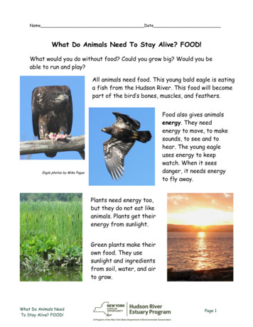
Common Diseases of Farm Animals, by R. A. CraigTitle: Common Diseases of Farm AnimalsAuthor: R. A. Craig, D. V. M.Release Date: July, 2005 [EBook #8502]Edition: 10Language: EnglishCharacter set encoding: ASCIIVisit: www.kashvet.orgCOMMON DISEASES OF FARM ANIMALSBy R. A. Craig, D.V.M.[Illustration: Frontispiece--INSANITARY DAIRY STABLE AND YARDS. DISEASEANDFINANCIAL LOSS ARE TO BE EXPECTED WHEN FARM ANIMALS ARE KEPTIN FILTHY,INSANITARY QUARTERS]PREFACEIn preparing the material for this book, the author has endeavored toarrange and discuss the subject matter in a way to be of the greatestservice and help to the agricultural student and stockman, and place attheir disposal a text and reference book.The general discussions at the beginning of the different sections andchapters, and the discussions of the different diseases are naturallybrief. An effort has been made to conveniently arrange the topics for bothpractical and class-room work. The chapters have been grouped under thenecessary heads, with review questions at the end of each chapter, and thebook divided into seven parts.The chapters on diseases of the locomotory organs, the teeth, surgicaldiseases and castration, although not commonly discussed in books of this
class, the writer believes will be of value for reference and instructionalwork.When used as a text-book, it will be well for the instructor to supplementthe text with class-room discussions.The writer has given special emphasis to the cause and prevention ofdisease, and not so much to the medicinal treatment. Stockmen are notexpected to practise the medicinal treatment, but rather the preventivetreatment of disease. For this reason it is not deemed advisable to give alarge number of formulas for the preparation of medicinal mixtures to beused for the treatment of disease, but such treatment is suggested in themost necessary cases.R. A. CRAIG.PURDUE UNIVERSITY, LaFayette, Ind. August, 1915.CONTENTSPART I.--INTRODUCTORY.I.II.III.GENERAL DISCUSSION OF DISEASEDIAGNOSIS AND SYMPTOMS OF DISEASETREATMENTPART II.--NON-SPECIFIC OR GENERAL XVI.XVII.DISEASES OF THE DIGESTIVE SYSTEMDISEASES OF THE LIVERDISEASES OF THE URINARY ORGANSDISEASES OF THE GENERATIVE ORGANSDISEASES OF THE RESPIRATORY APPARATUSDISEASES OF THE CIRCULATORY ORGANSDISEASES OF THE NERVOUS SYSTEMDISEASES OF THE SKINDISEASES OF THE EYEGENERAL DISEASES OF THE LOCOMOTORY APPARATUSSTRUCTURE OF THE LIMBS OF THE HORSEUNSOUNDNESSES AND BLEMISHESDISEASES OF THE FORE-LIMBDISEASES OF THE FOOT
XVIII. DISEASES OF THE HIND LIMBPART III.--THE TEETH.XIX.XX.DETERMINING THE AGE OF ANIMALSIRREGULARITIES OF THE TEETHPART IV.--SURGICAL DISEASES.XXI. INFLAMMATION AND WOUNDSXXII. FRACTURES AND HARNESS INJURIESXXIII. COMMON SURGICAL OPERATIONSPART V.--PARASITIC DISEASES.XXIV. PARASITIC INSECTS AND MITESXXV. ANIMAL PARASITESPART VI.--INFECTIOUS DISEASES.XXVI. HOG-CHOLERAXXVII. TUBERCULOSISXXVIII. INFECTIOUS DISEASES COMMON TO THE DIFFERENT SPECIES OFDOMESTICANIMALSXXIX. INFECTIOUS DISEASES OF THE HORSEXXX. INFECTIOUS DISEASES OF CATTLEXXXI. INFECTIOUS DISEASES OF POULTRYREFERENCE BOOKSILLUSTRATIONSFIG.(Frontispiece) Insanitary dairy stable and yards.1. Side and posterior view of bull showing conformation favorable to thedevelopment of disease.2. Insanitary yards.3. Showing where pulse of horse is taken.4. Auscultation of the lungs.5. Fever thermometer.6. Dose syringe.7. Hypodermic syringes.
8. Photograph of model of horse's stomach.9. Photograph of model of stomach of ruminant.10. Oesophageal groove.11. Dilated stomach of horse.12. Rupture of stomach of horse.13. Showing the point where the wall of flank and rumen are puncturedwith trocar and cannula in "bloat".14. Photograph of model of digestive tract of horse.15. Photograph of model of digestive tract of ruminant.16. A yearling colt that died of aneurism colic.17. Photograph of model of udder of cow.18. Photograph of model of uterus of cow containing foetus.19. Placenta of cow.20. A case of milk-fever.21. Milk-fever apparatus.22. A case of catarrhal cold.23. Photograph of model of horse's heart.24. Elephantiasis in horse.25. Photograph of model of horse's brain.26. Unilateral facial paralysis.27. Bilateral facial paralysis.28. Skeleton of horse.29. Photograph of model of stifle joint.30. Atrophy of the muscles of the thigh.31. Shoulder lameness.32. Shoe-boil.33. Sprung knees.34. Splints.35. Bones of digit.36. Photograph of a model of the foot.37. Foot showing neglect in trimming wall.38. A very large side bone.39. A case of navicular disease.40. An improperly shod foot.41. Toe-cracks.42. Quarter-crack caused by barb-wire cut.43. Changes occurring in chronic laminitis.44. Atrophy of the muscles of the quarter.45. String-halt.46. A large bone spavin.47. Normal cannon bone and cannon bone showing bony enlargement.48. Bog spavins.49. Thorough pin.50. Curbs.51. Head of young horse showing position and size of teeth.52. Longitudinal section of incisor tooth.
53. Cross-section of head of young horse, showing replacement of molartooth.54. Transverse section of incisor tooth55. Transverse sections of incisor tooth showing changes at differentages.56. Teeth showing uneven wear occurring in old horses.57. Fistula of jaw.58. A large hock caused by a punctured wound of the joint.59. A large inflammatory growth following injury.60. Fistula of the withers.61. Shoulder abscess caused by loose-fitting harness.62. A piece of the wall of the horse's stomach showing bot-fly larvaeattached.63. Biting louse.64. Sucking louse.65. Nits attached to hair.66. Sheep-tick.67. Sheep scab mite.68. Sheep scab.69. A severe case of mange.70. Liver flukes.71. Tapeworm larvae in liver.72. Tapeworms.73. Tapeworm larvae in the peritoneum.74. Thorn-headed worms.75. Large round-worm in intestine of hog.76. Lamb affected with stomach worm disease.77. Whip-worms attached to wall of intestine.78. Pin-worms in intestine.79. A hog yard where disease-producing germs may be carried overfrom year to year.80. Carcass of a cholera hog.81. Kidneys from hog that died of acute hog-cholera.82. Lungs from hog that died of acute hog-cholera.83. A piece of intestine showing intestinal ulcers.84. Cleaning up a hog lot.85. Hyperimmune hogs used for the production of anti-hog-cholera serum.86. Preparing the hog for vaccination.87. Vaccinating a hog.88. Koch's Bacillus tuberculosis .89. A tubercular cow.90. Tubercular spleens.91. The carcass of a tubercular cow.92. A section of the chest wall of a tubercular cow.93. A very large tubercular gland.94. A tubercular gland that is split open.
95. Caul showing tuberculosis.96. Foot of hog showing tuberculosis of joint.97. Staphylococcus pyogenes .98. Streptococcus pyogenes .99. Bacillus of malignant oedema, showing spores.100. Bacillus of malignant oedema.101. Bacillus bovisepticus .102. A yearling steer affected with septicaemia haemorrhagica.103. Bacillus anthracis .104. Bacillus necrophorus .105. Negri bodies in nerve-tissue.106. A cow affected with foot-and-mouth disease.107. Slaughtering a herd of cattle affected with foot-and-mouth disease.108. Disinfecting boots and coats before leaving a farm where cattle havebeen inspected for foot-and-mouth disease.109. Cleaning up and disinfecting premises.110. Bacillus tetani .111. Head of horse affected with tetanus.112. A subacute case of tetanus.113. Streptococcus of strangles.114. Bacillus mallei .115. Nasal septum showing nodules and ulcers.116. Streptococcus pyogenes equi .117. A case of "lumpy jaw".118. The ray fungus.119. Bacillus of emphysematous anthrax.120. Cattle tick (male).121. Cattle tick (female).122. Blood-cells with Piroplasma bigeminum in them.123. Bacillus avisepticus .Visit: www.kashvet.orgPART I.--INTRODUCTORYCHAPTER IGENERAL DISCUSSION OF DISEASEDisease is the general term for any deviation from the normal or healthycondition of the body. The morbid processes that result in either slight ormarked modifications of the normal condition are recognized by the
injurious changes in the structure or function of the organ, or group ofbody organs involved. The increase in the secretion of urine noticeable inhorses in the late fall and winter is caused by the cool weather and thedecrease in the perspiration. If, however, the increase in the quantity ofurine secreted occurs independently of any normal cause and is accompaniedby an unthrifty and weakened condition of the animal, it would thencharacterize disease. Tissues may undergo changes in order to adaptthemselves to different environments, or as a means of protectingthemselves against injuries. The coat of a horse becomes heavy and appearsrough if the animal is exposed to severe cold. A rough, staring coat isvery common in horses affected by disease. The outer layer of the skinbecomes thickened when subject to pressure or friction from the harness.This change in structure is purely protective and normal. In disease thedeviation from normal must be more permanent in character than it is in theexamples mentioned above, and in some way prove injurious to the bodyfunctions.CLASSIFICATION.--We may divide diseases into three classes: non-specific,specific and parasitic .Non-specific diseases have no constant cause. A variety of causes mayproduce the same disease. For example, acute indigestion may be caused by achange of diet, watering the animal after feeding grain, by exhaustion andintestinal worms. Usually, but one of the animals in the stable or herd isaffected. If several are affected, it is because all have been subject tothe same condition, and not because the disease has spread from one animalto another.Specific Diseases. --The terms infectious and contagious are used inspeaking of specific diseases. Much confusion exists in the popular use ofthese terms. A contagious disease is one that may be transmitted bypersonal contact, as, for example, influenza, glanders and hog-cholera. Asthese diseases may be produced by indirect contact with the diseased animalas well as by direct, they are also infectious . There are a few germdiseases that are not spread by the healthy animals coming in directcontact with the diseased animal, as, for example, black leg and southerncattle fever. These are purely infectious diseases. Infection is a morecomprehensive term than contagion, as it may be used in alluding to allgerm diseases, while the use of the term contagion is rightly limited tosuch diseases as are produced principally through individual contact.Parasitic diseases are very common among domestic animals. This class ofdisease is caused by insects and worms, as for example, lice, mites, ticks,flies, and round and flat worms that live at the expense of their hosts.They may invade any of the organs of the body, but most commonly inhabitthe digestive tract and skin. Some of the parasitic insects, mosquitoes,
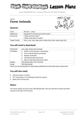
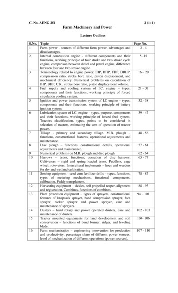
flies and ticks, act as secondary hosts for certain animal microorganismsthat they transmit to healthy individuals through the punctures or thebites that they are capable of producing in the skin.CAUSES.--For convenience we may divide the causes of disease into thepredisposing or indirect, and the exciting or direct.The predisposing causes are such factors as tend to render the body moresusceptible to disease or favor the presence of the exciting cause. Forexample, an animal that is narrow chested and lacking in the development ofthe vital organs lodged in the thoracic cavity, when exposed to the samecondition as the other members of the herd, may contract disease while theanimals having better conformation do not (Fig. 1). Hogs confined inwell-drained yards and pastures that are free from filth, and fed in pensand on feeding floors that are clean, do not become hosts for large numbersof parasites. Hogs confined in filthy pens are frequently so badly infestedwith lice and intestinal worms that their health and thriftiness areseriously interfered with. In the first case mentioned the predispositionto disease is in the individual, and in the second case it is in thesurroundings (Fig. 2).[Illustration: FIG. 1.--Side and posterior view of bull showingconformation favorable to the development of disease.]The exciting causes are the immediate causes of the particular disease.Exciting causes usually operate through the environment. With the exceptionof the special disease-producing germs, the most common exciting causes arefaulty food and faulty methods of feeding. The following predisposingcauses of disease may be mentioned:Age is an important factor in the production of disease. Young andimmature animals are more prone to attacks of infectious diseases than areold and mature animals. Hog-cholera usually affects the young hogs in theherd first, while scours, suppurative joint disease and infectious soremouth are diseases that occur during the first few days or few weeks of theanimal's life. Lung and intestinal parasites are more commonly found in theyoung, growing animals. Old animals are prone to fractures of bones anddegenerative changes of the body tissues. As a general rule, the young aremore subject to acute diseases and the old to chronic diseases.[Illustration: FIG. 2.--Insanitary yards.]The surroundings or environments are important predisposing factors. Adark, crowded, poorly ventilated stable lowers the animal's vitality, andrenders it more susceptible to the disease. A few rods difference in thelocation of stables and yards may make a marked difference in the health of
the herd. A dry, protected site is always preferable to one in the open oron low, poorly drained soil. The majority of domestic animals need butlittle shelter, but they do need dry, comfortable quarters during wet, coldweather.Faulty feed and faulty methods of feeding are very common causes ofdiseases of the digestive tract and the nervous system. A change from dryfeed to a green, succulent ration is a common cause of acute indigestion inboth horses and cattle. The feeding of a heavy ration of grain to horsesthat are accustomed to exercise, during enforced rest may cause liver andkidney disorders. The feeding of spoiled, decomposed feeds may causeserious nervous and intestinal disorders.One attack of a certain disease may influence the development ofsubsequent attacks of the same, or a different disease. An individual maysuffer from an attack of pneumonia that so weakens the disease-resistingpowers of the lungs as to result in a tubercular infection of these organs.In the horse, one attack of azoturia predisposes it to a second attack. Oneattack of an infectious disease usually confers immunity against thatparticular disease. Heredity does not play as important a part in thedevelopment of diseases in domestic animals as in the human race. A certainfamily may inherit a predisposition to disease through the faulty orinsufficient development of an organ or group of organs. The differentspecies of animals are affected by diseases peculiar to that particularspecies. The horse is the only species that is affected with azoturia.Glanders affects solipeds, while black leg is a disease peculiar to cattle.QUESTIONS1. What is disease?2. How are diseases classified? Give an example of the different classes.3. What is a predisposing cause? Exciting cause?4. Name the different predisposing and exciting causes of disease.CHAPTER IIDIAGNOSIS AND SYMPTOMS OF DISEASE
The importance of recognizing or diagnosing the seat and nature of themorbid change occurring in an organ or group of organs cannot beoverestimated. Laymen do not comprehend the difficulty or importance ofcorrectly grouping the signs or symptoms of disease in such a way as toenable them to recognize the nature of the disease. In order to be able tounderstand the meaning of the many symptoms or signs of disease, we mustpossess knowledge of the structure and physiological functions of thedifferent organs of the body. We must be familiar with the animal when itis in good health in order to be able to recognize any deviation from thenormal due to disease, and we must learn from personal observation thedifferent symptoms that characterize the different diseases. Stockmenshould be able to tell when any of the animals in their care are sick assoon as the first symptom of disease manifests itself, by changes in thegeneral appearance and behavior. But in order to ascertain the exactcondition a general and systematic examination is necessary. The examiner,whether he be a layman or a veterinarian, must observe the animalcarefully, noting the behavior, appearance, surroundings, and general andlocal symptoms.Before making a general examination of the animal it is well, if theexaminer is not already acquainted with the history of the case (care, feedand surroundings), to learn as much about this from the attendant as ispossible. Inquiry should be made as to the feeding, the conditions underwhich the animal has been kept, the length of time it has been sick, itsactions, or any other information that may be of assistance in forming thediagnosis and outlining the treatment.The general symptoms inform us regarding the condition of the differentgroups of body organs. A careful study of this group of symptoms enables uscorrectly to diagnose disease and inform ourselves as to the progress oflong, severe affections. These symptoms occur in connection with the pulse,respirations, body temperature, skin and coat, visible mucous membranes,secretions and excretions, and behavior of the animal.The local symptoms are confined to a definite part or organ. Swelling,pain, tenderness and loss of function are common local symptoms. A directsymptom may also be considered under this head because of its directrelation to the seat of disease. It aids greatly in forming the diagnosis.Other terms used in describing symptoms of disease are objective , whichincludes all that can be recognized by the person making the examination;indirect , which are observed at a distance from the seat of the disease;and premonitory , which precede the direct, or characteristic symptoms.The subjective symptoms include such as are felt and described by thepatient. These symptoms are available from the human patient only.
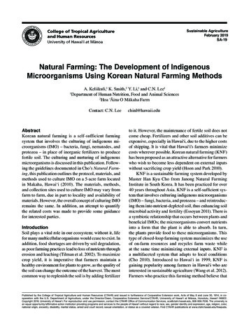
Pulse.--The character of the intermittent expansion of the arteries, calledthe pulse, informs us as to the condition of the heart and blood-vessels.The frequency of the pulse beat varies in the different species of animals.The smaller the animal the more frequent the pulse. In young animals thenumber of beats per minute is greater than in adults. Excitement or fear,especially if the animal possesses a nervous temperament, increases thefrequency of the pulse. During, and for a short time after, feeding andexercise, the pulse rate is higher than when the animal is standing atrest.The following table gives the normal rate of the pulse beats per minute:Horse 36 to 40 per minuteOx 45 to 50 per minuteSheep 70 to 80 per minutePig 70 to 80 per minuteDog 90 to 100 per minuteIn sickness the pulse is instantly responsive. It is of the greatest aidin diagnosing and in noting the progress of the disease. The followingvarieties of pulse may be mentioned: frequent, infrequent, quick, slow,large, small, hard, soft and intermittent . The terms frequent andinfrequent refer to the number of pulse beats in a given time; quick andslow to the length of time required for the pulse wave to pass beneath thefinger; large and small to the volume of the wave; hard and soft to itscompressibility; and intermittent to the occasional missing of a beat. Apulse beat that is small and quick, or large and soft, is frequently metwith in diseases of a serious character.[Illustration: FIG. 3.--The X on the lower border of the jaw indicates theplace where the pulse is taken.]The horse's pulse is taken from the submaxillary artery at a pointanterior to, or below the angle of the jaw and along its inferior border(Fig. 3). It is here that the artery winds around the inferior border ofthe jaw in an upward direction, and, because of its location immediatelybeneath the skin, it can be readily located by pressing lightly over theregion with the fingers.Cattle's pulse is taken from the same artery as in the horse. The arteryis most superficial a little above the border of the jaw. It is moredifficult to find the pulse wave in cattle than it is in horses, because ofthe larger amount of connective tissue just beneath the skin and theheavier muscles of the jaw. A very satisfactory pulse may be found in thesmall arteries located along the inferior part of the lateral region of thetail and near its base.
The sheep's pulse may be taken directly from the femoral artery byplacing the fingers over the inner region of the thigh. By pressing withthe hand over the region of the heart we may determine its condition.The hog's pulse can easily be taken from the femoral artery on theinternal region of the thigh. The artery crosses this region obliquely andis quite superficial toward its anterior and lower portion.The dog's pulse is usually taken from the brachial artery. The pulse wavecan be readily felt by resting the fingers over the inner region of the armand just above the elbow. The character of the heart beats in dogs may bedetermined by resting the hand on the chest wall.RESPIRATION.--The frequency of the respirations varies with the species.The following table gives the frequency of the respirations in domesticanimals:Horse 8 to 10 per minuteOx12 to 15 per minuteSheep 12 to 20 per minuteDog 15 to 20 per minutePig 10 to 15 per minuteThe ratio of the heart beats to the respirations is about 1:4 or 1:5. Thisratio is not constant in ruminants. Rumination, muscular exertion andexcitement increase the frequency and cause the respirations to becomeirregular. In disease the ratio between the heart beats and respirationsis greatly disturbed, and the character of the respiratory sounds andmovements may be greatly changed (Fig. 4).[Illustration: FIG. 4.--Auscultation of the lungs can be practised to anadvantage over the outlined portion of the chest wall, only.]Severe exercise and diseased conditions of the lungs cause the animal tobreathe rapidly and bring into use all of the respiratory muscles. Suchforced or labored breathing is a common symptom in serious lung diseases,"bloat" in cattle, or any condition that may cause dyspnoea. Horsesaffected with "heaves" show a double contraction of the muscles in theregion of the flank during expiration. In spasm of the diaphragm or"thumps" the expiration appears to be a short, jerking movement of theflank. In the abdominal form of respiration the movements of the walls ofthe chest are limited. This occurs in pleurisy. In the thoracic form ofrespiration the abdominal wall is held rigid and the movement of the chestwalls make up for the deficiency. This latter condition occurs inperitonitis.
A cough is caused by irritation of the membrane lining the air passages.The character of the cough may vary according to the nature of the disease.We may speak of a moist cough when the secretions in the air passages aremore or less abundant. A dry cough occurs when the lining membrane of theair passages is dry and inflamed. This may occur in the early stage of theinflammation, or as a result of irritation from dust or irritating gases.Chronic cough occurs when the disease is of long duration or chronic. Inpleurisy the cough may be short and painful, and in broken wind, deep andsuppressed. In parasitic diseases of the air passages and lungs, theparoxysm of coughing may be severe and "husky" in character.The odor of the expired air, the character of the discharge and therespiratory sounds found on making a careful examination are important aidsin arriving at a correct diagnosis, and in studying the progress of thedisease.[Illustration: FIG. 5.--Fever thermometer.]Body Temperature.--The body temperature of an animal is taken by insertingthe fever thermometer into the rectum. In large animals a five-inch, and insmall animals a four-inch fever thermometer is used. It should be insertedfull length and left in position from one and one-half to three minutes,depending on the rapidity with which it registers (Fig. 5).The average normal body temperatures of domestic animals are as follows:Horses 100.5\260 F.Cattle 101.4\260 F.Sheep 104.0\260 F.Swine 103.0\260 F.Dog 101.4\260 F.There is a wide variation in the body temperatures of domesticanimals. This is especially true of cattle, sheep and hogs. In order todetermine the normal temperature of an animal, it may be necessary to taketwo or more readings at different times, and compare them with the bodytemperatures of other animals in the herd that are known to be healthy.Exercise, feeding, rumination, excitement, warm, close stables, exposure tocold and drinking ice cold water are common causes of variations in thebody temperatures of domestic animals.Visible Mucous Membranes.--The visible mucous membranes, as they aretermed, are the lining membranes of the eyelids, nostrils and nasalcavities, and mouth. In health they are usually a pale red, excepting when
the animal is exercised or excited, when they appear a brighter red andsomewhat vascular. In disease the following changes in color and appearancemay be noted: When inflamed, as in cold in the head, a deep red; inimpoverished or bloodless conditions of the body and in internalhaemorrhage, pale; in diseases of the liver, sometimes yellowish, or darkred; in diseases of the digestive tract (buccal mucous membrane), coated;if inflamed, dry at first, later excessively moist; and in certain germdiseases a mottled red, or showing nodules, ulcers and scars.Surface of the Body.--When a horse is in a good condition and well caredfor, the coat is short, fine, glossy and smooth and the skin pliable andelastic. Healthy cattle have a smooth, glossy coat and the skin feelsmellow and elastic. The fleece of sheep should appear smooth and haveplenty of yolk, the skin pliable and light pink in color. When the coatloses its lustre and gloss and the skin becomes hard, rigid, thickened anddirty, it indicates a lack of nutrition and an unhealthy condition of thebody. In sheep, during sickness, the wool may become dry and brittle andthe skin pale and rigid. When affected with external parasites, the hair orwool becomes dirty and rough, a part of the skin may be denuded of hair,and it appears thickened, leathery and scabby, or shows pimples, vesiclesand sores.During fever, the temperature of the surface of the body is very unequal.In serious diseases or diseases that are about to terminate fatally, theskin feels cold and the hair is wet with sweat.When animals are allowed to "rough it" during the cold weather, the coat ofhair becomes heavy and rough. This is a provision of nature and enablesthem, as long as the coat is dry, to withstand severe cold.Horses that are in a low physical condition, or when accustomed to hardwork, if then kept in a stall for a few days without exercise, commonlyshow a filling of the cannon regions of the posterior extremities. Thiscondition also commonly occurs in disease and in mares that have reachedthe latter period of pregnancy. Sheep that are unthrifty and in a poorphysical condition, especially if this is due to internal parasites,frequently develop dropsical swellings in the region of the jaw, or neck.Body Excretions.--The character of the body excretions, faeces and urinemay become greatly changed in certain diseases. It is important that thestockman or veterinarian observe these changes, and in certain diseasesmake an analysis of the urine. This may be necessary in order properly todiagnose the case.Behavior of the Animal.--When the body temperature is high, the animal mayappear greatly depressed. If suffering severe pain, it may be restless. In
diseases of the nervous system, the behavior of the animal may be greatlychanged. Spasms, convulsions, general local paralysis, stupid condition andunconsciousness may occur as symptoms of this class of disease.QUESTIONS1. What information is necessary in order to be able to recognize ordiagnose disease? 2. What are the general symptoms of disease?3. What are the subjective symptoms of disease?4. Describe method of taking the pulse beat in the different animals andits character in health and disease.5. Give the ratio of the heart beats to the respirations in the differentspecies of animals.6. What are the normal body temperatures in the different domestic animals?7. What are the visible mucous membranes?8. Is the condition of the coat and skin any help in the recognition ofdisease?CHAPTER IIITREATMENTPreventive Treatment.--The subject of preventive medicine becomes moreimportant as our knowledge of the cause of disease advances. A knowledge offeeds, methods of feeding, care, sanitation and the use of such biologicalproducts as bacterins, vaccines and protective serums is of the greatestimportance to the farmer and veterinarian. We are beginning to realize thatone of the most important secrets of profitable and successful stockraising is the prevention of disease; that the agricultural colleges aredoing a great work in helping to teach farmers that there are right andwrong methods of feeding and caring for animals; that the practice ofsanitation in caring for animals is the cheapest method of treatingdisease; and that it is advisable to practise radical methods of control,when necessary, in order to rid the herd of an infectious disease.
The ration fed and the method of feeding are not only important inconsidering the causes of diseases of the digestive tract, but diseases ofother organs as well. The feeding of an excessive, or insufficient quantityof feed, or a ration that is too concentrated, bulky and innutritious, poorin quality, or spoiled may produce disease.An impure water supply is a common cause of disease. A deep well that isclosed in properly and does not permit of contam
COMMON DISEASES OF FARM ANIMALS By R. A. Craig, D.V.M. [Illustration: Frontispiece--INSANITARY DAIRY STABLE AND YARDS. DISEASE AND FINANCIAL LOSS ARE TO BE EXPECTED WHEN FARM ANIMALS ARE KEPT IN FILTHY, INSANITARY QUARTERS] PREFACE In preparing the material for