Transcription
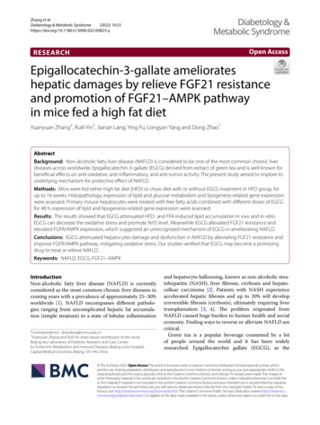
Zhang et al.Diabetology & Metabolic Syndrome(2022) betology &Metabolic SyndromeOpen AccessRESEARCHEpigallocatechin‑3‑gallate ameliorateshepatic damages by relieve FGF21 resistanceand promotion of FGF21–AMPK pathwayin mice fed a high fat dietYuanyuan Zhang†, Ruili Yin†, Jianan Lang, Ying Fu, Longyan Yang and Dong Zhao*AbstractBackground: Non-alcoholic fatty liver disease (NAFLD) is considered to be one of the most common chronic liverdiseases across worldwide. Epigallocatechin-3-gallate (EGCG) derived from extract of green tea and is well known forbeneficial effects on anti-oxidative, anti-inflammatory, and anti-tumor activity. The present study aimed to implore itsunderlying mechanism for protective effect of NAFLD.Methods: Mice were fed either high fat diet (HFD) or chow diet with or without EGCG treatment in HFD group, forup to 16 weeks. Histopathology, expression of lipid and glucose metabolism and lipogenesis-related gene expressionwere assessed. Primary mouse hepatocytes were treated with free fatty acids combined with different doses of EGCGfor 48 h, expression of lipid and lipogenesis-related gene expression were assessed.Results: The results showed that EGCG attenuated HFD- and FFA-induced lipid accumulation in vivo and in vitro.EGCG can decrease the oxidative stress and promote Nrf2 level. Meanwhile EGCG alleviated FGF21 resistance andelevated FGFR/AMPK expression, which suggested an unrecognized mechanism of EGCG in ameliorating NAFLD.Conclusions: EGCG attenuated hepatocytes damage and dysfunction in NAFLD by alleviating FGF21 resistance andimprove FGFR/AMPK pathway, mitigating oxidative stress. Our studies verified that EGCG may become a promisingdrug to treat or relieve NAFLD.Keywords: NAFLD, EGCG , FGF21–AMPKIntroductionNon-alcoholic fatty liver disease (NAFLD) is currentlyconsidered as the most common chronic liver diseases incoming years with a prevalence of approximately 25–30%worldwide [1]. NAFLD encompasses different pathologies ranging from uncomplicated hepatic fat accumulation (simple steatosis) to a state of lobular inflammation*Correspondence: zhaodong@ccmu.edu.cn†Yuanyuan Zhang and Ruili Yin share equal contribution to the studyBeijing Key Laboratory of Diabetes Research and Care, Centerfor Endocrine Metabolism and Immune Diseases, Beijing Luhe Hospital,Capital Medical University, Beijing 101149, Chinaand hepatocyte ballooning, known as non-alcoholic steatohepatitis (NASH), liver fibrosis, cirrhosis and hepatocelluar carcinoma [2]. Patients with NASH experienceaccelerated hepatic fibrosis and up to 20% will developirreversible fibrosis (cirrhosis), ultimately requiring livertransplantation [3, 4]. The problem originated fromNAFLD caused huge burden to human health and socialeconomy. Finding ways to reverse or alleviate NAFLD arecritical.Green tea is a popular beverage consumed by a lotof people around the world and it has been widelyresearched. Epigallocatechin gallate (EGCG), as the The Author(s) 2022. Open Access This article is licensed under a Creative Commons Attribution 4.0 International License, whichpermits use, sharing, adaptation, distribution and reproduction in any medium or format, as long as you give appropriate credit to theoriginal author(s) and the source, provide a link to the Creative Commons licence, and indicate if changes were made. The images orother third party material in this article are included in the article’s Creative Commons licence, unless indicated otherwise in a credit lineto the material. If material is not included in the article’s Creative Commons licence and your intended use is not permitted by statutoryregulation or exceeds the permitted use, you will need to obtain permission directly from the copyright holder. To view a copy of thislicence, visit http:// creat iveco mmons. org/ licen ses/ by/4. 0/. The Creative Commons Public Domain Dedication waiver (http:// creat iveco mmons. org/ publi cdoma in/ zero/1. 0/) applies to the data made available in this article, unless otherwise stated in a credit line to the data.
Zhang et al. Diabetology & Metabolic Syndrome(2022) 14:53main active ingredient of green tea, has various biological effects such as scavenging free radicals, anti-lipid peroxidation, anti-inflammatory, anti-viral, and enhancingthe immune function of the body. It has obvious pharmacological effects in preventing obesity, diabetes, atherosclerosis and other diseases [5, 6]. A lot of researchhad shown EGCG could attenuate high-fat diet- (HFD-)induced NAFLD in rats and mice [7, 8]. Nevertheless, therelated mechanisms of EGCG to ameliorate fat accumulation or NAFLD are not clearly understood yet.Fibroblast growth factor-21 (FGF21), as a member ofthe fibroblast growth factor family, is involved in manymetabolic processes including insulin sensitivity, glucose and lipid metabolism, and energy homeostasis. AsFGF21 is a liver derived, metabolically active hormone, itis conceivable that FGF21 is implicated in the pathobiology of NAFLD. There is an abundance of preclinical evidence demonstrating aberrant FGF21 signaling is, at leastpartially, responsible for the pathogenesis and progression of NAFLD [9, 10]. In human, observational studieshave demonstrated circulating FGF21 levels are elevatedin subjects with NAFLD [11]; animal experiments haveshown that FGF21 mRNA is increased in the livers ofmice with NAFLD [12]. Recent studies demonstrated thatactivation of peroxisome proliferator-activated receptorα (PPARα) increases FGF21 production in WAT and livers [12, 13].AMP-dependent protein kinase (AMPK) has been considered as the fuel gauge of the cells or the natural energysensor that regulates lipid metabolism [14]. Recent studies have revealed FGF21 can stimulate AMPK signalingin several tissues [15, 16]. Studies shown FGF21 regulatedmitochondrial activity and lipid metabolism throughstimulation of LKB-AMPK pathway in adipocytes and inwhite adipose tissue [17, 18]. Chau et al. demonstratedthat LKB1 might be is one of the phosphorylation targetsof FGFR.In the study, to delineate the potential therapeutic effects of EGCG in preventing NAFLD, we givenc57BL/6 mice an HFD diet with or without EGCG supplementation treatment until 16 weeks. We explored theunderlying mechanism and particularly focused on thePPARα–FGF21–AMPK signaling and oxidative stress inprevention of NAFLD for EGCG.Materials and methodsAnimals. Experimental protocols were approved bythe Animal Care and Use Committee of Beijing LuheHospital, Capital Medical University and conducted inaccordance with the NIH Guide for the Care and Use ofLaboratory Animals. All efforts were made to minimizesuffering of experimental mice in this research. Animalstudies are reported in compliance with the ARRIVEPage 2 of 11guidelines. Animals were randomly divided into threegroups after a week of adaptive feeding. The HFD composed of 45% energy as fat, 20% protein, and 35% carbohydrates (Medicience Biotechnology Co., Ltd, Jiangsu,China) was used to induce non-alcoholic fatty liver. Micewere divided three groups: a normal diet group (CON)(n 10), a high-fat diet (HFD) (n 12) and a high-fat dietsupplemented with 1% EGCG (HFD EGCG) (n 12).EGCG was administered at dose of 100 mg/kg for16 weeks. All mice were maintained on a 12 h light/darkcycle and allowed access to food and water ad libitum.Food/water intakes and body weights of the mice weremeasured. After mice were anesthetized using ketamineand xylazine, livers were harvested. Blood was collectedfrom the heart, immediately frozen with liquid nitrogen,and stored at 80 C.Biochemical analysisThe concentrations of triglyceride (TG), total cholesterol(TC), alanine aminotransferase (ALT), and aspartate aminotransferase (AST) in the plasma were examined byan automated hematology analyzer (BC-6900, Mindray,Shenzhen, Guangdong, China). The contents of TG andTC in hepatocytes and liver tissues in liver tissues weredetected by commercial enzyme-linked immunosorbentassay kits (Jiancheng Bioengineering Institute, Nanjing,Jiangsu, China).Oil red O stainingPrimary mouse hepatocytes (C57BL/6J, 6–8 weeks old)were plated into six-well plates and exposed to FFA (PAOA 1:2) with or without EGCG for 48 h (n 4). Afterfixation with 4% paraformaldehyde for 10 min, fixed cellswere stained in ORO solution for 15 min, and then theirnucleus was stained by hematoxylin for 3 min. Oil red Ostaining was used to visualize lipid droplets in the liver.Level of FGF21 expressionBlood samples were collected from the mice, and theplasma was isolated. Circulating FGF21 was measuredusing an enzyme-linked immunosorbent assay (ELISA)kit purchased from Jonln Bioscience (Shanghai, China).The cell culture medium was collected, and the level ofFGF21 protein expression was measured using an ELISAkit and conducted following the manufacturer’s protocol.Measurement of oxidative stress markers and antioxidantenzymesThe level of liver lipid peroxidation product MDA wasmeasurement by using a commercial kit (Applygen Technologies, Beijing, China). The activities of antioxidantdefense enzymes including SOD were measured by Elisakit (Enzyme-linked Biotechnology, Shanghai, China).
Zhang et al. Diabetology & Metabolic Syndrome(2022) 14:53Hematoxylin and eosin (HE) stainingLiver tissues were fixed in 4% paraformaldehyde, embedded in paraffin wax, Serial sections were sliced at 5 μmand stained with hematoxylin–eosin (H&E) to observethe damage.Real‑time quantitative PCRTotal RNA from liver tissues and primary hepatocyteswas extracted with TRIzol reagent (Invitrogen, USA) andthen reverse-transcribed using a cDNA Synthesis Kit(Bio-Rad, USA, Cat: 1708891). Primers were designedwith Primer Premier 6.0 software and chemically synthesized by Sangon Biotech (Shanghai, China) Co., Ltd. Theforward and reverse primer sequences for mouse genesare listed in Table 1. Real-time PCR was conducted withthe SYBR Green PCR kit (Bio-Rad, USA, Cat: 1725151),and quantification was achieved by normalization using18 s. The primer sequences are provided in Table 1.Western blotWestern blot assay was applied to determine the expression levels of proteins from cultured cells and frozen liversamples. The primary antibodies were as follows: FGF21(Abcam, UK, Cat: ab171941), phospho-Thr172 AMPKα(CST, USA, Cat: 2535), AMPKα (CST, USA, Cat: 2532),ACC (Santa Cruz, USA, Cat: sc137104), FASn (Abcam,UK, Cat: ab128870), Nrf2 (Abcam, UK, Cat: ab137550),Actin (Zsbio, China, Cat: TA-09). Band intensities wereanalyzed by densitometry using ImageJ software.Primary mouse hepatocytes culture and treatmentPrimary mouse hepatocytes were cultured as Yang et al.reported previously PMID: 28411267. Firstly, perfuse themouse liver with 50 mL Kreb solution, followed by 30 mLPage 3 of 11Kreb solution with 0.2 mL liberase TM (Roche, Germany)to digest the liver. The cells were filtered with a disposable cell strainer (Corning, USA) using 1640 medium containing 10% FBS; then washed and centrifuged at 50 g at4 C for three times. The cells were cultured with 1640containing 10% FBS for 6 h at 37 C with 5% C O2 beforetreating.Immunofluorescence analysisPrimary mouse hepatocytes were seeded onto glass coverslips in six-well plates and treated with FFA and EGCGfor 48 h, washed with cold PBS, and fixed in 4% paraformaldehyde in PBS at room temperature. The fixed cellswere washed three times with PBS and kept in chilledmethanol for 10 min at 20 C.The cells were washedthree times and incubated with the primary antibodyand secondary antibody (AlexaFluor goat anti-mouse488, 1:500).Then, the nucleus was counter-stained bluewith DAPI (4’,6-diamidino-2-phenylindole) and mountedwith anti-fluorescence quenching sealant. The slips wereimaged using an inverted fluorescence microscope.CCK8 assayPrimary mouse hepatocytes were plated at a density of5 104 cells/mL into 96-well plates (100 µL medium/well) with five replicates. The hepatocytes cells werethen treated with different doses of EGCG and FFA.After seeding for 48 h, cell viability was measured using aCCK8 assay according to the manufacturer’s instructions.StatisticsTable 1 Sequences of primer pairs used for amplification ofmRNA by real time PCRAll data were presented as the mean SD. Differencesbetween the two groups were determined by the twotailed Student’s t-test and one-way analysis of varianceusing GraphPad Prism 6 software. P 0.05 was considered to indicate a statistically significant difference.TargetForward primerReverse primerResultsFGFR2GCC TCT CGA ACA GTA T TC TCCT ACA GGG T TC ATA AGG CAT GGG EGCG decreased hyperlipidemia and glucose in HFD miceFGFR3TGG ATC AGT GAG AAT GTG GAGG CCT ATG AAA T TG GTG GCT CGAC FGFR4GCT CGG AGG TAG AGG TCT TGT CCA CGC TGA C TG GTA GGA ASREBP1ACT TCT GGA GAC ATC GCA AAC GGT AGA CAA CAG CCG CAT CACC1TGG TCG TGA C TG C TC TGT GCGTA GCC GAG GGT TCA GTT CCSCD1ATG TGC CAG AGG AGC TGA GTTGA TCC ACT GTT GCT TCT GCActinCGT AAA GAC C TC TAT GCC AACA CGG ACT CAT CGT ACT CCT GCT FASGCT GCG GAA ACT TCA GGA AAT AGA GAC GTG TCA C TC C TG GACTT AMPKTAC TCA ACC GGC GAG CGAGA CGG CGG C TT TCC T TT TPPARαAGG AAG CCG T TC TGT GAC ATTTG AAG GAG C TT TGG GAA GALKB1TTC AAC CGA GTG GGT TAT TTTGT TAC TGG GCA TCT GGG GTG TAG ActinCGT AAA GAC C TC TAT GCC AACA CGG ACT CAT CGT ACT CCT GCT Firstly, compared with normal group, the HFD EGCGmice had a less body weight (Fig. 1B). We estimated theeffects of EGCG on the lipid and glucose metabolismafter EGCG administration. Livers were analyzed histologically using H&E and oil red staining. Comparedwith the control, HFD mice has shown a mount numberof lipid droplets in hepatocytes. But these lipid dropletswere decreased in both size and number in EGCG HFDgroup (Fig. 1K, L). In according with the morphological data, TG and TC level in liver tissue (Fig. 1F, G) andin plasma (Fig. 1D, E) were also obviously decreased byEGCG administration. Serum levels of ALT and ASTare typical indicators of biochemical parameters of liverinjury, correlated well with the degree of steatosis. The
Zhang et al. Diabetology & Metabolic Syndrome(2022) 14:53Page 4 of 11Fig. 1 Effects of EGCG on body mass, biochemical levels in HFD-fed C57BL/6 mice. A Experimental flow chart; B effects of EGCG on body weight; Ceffects of EGCG on glucose levels; D, E effects of EGCG on serum TG and TC, F, G effects of EGCG on TG and TC in liver tissue, H, I serum ALT and ASTlevels, J IPGTT-AUC (area under the curve); K HE ( 200) of livers, L oil red O staining ( 200) of livers. #p 0.05, ##p 0.01, ###p 0.001, ####p 0.0001vs CON; *p 0.05, **p 0.01, ***p 0.001, ****p 0.0001 vs HFD (the CON, n 10), HFD (n 12) and HFD EGCG (n 12)
Zhang et al. Diabetology & Metabolic Syndrome(2022) 14:53results of serum ALT and AST are shown long time HFDadministration elevated the levels of ALT and AST, whichcould be reversed by EGCG (Fig. 1H, I). By contrast, theglucose response during intraperitoneal glucose tolerancetest (IPGTT) significantly improved in the HFD EGCGgroup (Fig. 1C). After 16 weeks of treatment, the IPGTTAUC (area under the curve) showed the HFD EGCGgroup was significantly lower than HFD group (Fig. 1J).EGCG downregulates lipogenesis‑related genes in HFD‑fedmiceTo illuminate the mechanism of EGCG exerted its antisteatotic, hypolipidemic effects on HFD, we analyzedgenes levels of de novo lipogenesis-related genes inthe liver. The levels of ACC, fatty acid synthase (FAS),LKB1, LXR, PPARα, stearoyl-CoA desaturase-1 (SCD-1),CHERBP1 and SREBP-1 were higher in the HFD groupthan the normal, but EGCG administration significantlydecreased the levels of these genes except for CHERBP1gene (Fig. 2F). In consistence with the vivo, the genes ofACC1, FASn, SREBP1, PPARα and SCD1 mRNA were allincreased with FFA in primary mouse hepatocyte, butthe levels were reversed in EGCG administration in vitro(Fig. 3D).EGCG inhibited oxidative stress and increased the levelof antioxidant enzymes (SOD) in HFD‑fed miceTo better understand the effect of EGCG on oxidative stress in HFD-Fed Mice, an oxidative stress markermalondialdehyde (MDA), the level of Nrf2 and the levelof antioxidant enzymes (SOD) in HFD-induced micewith or without EGCG treatment were measured. Wefound that the levels of MDA were increased, whereasthe antioxidant enzymes SOD and Nrf2 levels were significantly decreased in HFD-induced mice (Figs. 2D, 4A,B). Treatment with EGCG significantly improved HFDinduced hepatic oxidative stress.EGCG inhibited lipid accumulation and FFA‑induced injuryin primary mouse hepatocytesTo further study the beneficial mechanism of EGCG inNAFLD, we treated the primary mouse hepatocyteswith FFA. We treated the mouse hepatocytes with different doses of FFAs and EGCG for 48 h to measure theeffect of FFAs and EGCG using an CCK8 assay (Fig. 3A,B). The cell viability rate is dose-dependent with EGCGadministration, the beneficial effects of EGCG was in adose-dependent manner on hepatocytes. After analyzingthe variation tendency, we found EGCG showed the besteffect at 0.5 mM. Thus, EGCG was used at the doses of0.5 mM to treat the cell hepatocytes. The oil red staining results showed that EGCG stimulation could obviously reduce the lipid concentration (Fig. 3C) and TGPage 5 of 11levels (Fig. 5A) in FFA-treated hepatocytes. These resultsshown EGCG could protect primary hepatocytes againstFFA-induced injury.Hfd‑fed mice and FFA induced lipid accumulation in cellsis inhibited by EGCG through regulation the FGF21–AMPKaxisBased on the beneficial effects of FGF21 mentionedabove, we speculated the protective effects of EGCGwere closely related to FGF21–FGFR–AMPK axis, so wedetected the changes both in vivo and in vitro. Plasmacirculating FGF21 levels in HFD-fed mice were increased.However, EGCG can alleviate this increase (Fig. 2B).Meanwhile in liver tissue the FGF21 levels also decreasedin immunohistochemistry in EGCG administration(Fig. 2G). EGCG treatment can down-regulate FGF21expression and up-regulate the FGFR2 and FGFR3 levels not only in liver tissue (Fig. 2A) but also in hepatocytes (Fig. 5D), accordingly activating P-AMPK/AMPKto protect liver against HFD-induced (Fig. 2D) and FFAinduced injury (Fig. 5B). Together, these results suggested that the activation of the FGF21–AMPK pathwayis involved in EGCG-induced suppression of lipogenesis.DiscussionEGCG plays important biological and pharmacologicalroles in mammals. Nevertheless, the beneficial effect andmechanism of EGCG in the process of NAFLD are not yetexplained. We found that systemic administration of EGCGin HFD group can ameliorate the glucose tolerance, TGand TC levels; accompanied with down-regulating the oxidative stress and the levels of FGF21which alleviated theFGF21 resistance, at the same time increased the levels ofFGFR, simultaneously activating the p-AMPKα/AMPKαand downstream genes associated with lipogenesis. Thesedata together indicated that EGCG may become a promisingdrug to treat or relieve NAFLD (Fig. 6).Firstly, EGCG administration not only decreased bodyweight and the levels of TC, TG in plasma and in liver, butalso reduced serum glucose level in mice. At the same time,serum levels of ALT and AST are typical indicators of biochemical parameters of liver injury, correlated well withthe degree of steatosis. The levels of ALT and AST weredecreased in EGCG administration. The results of oil redstaining showed EGCG administration alleviated the formation of lipid droplets. In consistent with previous literature,EGCG treatment can inhibit HFD-induced weight comparewith HFD group [8]. Our study indicated administrationof EGCG significantly alleviated metabolic profile and prevented hepatic fat accumulation in HFD-fed mice.FGF21 is a hormone-like FGF commonly expressedin the liver, adipose tissue and pancreas [19]. Circulating FGF21 is predominantly liver-derived [19]. As we all
Zhang et al. Diabetology & Metabolic Syndrome(2022) 14:53Page 6 of 11Fig. 2 Effects of EGCG on the expression of lipogenesis-related genes and FGF21/AMPK protein in liver tissue. A The levels of FGFR 2 and FGFR3.B Serum FGF21 levels. C TG levels in liver tissue. D Expression of PPAR–FGF21–AMPK by Western blotting. E Protein densities were measured byWestern blotting, F Expression levels of lipogenesis-related genes mRNA were determined in liver tissue. G Immunohistochemical assay FGF21 ofliver; #p 0.05, ##p 0.01, ###p 0.001, ####p 0.0001 vs CON; *p 0.05, **p 0.01, ***p 0.001, ****p 0.0001 vs HFD. (the CON, n 10), HFD (n 12)and HFD EGCG (n 12)
Zhang et al. Diabetology & Metabolic Syndrome(2022) 14:53Page 7 of 11Fig. 3 EGCG suppressed FFAs-induced injury in hepatocytes. A, B CCK8 assay; C Immunofluorescence for lipid( 20); D Relative expression oflipogenesis related genes in hepatocytes; Data were presented as mean SEM. #p 0.05, ##p 0.01, ###p 0.001, ####p 0.0001 vs BSA; *p 0.05,**p 0.01, ***p 0.001, ****p 0.0001 vs FFA. (n 4)
Zhang et al. Diabetology & Metabolic Syndrome(2022) 14:53Page 8 of 11Fig. 4 Effects of EGCG on the gene expression and protein in FFAs-induced hepatocytes. A TG levels in hepatocytes; B effects of EGCG on proteinexpression of ACC1, FASn, PPAR–FGF21–AMPK; C quantification of Fig. B; D levels of FGFR2,FGFR3 and FGFR4 in hepatocytes; #p 0.05, ##p 0.01,###p 0.001, ####p 0.0001 vs BSA; *p 0.05, **p 0.01, ***p 0.001, ****p 0.0001 vs FFAknown, FGF21 plays important role in many metabolicprocesses including insulin sensitivity, glucose and lipidmetabolism by binding to mainly fibroblast growth factor receptors (FGFRs) and co-receptor β-Klotho, a transmembrane glycoprotein. There are four FGFRs: FGFR1-4[20, 21]. Recent studies demonstrated FGF21 is regulatedby peroxisome proliferator-activated receptor α (PPARα)in WAT and liver [12, 13]. But the observational studiesin humans have shown circulating FGF21 levels are elevated in subjects with NAFLD compare with the control[11, 22]. Consistent with the notion the ‘FGF21 resistance’ proposed by Yang et al. to highlight this indicateda state of NAFLD [23]. In the setting of NAFLD, PPARαis activated by higher intrahepatic fatty acids [24] andsustained activation of this dysfunctional PPARα signaling pathway results in the elevated FGF21 levels that arecharacteristic of individuals with NAFLD [25].Mechanistically, it appears that the elevation of FGF21levels in NAFLD patients is the product of dysfunctionalPPARα signaling. Mice with NAFLD have an augmentedresponse to PPARα agonism, resulting in enhancedFGF21 expression when compared with non-NAFLDcontrols. In according with other study, our study alsohas shown the same results that high PPARα, FGF21level, and lower FGF receptors level in HFD-fed mice andin cells, indicating the dysfunction of beneficial metaboliceffects about FGF21. This phenomenon was reversedafter administration of EGCG not only in vivo but also
Zhang et al. Diabetology & Metabolic Syndrome(2022) 14:53Page 9 of 11Fig. 6 Schematic of the mechanism of EGCG in attenuating NAFLD.Lipotoxicity induced from HFD can induce ROS, and activating ROSand chronic inflammation suppress FGFR expression, leading toa compensatory increase in FGF21 synthesis and secretion. It caninhibit AMPK pathway and Nrf2 pathway. The damage was reversedafter administration of EGCG Fig. 5 Effects of EGCG on the SOD (A) and MDA (B) levels in livers.#p 0.05, ##p 0.01, ###p 0.001, ####p 0.0001 vs CON; *p 0.05,**p 0.01, ***p 0.001, ****p 0.0001 vs HFD. the CON ( n 10), HFD(n 12) and HFD EGCG (n 12)in cells. Meanwhile EGCG can increase the expression ofFGF receptors to improve the anti-NAFLD function ofFGF21.It has been shown that AMP-dependent protein kinaseα (AMPKα) has been considered as the fuel gauge ofthe cells or the natural energy sensor that regulates lipidmetabolism [14]. Liver kinase B1 (LKB1) accounts for thephosphorylation and activation of AMPK and also is themain upstream kinase. AMPKα plays important roles inregulating de novo lipogenesis in livers and it is acceptable that inhibiting of hepatic lipogenesis for preventionof NAFLD progression has been confirmed as a potentialtherapeutic target [26]. Usually activated AMPK possessthe ability by the phosphorylation and inactivate Acetylcoenzyme A carboxylase (ACC) [27] to increase fatty acidoxidation in the liver, with simultaneous inhibition ofhepatic lipogenesis and cholesterol synthesis [28]. Meanwhile, research has shown activation of AMPKα candecrease the sterol element-binding protein 1c (SREBP1C) carbohydrate response element-binding protein(ChREBP) as a negative regulator [29]. As we all known,SREBP1 is a master transcription factor regulating theexpression of genes that control fatty acid and cholesterol synthesis, including acetyl CoA carboxylase (ACC),fatty acid synthase (FAS) and stearoyl-CoA desaturase-1(SCD1) [30–32]. In the study, AMPK downstream genes(ACC, FASn, SREPB1, ChREBP and SCD1) involved inthe de novo lipogenesis pathway also were investigated.These genes in our study has shown EGCG administration can improve lipid metabolism by regulating thelipogenic genes, which in turn alleviate the lipid accumulation of NAFLD. Li et al. EGCG can reduced obesity andwhite adipose tissue through AMPK activation. Therefore, we examined the activity of LKB-AMPKα pathwayin liver tissue. Our results shown that P-AMPKα andLKB were inhibited in HFD-fed group and administration of EGCG can increase p-AMPKα level. This is consistent with another study, which shown that supplementwith EGCG stimulates AMPK activation and regulatesessential enzymes involved in the de novo lipogenesispathway in HFD-fed mice.EGCG is principally claimed as a hepatoprotectant andcrucially affects the hepatic system of animals upon oxidative stress. NRF2 is a key factor to limit oxidative stressand it can also alleviate NAFLD through multiple mechanisms, including regulating the expressions of genesregarding pro-inflammatory response and lipid metabolism and mitigating oxidative stress. So we also assessedthe antioxidant effect including Nrf2 and antioxidant
Zhang et al. Diabetology & Metabolic Syndrome(2022) 14:53enzymes SOD in livers. Our study showed HFD can promote the oxidative stress and inhibit Nrf2 protein. Andthey were reversed after administration of EGCG.Page 10 of 117.8.ConclusionTaken together, our data suggested that EGCG can alleviate HFD-induced NAFLD through reverse of FGF21resistance and promotion of FGF21–AMPK pathway andNrf2-antioxidative roles. EGCG seemed be as an promising options for the treatment of NAFLD.9.AcknowledgementsWe would like to thank the author in the study.11.Author contributionsYF and LYY conceived and designed the study. RLY, DZ and JNL collected,managed, and analyzed the data. YYZ drafted the manuscript. All authors readand approved the final manuscript.12.FundingThis research was supported by the capital funds for Capital’s Funds forHealthy Improvement and Research (Grant #2020-4-7082) and OutstandingProject Talents in Beijing (2018000082595G486).10.13.14.Availability of data and materialsData are available upon reasonable request. Requests to access the datasetsshould be directed to zhaodong@ccmu.edu.cn.15.Declarations16.Ethics approval and consent to participateThe studies were reviewed and approved by Beijing Luhe Hospital Affiliatedto Capital Medical University. QueryThe animal study was reviewed andapproved by Beijing Luhe Hospital Affiliated to Capital Medical University.17.Consent for publicationNot applicable.18.Competing interestsThe authors have no competing interests.19.Received: 10 December 2021 Accepted: 1 April 202220.21.References1. Ahmed MH, Noor SK, Bushara SO, Husain NE, Elmadhoun WM, GinawiIA, Osman MM, Mahmoud AO, Almobarak AO. Non-alcoholic fatty liverdisease in africa and middle east: an attempt to predict the presentand future implications on the healthcare system. Gastroenterol Res.2017;10:271–9.2. Li S, Hong M, Tan HY, Wang N, Feng Y. Insights into the role and interdependence of oxidative stress and inflammation in liver diseases. OxidMed Cell Longev. 2016;2016:4234061.3. Chalasani N, Younossi Z, Lavine JE, Charlton M, Cusi K, Rinella M, HarrisonSA, Brunt EM, Sanyal AJ. The diagnosis and management of nonalcoholicfatty liver disease: practice guidance from the American Association forthe study of liver diseases. Hepatology. 2018;67:328–57.4. Fazel Y, Koenig AB, Sayiner M, Goodman ZD, Younossi ZM. Epidemiology and natural history of non-alcoholic fatty liver disease. Metabolism.2016;65:1017–25.5. Reygaert WC. The antimicrobial possibilities of green tea. Front Microbiol.2014;5:434.6. Recio MC, Andujar I, Rios JL. Anti-inflammatory agents from plants:progress and potential. Curr Med Chem. 2012;19:2088–103.22.23.24.25.26.27.Wu D, Liu Z, Wang Y, Zhang Q, Li J, Zhong P, Xie Z, Ji A, Li Y. Epigallocatechin-3-gallate alleviates high-fat diet-induced nonalcoholic fatty liver disease via inhibition of apoptosis and promotion of autophagy through theROS/MAPK signaling pathway. Oxid Med Cell Longev. 2021;2021:5599997.Bae UJ, Park J, Park IW, Chae BM, Oh MR, Jung SJ, Ryu GS, Chae SW,Park BH. Epigallocatechin-3-gallate-rich green tea extract amelioratesfatty liver and weight gain in mice fed a high fat diet by activating thesirtuin 1 and AMP activating protein kinase pathway. Am J Chin Med.2018;46:617–32.Katsiki N, Mantzoros C. Fibroblast growth factor 21: a role in cardiometabolic disorders and cardiovascular risk prediction? Metabolism.2019;93:iii–v.Liu J, Xu Y, Hu Y, Wang G. The role of fibroblast growth factor 21 in thepathogenesis of non-alcoholic fatty liver disease and implications fortherapy. Metabolism. 2015;64:380–90.He L, Deng L, Zhang Q, Guo J, Zhou J, Song W, Yuan F. Diagnostic valueof CK-18, FGF-21, and related biomarker panel in nonalcoholic fattyliver disease: a systematic review and meta-analysis. Biomed Res Int.2017;2017:9729107.Badman MK, Pissios P, K
488, 1:500).en, the nucleus was counter-stained blue with DAPI (4',6-diamidino-2-phenylindole) and mounted with anti-uorescence quenching sealant. e slips were imaged using an inverted uorescence microscope. CCK8 assay Primary mouse hepatocytes were plated at a density of 5 10 4 cells/mL into 96-well plates (100 µL medium/