Transcription
View metadata, citation and similar papers at core.ac.ukbrought to you byCOREprovided by Elsevier - Publisher ConnectorResearch ArticleHepatic but not brain iron is rapidly chelated by deferasirox inaceruloplasminemia due to a novel gene mutationArmin Finkenstedt1, Elisabeth Wolf2, Elmar Höfner2, Bethina Isasi Gasser1, Sylvia Bösch2,Rania Bakry3, Marc Creus4, Christian Kremser5, Michael Schocke5, Milan Theurl6, Patrizia Moser7,Melanie Schranz1, Guenther Bonn3, Werner Poewe2, Wolfgang Vogel1,Andreas R. Janecker8,9, Heinz Zoller1, 1Department of Medicine II Gastroenterology and Hepatology, Medical University of Innsbruck, Innsbruck, Austria; 2Department ofNeurology, Medical University of Innsbruck, Innsbruck, Austria; 3Department of Analytical Chemistry & Radiochemistry, Leopold FranzensUniversity of Innsbruck, Innsbruck, Austria; 4Department of Chemistry, University of Basel, Basel, Switzerland; 5Department of RadiologyI, Medical University of Innsbruck, Innsbruck, Austria; 6Department of Ophthalmology, Medical University of Innsbruck, Innsbruck, Austria;7Department of Pathology, Medical University of Innsbruck, Innsbruck, Austria; 8Department of Pediatrics II, Medical University of Innsbruck,Innsbruck, Austria; 9Department of Human Genetics, Medical University of Innsbruck, Innsbruck, AustriaBackground & Aims: Aceruloplasminemia is a rare autosomalrecessive neurodegenerative disease associated with brain andliver iron accumulation which typically presents with movementdisorders, retinal degeneration, and diabetes mellitus. Ceruloplasmin is a multi-copper ferroxidase that is secreted into plasmaand facilitates cellular iron export and iron binding to transferrin.Results: A novel homozygous ceruloplasmin gene mutation,c.2554 1G T, was identified as the cause of aceruloplasminemiain three affected siblings. Two siblings presented with movementdisorders and diabetes. Complementary DNA sequencing showedthat this mutation causes skipping of exon 14 and deletion ofamino acids 809–852 while preserving the open reading frame.Western blotting of liver extracts and sera of affected patientsshowed retention of the abnormal protein in the liver. Aceruloplasminemia was associated with severe brain and liver ironoverload, where hepatic mRNA expression of the iron hormonehepcidin was increased, corresponding to the degree of iron overload. Hepatic iron concentration normalized after 3 and 5 monthsof iron chelation therapy with deferasirox, which was also associated with reduced insulin demands. During short term treatment there was no clinical or imaging evidence for significanteffects on brain iron overload.Conclusions: Aceruloplasminemia can show an incomplete clinical penetrance but is invariably associated with iron accumulation in the liver and in the brain. Iron accumulation inKeywords: Metabolic liver disease; Iron chelation; Iron overload; Fibrosis;Neurodegeneration.Received 11 March 2010; received in revised form 27 April 2010; accepted 28 April2010; available online 4 August 2010 Corresponding author. Address: Department of Medicine II Gastroenterologyand Hepatology, Medical University of Innsbruck, Anichstrasse 35, 6020 Innsbruck, Austria. Tel.: 43 512 504 23255; fax: 43 512 504 23309.E-mail address: heinz.zoller@i-med.ac.at (H. Zoller).Abbrevations: ACP, aceruloplasmin; CP, ceruloplasmin; GPI, glycosylphosphatidylinositol; PCR, polymerase chain reaction; RT-PCR, reverse transcription PCR;MRI, magnetic resonance imaging; NAFLD, non alcoholic fatty liver disease; DMS,dysmetabolic siderosis; Bp, basepairs.aceruloplasminemia is a result of defective cellular iron export,where hepcidin regulation is appropriate for the degree of ironoverload. Iron chelation with deferasirox was effective in mobilizing hepatic iron but has no effect on brain iron.Ó 2010 European Association for the Study of the Liver. Publishedby Elsevier B.V. All rights reserved.IntroductionAceruloplasminemia (ACP) is a rare autosomal recessive disorderof iron metabolism, where affected patients typically presentwith a triad of diabetes mellitus, retinal degeneration, and neurological symptoms [1]. Absence of ceruloplasmin (CP) in plasma incombination with mild anemia, hypoferremia, and hyperferritinemia is a diagnostic finding.The prevalence of ACP is highest in Japan where it is estimatedto be 1 in 2 million [2]. ACP can be caused by nonsense, missense,splice-site or frame shift mutations in the CP gene, where a totalof about 40 different mutations has been described [3,4]. One ACPpatient, heterozygous for only one missense CP mutationp.R701W, has been reported suggesting a dominant negativeeffect of this mutation [5].CP is mainly expressed in hepatocytes and glial cells, where thehepatic splicing variant includes exons 1–19 and encodes a soluble,1040 amino acid protein [6]. Astrocytes express a membranebound glycosylphosphatidylinositol (GPI) anchored form of CP,resulting from alternative splicing, where a cryptic splice sitewithin exon 18 is spliced to exon 20 and exon 19 is skipped [6].Hepatic CP is secreted into the plasma as a glycosylated metalloprotein that binds 90% of the copper in plasma. CP is a multi-copper ferroxidase which facilitates iron binding to transferrin andpromotes iron export from hepatocytes, macrophages, and glialcells [7–9]. Iron export from macrophages is required for the recycling of heme–iron from senescent red blood cells. In contrast, ironexport from hepatocytes supplies iron from stores during ironJournal of Hepatology 2010 vol. 53 j 1101–1107
Research Articledeprivation or iron loss [10]. The only known iron-exporter is ferroportin, which is stabilized by GPI-anchored CP in glial cells [9].How soluble CP in plasma promotes iron export from hepatocytesremains unknown. Ferroxidase activity and stabilization of ferroportin at the plasma-membrane require copper in the active siteof CP [9]. Supply of copper to the hepatocellular Golgi apparatusfor correct CP assembly and folding requires the activities ofATP7A, which is implicated in intestinal copper uptake, as wellas the activity of ATP7B, which is an intracellular copper transportprotein [11,12]. Menkes disease and Wilson’s disease, which areassociated with genetic defects in ATP7A and ATP7B, respectively,are characteristically associated with low serum CP concentrationsresulting from reduced copper supply to the Golgi apparatus [4].Iron accumulation in ACP affects the liver, pancreas, and central nervous system, where the retina, striatum, thalamus, anddentate nucleus are primarily affected. Brain iron accumulationcan be associated with retinal degeneration, extrapyramidalsymptoms, cerebellar symptoms, and mental dysfunction whichindicate neurodegeneration [3]. In contrast, hepatic iron overloadis not associated with liver disease in ACP but diabetes may bethe result of iron-induced damage of pancreatic islet cells [13].Disease onset typically occurs in the 4th–5th decade of life andACP has a slowly progressive course, with deterioration of neurological symptoms that can cause typical complications and deathin most patients in the 6th decade of life [3].Iron chelation therapy with deferoxamine or deferiprone hasbeen reported to reduce hepatic iron stores in ACP and transfusion of fresh plasma has been shown to partially reconstituteplasma ferroxidase activity [14–18]. Brain iron concentrationsand neurological symptoms did not improve on iron chelators,which may be a result of the limited permeability of the blood–brain barrier for these drugs [14,15].Herein, we report the clinical presentation of ACP in three siblings and the biochemical consequences of a novel CP mutation ina canonical splice-site. Further, the clinical, biochemical, andradiological responses to iron chelation therapy with deferasiroxare presented.To screen 100 chromosomes from healthy controls for the presence of aneventually identified c.2554 1G T mutation, a restriction fragment length polymorphism assay was set up. The newly identified c.2554 1G T mutation createsan additional RsaI cleavage cut site in the corresponding PCR product, which canbe used for high throughput screening. PCR products were analyzed after RsaIdigestion by HPLC using a WAVE DHPLC system (Transgenomic, Glasgow, UK).Liver biopsy processing and MRI studiesSections of formalin-fixed paraffin-embedded liver tissue were stained withhematoxylin and eosin and with Perls’ reagents according to standard methods.Tissue iron and copper concentrations were determined in an unfixed and airdried liver biopsy sample by atomic absorption spectroscopy.For the non-invasive quantification of liver iron, T2* relaxation times werecalculated from a fat-saturated multi-echo gradient-echo MR sequence. For thequantification of liver T1-values a fast T1-mapping sequence based on an inversion recovery snapshot FLASH sequence was used which allowed the acquisitionof a single T1 map during one breath hold [20,21]. T1 and T2* acquisitions wereobtained for identical slice positions and slice parameters. T2* and T1 maps werecalculated off-line using ImageJ software (NIH, MD, USA). The parameter mapswere analyzed by the placement of region of interests into different hepatic lobesegments, whereby identical regions of interests were used in T1 and T2* maps.Care was taken to avoid the inclusion of large vessels.Immunoblotting of the liver extractsProtein extracts from liver biopsies were made by the addition of radio-immunoprecipitation buffer-containing protease inhibitors to a 1 mm3 snap-frozen pieceof liver tissue. After reduction and heat denaturation, protein extracts were separated on an 8% discontinuous SDS gel. After blotting onto a PVDF membrane, CPimmunoreactivity was detected by incubation of blocked (in 1% skim milk in PBScontaining 0.1% Tween) membranes in goat anti human CP polyclonal antibody(C0911-Sigma Aldrich, Vienna, Austria) diluted 1:100 in blocking buffer. Immunecomplexes were visualized by ECL after incubation of washed membranes in arabbit anti goat peroxidase conjugate at a 1:1000 dilution.Homology modellingA 3D homology model of truncated CP protein was made based on the PDB template 2J5WA using the Swiss-Model workspace [22]. Models were analyzed andfigures were prepared with Accelrys DS Visualizer v2.0.ResultsMaterial and methodsCase reportSerum iron parameters, copper, ceruloplasmin, and ferroxidase determinationThe index case (IV.2) was a 47 year old male who complained of aprogressive gait disorder with falls which had deteriorated overthe last 3–4 years. In the neurological examination the patientpresented with a cerebellar syndrome with a slurred speech, hypometric saccades, and an intention tremor in the right hand.When standing he was unsteady with a reduced postural stabilityand a pronounced gait ataxia. The patient also had a left-sidedspastic hemiparesis following a traumatic brain injury at theage of 23 years. Insulin-dependent diabetes mellitus was diagnosed at the age of 26 years.Blood tests showed marked hyperferritinemia and hypoferremia with normal transferrin but decreased transferrin saturation(Table 1). Serum CP and ferroxidase activity were undetectable.Serum copper was 2.1 lM (reference range 11–22 lM). No hematological or other biochemical abnormalities were found.This study was approved by our local ethics committee and informed consent wasobtained from all patients.Serum iron parameters, serum copper concentration, serum CP concentrations, and hematological parameters were determined using routine clinical biochemical methods.Serum ferroxidase activity was determined using p-Phenylenediamine(Sigma Aldrich, Vienna, Austria) as artificial substrate according to a previouslypublished method [19].CP gene analysisFor cDNA sequencing, total liver RNA extracted from a liver biopsy specimen ofthe index case was used as a PCR template after reverse transcription. PCR products were directly sequenced using the BigDye Terminator Kit v.3.2 (Applied Biosystems, Vienna, Austria) and an ABI Prism 3130 Genetic Analyzer (AppliedBiosystems, Vienna, Austria). Primers and annealing temperatures for RT-PCRare listed in Supplementary Table 1.Exons 11–17 and the exon–intron boundaries of the CP gene were sequencedfrom genomic DNA. Primer sequences are listed in Supplementary Table 2.Sequence analysis was carried out with the Sequencher v4.9 software (Genecodes, Ann Arbor, MI) using the Ensemble entries for human CP as reference(ENSG00000047457, ENST00000264613).1102MRI studies and liver histologyTo assess tissue iron concentrations, MRI of the abdomen and thebrain with T1 and T2* relaxation time-mapping was carried out.Journal of Hepatology 2010 vol. 53 j 1101–1107
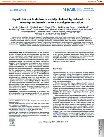
JOURNAL OF HEPATOLOGYAICIIIIIIII.1IVIV.1IV.2IV.3IV.4IV.5Fig. 1. Pedigree of the affected family. Three children of a consanguineousmarriage are affected by ACP, one sibling and the mother are heterozygouscarriers of c.2554 1G T. The index case is marked by an arrow.As shown in Fig. 2A, iron accumulation was found in the liver, thebasal ganglia, and the dentate nuclei. Liver histology showed anormally structured liver with severe hepatocellular iron overload but no stainable iron in macrophages (Fig. 3). The resultsfrom hepatic iron quantification are shown in Table 1. Based onthese findings the clinical diagnosis of ACP was made in the proband and family studies were initiated.BD1200Family studies showed a consanguineous loop and the family history revealed diabetes in a sister (IV.4) of the proband (Fig. 1). Asshown in Table 1 hyperferritinemia, hypoferremia, and undetectable serum CP in two sisters indicated ACP. A movement disorderwith a hypomimia and a postural tremor of both arms were present in the older sister (IV.4), whereas all other family memberswere clinically unaffected. Ophthalmological investigation in allindividuals showed retinal pigment epithelium changes with acentral depigmentation and a bulls-eye like macula but a normalvision and perimetry in patients IV.4 and IV.5. MR imaging andliver histology in the biochemically affected sisters showed ironaccumulation in the basal ganglia, dentate nuclei, and the liver.The results from family studies further supported the diagnosisof autosomal recessive inheritance of ACP in this pedigree andmolecular genetic testing was initiated.1000Molecular genetics: detection of a CP splice-site mutationTo identify the genetic cause of ACP, hepatic cDNA analysis wascarried out and RT-PCR did not show the expected full lengthproduct but showed an aberrant fragment, 100 bp shorter thanthe expected for the PCR product spanning from exons 13 to 16(Supplementary Fig. 1). Sequencing of the aberrant fragmentshowed absence of exon 14 in all biochemically affected familymembers. No other mutations were detected.Direct sequencing of genomic DNA revealed homozygosity forc.2554 1G T in intron 14 of the CP gene in all biochemicallyaffected family members. The mother (III.1) and one unaffectedbrother (IV.3) were heterozygous carriers, whereas the otherbrother had the wild-type sequence on both alleles. Thec.2554 1G T mutation of the CP gene was not found in 100 control chromosomes by PCR amplification, restriction fragmentserum ferritin µg/LFamily 40270300follow-up (days)first deferasirox doseFig. 2. Iron accumulation and results from deferasirox treatment. (A) T2 brainMRI of patient IV.4 shows a marked hypodensity in the area of the basal gangliaindicating severe iron accumulation (arrows). (B) T2* maps of the brain confirmthe significant iron accumulation in the basal ganglia and show iron loading inthe dentate nucleus (red colour indicates areas with a high and dark blue colourthose with a low tissue iron concentration). (C) Abdominal T2* maps of patientIV.4 show liver iron overload of approximately 250 lmol Fe/g liver tissue beforedeferasirox treatment. After 2 months of treatment a reduction in liver ironconcentration was found and no visible liver iron remained after 5 months oftreatment (25 lmol Fe/g liver tissue). (D) Serum ferritin concentrations rapidlydecreased during deferasirox treatment. Arrows indicate the first visit aftercessation of iron chelation therapy.length polymorphism, and D-HPLC (Supplementary Fig. 2).Genetic and biochemical findings in this family show segregationof this novel mutation with ACP.Functional consequencesThe c.2554 1G T mutation, which was shown to cause skippingof exon 14 is predicted to preserve the open reading frame andJournal of Hepatology 2010 vol. 53 j 1101–11071103
Research ArticleCourse on iron chelation treatmentFig. 3. Liver histology shows a normally structured liver with severe hepatocellular iron over-load. (A) Perls’ stained liver biopsy of the index case withgrade III iron accumulation in hepatocytes and (B) an iron free focus. (C) HEstained liver biopsy of the index case showing normally structured liver lobeswithout steatosis or inflammation but with hemosiderin pigment in hepatocytes.(D) Reticulin-stained liver biopsy shows minimal fibrosis (grade I). All pictureswere captured using an Olympus BH2 microscope (Olympus, Vienna, Austria)with a Jenoptik Progress C12 digital camera and Progress Capture Pro 2.5 software(Jenoptik, Jena, Germany). Panels A and D 100 , panels B and C 200 .To treat iron overload associated with ACP, iron chelation therapywith deferasirox was started in all three homozygotes for the CPsplice-site mutation with initial doses of 17–20 mg/kg bodyweight per day. Treatment duration ranged from 1 week to5 months. In the index case deferasirox dose was reduced from20 to 15 mg/kg/day after 4 weeks due to diarrhea and after3 months treatment was stopped because of anemia (hemoglobin9.9 g/dl). Patient IV.4 also developed anemia (hemoglobin 10 g/dl) and treatment was stopped after 5 months due to a gradualincrease in creatinine up to 1.3 mg/dl. Treatment was discontinued after one week in patient IV.5 because of a skin rash.Serum ferritin concentrations decreased in all patientswhereas serum iron, transferrin, and transferrin saturationremained unchanged during deferasirox therapy (Fig. 2D). Noninvasive iron quantification by MRI showed a marked decreasein liver iron concentration after 3 months in the index case andin patient IV.4. After 5 months of treatment T1 and T2* relaxationtimes in patient IV.4 increased from 390 to 513 ms (normal570 40 ms) and from 1.8 to 10.9 ms (normal 24 6 ms), respectively. However, there was no imaging evidence for significanteffects on brain iron overload during iron chelation therapy inboth patients. Accordingly, neurological symptoms remained stable during the follow up. Both patients reported a transientreduction in their insulin demands on deferasirox. Iron chelationtherapy with deferasirox is effective in reducing hepatic iron concentrations and improves endogenous insulin production, but hasno short term effect on brain iron accumulation.Discussionresulted in the loss of 43 amino acids spanning residues G809–G852 with a molecular weight of 4.6 kDa. This was confirmedby SDS–PAGE and Western blotting of proteins extracted fromliver biopsies of genetically affected family members and ahealthy control, which showed major immunoreactive bands atthe expected molecular weights (Fig. 4A).A structural homology model of the mutant protein thatappears to be expressed in the liver of genetically affected individuals was constructed (Supplementary Fig. 3). Our model suggests that the missing region from G809–G852 is located withindomain 5 (D724–P903) of CP, which contains neither a copperbinding site nor the ferroxidase site [23]. Analysis of the modelsuggests that rearrangement of the termini flanking the deletedresidues would require a cumulative shift in their positions ofapproximately 20 Å. The abnormal CP, where the open readingframe and all copper-binding sites are putatively preserved, isexpressed in hepatocytes, but undetectable in plasma.Hepcidin in ACPTo study the pathogenesis of iron overload in ACP, mRNA of thekey regulator of iron metabolism hepcidin was quantified by realtime PCR in liver biopsies. To determine if the observed increasein hepcidin expression was appropriate for the degree of ironoverload, hepcidin/log (ferritin) ratios were calculated. As shownin Fig. 4B mean hepcidin mRNA levels and hepcidin/log (ferritin)ratios are higher in ACP, dysmetabolic siderosis, and patientswith fatty liver disease as compared to patients with C282Yhomozygous hemochromatosis or viral hepatitis.1104CP is a multi-copper ferroxidase essential for iron homeostasiswhich facilitates cellular iron efflux [7,24]. Lack of functional CPis typically associated with iron overload in the liver, pancreas,and the basal ganglia as illustrated in the reported cases. Retinaldegeneration has a penetrance ranging from 75% to 93% inpatients with ACP and retinal pigment epithelium changes werepresent in patients IV.4 and IV.5 [4]. It is unclear if the incompletepenetrance of neurological symptoms and diabetes in ourpatients is a result of disease modifiers or reflects the naturalcourse of ACP. Both clinically affected siblings are diabetic sincetheir twenties, whereas the youngest genetically affected siblingis asymptomatic and not diabetic at the age of 37, which suggeststhe presence of modifiers that affect disease expression.In agreement with previous reports of ACP, none of the genetically affected individuals had evidence of impaired biochemicalliver function or advanced fibrosis despite the significant hepatocellular iron overload that exceeded the thresholds for hepaticfibrosis established in hemochromatosis cohorts [13,25,26]. Thisobservation suggests that iron is only a cofactor in hepatic fibrogenesis [27].Liver histology showed the presence of iron in hepatocytes,whereas no iron was found in macrophages. Moreover, magneticresonance imaging also showed the absence of iron overload inthe spleen, suggesting that iron overload in Kupffer cell is a lateevent in ACP [15]. This observation suggests that CP is importantfor hepatocellular iron efflux, whereas CP-independent mechanisms for iron export from macrophages exist. If CP was indispensable for iron export from recycling macrophages, oneJournal of Hepatology 2010 vol. 53 j 1101–1107
JOURNAL OF HEPATOLOGYTable 1. Demographic, hematological, serum iron parameters, hepatic, and genetic data of members of the affected family at the time of first .515.43.74.9Transferrin(mg/dl)30-300/15-150 C(mg/dl)16-4516-36n.d.n.d. 2.118.6 2.1 2.12892088HICHC(µmol/g) (µmol/g) 25 1.6409.20.60139.4198.50.610.32CP - genotypeReference range (male/female)Heterozygous c.2554 1G TWild Type both allelesHomozygous c.2554 1G THeterozygous c.2554 1G THomozygous c.2554 1G THomozygous c.2554 1G TAbbreviations: f, female; m, male; Hb, hemoglobin; TfS, transferrin saturation; SC, serum ceruloplasmin; HC, hepatic copper concentration; HIC, hepatic iron concentration;n.d., not kD75kDBHepcidin / log (ferritin) ratiowould expect more pronounced anemia and even higher sTfRconcentrations.In our ACP patients, stainable iron was restricted to hepatocytes, in comparison to patients with advanced hemochromatosisor transfusional iron overload where the iron storage typicallyaffects parenchymal cells and macrophages. The decline of serumferritin on deferasirox treatment in ACP patients was faster thanin patients with ß-thalassemia, which suggests that iron-loadedhepatocytes are the main source of serum ferritin and hepatocellular iron is more accessible to deferasirox than macrophagealiron. The prompt increase in serum ferritin after cessation of deferasirox treatment may indicate a redistribution of iron, since there-accumulation of food iron in such a short time appearsunlikely.Hepatocellular iron overload induces expression of the peptide hormone hepcidin in normal individuals, which causes ferroportin degradation. This results in cellular iron retention andreduced iron absorption [28,29]. The observation that hepatichepcidin mRNA expression in ACP patients and in dysmetabolicsiderosis patients are comparable, but low in HFE hereditaryhemochromatosis, suggests that CP but not HFE is dispensablefor iron-mediated hepcidin regulation [30]. We found higher hepcidin mRNA expression in ACP than in viral hepatitis patientswhere hepcidin expression is suppressed [31], which also suggests that hepcidin responds normally to iron in ACP. Normalhepcidin regulation in ACP supports the concept that regionaliron accumulation in affected organs results from disturbed cellular iron metabolism rather than from alterations in total bodyiron homeostasis.The functional consequence of the splice-site mutationc.2554 1G T is the skipping of exon 14, which preserves theopen reading frame. The structural model suggests that theabnormal protein folds into a potentially functional protein, butsuch a significant structural change within domain 5 of CP islikely to affect the stability of the variant protein, resulting inintracellular retention. The phenotypic similarity with CP nonsense mutations further suggests that this variant is dysfunctional [32].Skipping of exon 14 is also predicted to affect the alternativetranscript in the brain, where activation of a cryptic splice-siteresults in direct splicing of exons 18–20 [6]. Iron accumulationin the basal ganglia of c.2554 1G T homozygotes suggests theabsence of functional GPI-anchored CP and supports the importance of correct protein folding for the activity and stability ofGPI-anchored CP. Ferroxidase activity is required to stabilizethe iron export pump ferroportin in cells expressing GPIanchored CP, which provides a molecular explanation for cellulariron retention in ACP [9].300250200150100500n 3n 10n 2n 6n 4ACPHBV/HCVHFEHHNAFLDlow ironDMSFig. 4. Proteins extracted from liver biopsies of genetically affected familymembers. (A) Western blotting of protein extracts from a control liver shows animmunoreactive band at 150 kDa. In protein extracts from liver biopsies ofgenetically affected family members the strongest signal has an apparentmolecular weight of 145 kDa and is slightly lower than the major band in thecontrol liver extract. The difference corresponds to the predicted loss of 4.6 kDa inmolecular weight of the protein translated from the abnormal transcript inc.2554 1G T homozygotes. (B) Hepcidin mRNA concentrations in liver biopsies ofthe three ACP patients are comparable to those in patients with non alcoholicfatty liver disease (NAFLD) or dysmetabolic siderosis (DMS) but higher than inpatients with HFE hemochromatosis (HFE HH) or viral hepatitis.Iron-mediated lipid peroxidation and oxidative stress are themain causes of neurodegeneration secondary to brain iron accumulation [33,34]. To reduce iron toxicity chelation therapy wasintroduced in ACP. The iron chelator deferoxamine was effectivein reducing hepatic iron overload and leading to a partialimprovement of neurological symptoms and brain iron accumulation, as reported in a single case report [16]. The positive effectof deferoxamine on central nervous symptoms and signs was notJournal of Hepatology 2010 vol. 53 j 1101–11071105
Research Articlefound in subsequent studies [14,15], which highlights the lowpermeability of the blood–brain barrier for deferoxamine. Despitethe lower molecular weight and more lipophilic properties of deferiprone, this orally bioavailable iron chelator had no beneficialeffect in ACP in a previous report [14]. In agreement, we foundno improvement of neurological symptoms or brain iron accumulation during deferasirox treatment over a period of severalmonths. Deferasirox could still be an effective brain iron chelator,whose clinical benefit might be missed after a short term treatment. By comparison, the neurological improvement on zinc-sulphate was found after 15 months of treatment in a patient withACP [35]. In contrast, hepatic iron concentration and insulinrequirements improved in both patients who were treated for 3and 5 months, respectively. Taken together, short term deferasirox is effective in reducing hepatic iron overload, but ineffectivefor the treatment of brain iron accumulation. In all patients, sideeffects prohibited the long term therapy that may be required tomobilize iron from the brain. Given the high efficacy of a highdose of deferasirox, a low dose of the iron chelator may have amore favourable side effect profile, which in turn may allow fora long term and combination therapy. Recent studies in an animalmodel of Friedreich’s ataxia suggest that the combined use ofpyridoxal isonicotinoyl hydrazone and deferoxamine is an effective treatment of iron accumulation in glial cells [36]. Furtherwork is required to determine if similar strategies can be developed for patients with ACP and other neurodegenerative diseasesthat are associated with brain iron accumulation.Financial disclosureThis work was supported by the Austrian Science Fund Project19579 to H.Z. The authors report no commercial relationshipsthat might pose a conflict of interest in connection with thismanuscript.AcknowledgementsThe authors thank Anna Schlögl, Gisela Egg, and Nadja Baumgartner for excellent technical assistance and PD Dr. Philip Eller forassisting genealogical studies.Supplementary dataSupplementary data associated with this article can be found, inthe online version, at doi:10.1016/j.jhep.2010.04.039.References[1] Xu X, Pin S, Gathinji M, Fuchs R, Harris ZL. Aceruloplasminemia: an inheritedneurodegenerative disease with impairment of iron homeostasis. Ann NYAcad Sci 2004;1012:299–305.[2] Miyajima H, Kohno S, Takahashi Y, Yonekawa O, Kanno T. Estimation of thegene frequency of aceruloplasminemia in Japan. Neurology 1999;53:617–619.[3] McNeill A, Pandolfo M, Kuhn J
load. Hepatic iron concentration normalized after 3 and 5 months of iron chelation therapy with deferasirox, which was also asso-ciated with reduced insulin demands. During short term treat-ment there was no clinical or imaging evidence for significant effects on brain iron overload. Conclusions:Aceruloplasminemia can show an incomplete clini-