Transcription
Dig Dis Sci (2014) 59:976–985DOI 10.1007/s10620-013-2966-5ORIGINAL ARTICLEEpigallocatechin Gallate Does Not Accelerate the Early Phaseof Liver Regeneration After Partial Hepatectomy in RatsVojtěch Mezera Otto Kučera Alena MoravcováEva Peterová Zuzana Červinková Received: 1 July 2013 / Accepted: 15 November 2013 / Published online: 8 December 2013Ó The Author(s) 2013. This article is published with open access at Springerlink.comAbstractBackground Two-thirds partial hepatectomy (PHx) is anestablished model for the study of liver regeneration afterresection. This process is accompanied by oxidative stress.Aims In our study, we tested the effect of epigallocatechin gallate (EGCG), a green tea antioxidant, on the earlyphase of liver regeneration after PHx.Methods Male Wistar rats were divided into five groups:(I) laparotomy ? water for intraperitoneal injections, (II)laparotomy ? EGCG 50 mg/kg body weight, (III)PHx ? water for injections, (IV) PHx ? EGCG 20 mg/kgand (V) PHx ? EGCG 50 mg/kg, for 3 consecutive days.The rats were killed 24 h after surgery. Biochemical analysis of rat sera was performed. Histological samples werestained with hematoxylin & eosin and bromodeoxyuridine(BrdU). In hepatectomized rats, we also measured plasmamalondialdehyde, tissue malondialdehyde, glutathione andcytokines levels, the activity of caspases 3/7, expression ofV. Mezera (&) O. Kučera A. Moravcová Z. ČervinkováDepartment of Physiology, Faculty of Medicine in HradecKralove, Charles University in Prague, Simkova 870,500 38 Hradec Kralove 1, Czech Republice-mail: mezev5ar@lfhk.cuni.czO. Kučerae-mail: kucerao@lfhk.cuni.czA. Moravcováe-mail: moravca2@lfhk.cuni.czZ. Červinkováe-mail: wolff@lfhk.cuni.czE. PeterováDepartment of Biochemistry, Faculty of Medicine in HradecKralove, Charles University in Prague, Hradec Kralove 1, CzechRepublice-mail: peterovae@lfhk.cuni.cz123Nqo-1 and HO-1 genes at the mRNA level, and expressionof p21, p-p27 and p-p53 genes at the protein level.Results We observed lower accumulation of BrdU ingroup V when compared to groups III and IV. The activityof caspases 3/7 and expression of p-p53 were lower ingroup V than in groups III and IV. Tissue levels of IL-6were lower in group V when compared to group III. Significant differences were not noted in other parameters.Conclusions Administration of EGCG did not stimulateearly phase liver regeneration in rats after PHx. There waseven lower DNA synthesis in the group treated with a highdose of EGCG.Keywords Antioxidants Epigallocatechin gallate Hepatectomy Liver regeneration Oxidative stressAbbreviationsAIWater for injectionsALTAlanine aminotransferaseALPAlkaline phosphataseASTAspartate allocatechin gallateEGCG20 Epigallocatechin gallate 20 mg/kgweightEGCG50 Epigallocatechin gallate 50 mg/kgweightGSHGlutathione (reduced form)GSSGGlutathione (oxidized form)HO-1Heme oxygenase-1HRRHepatic regeneration rate [25]IL-6Interleukin-6LAPLaparotomyLRRLiver regeneration rate [26]MDAMalondialdehydebodybody
Dig Dis Sci (2014) ne-oxidoreductaseNF-E2-related factor 2Two-thirds partial hepatectomyTransforming growth factor beta1Tumor necrosis factor alphaIntroductionLiver resection is a surgical procedure performed for avariety of malignant and benign focal liver lesions. It is oneof the few methods that gives the patient a chance forcomplete recovery. This is in both primary liver tumors,especially hepatocellular carcinoma [1], and secondarytumors, notably liver metastases from colorectal carcinoma[2].Vascular occlusion, used during liver resection to prevent hemorrhage, causes ischemia–reperfusion injury.Reperfusion of the liver enhances cell injury by oxidativestress and an inflammatory reaction [3]. In addition, liverregeneration itself was found to be a state of increasedoxidative stress [4, 5]. Some surgical procedures for preventing the ischemia–reperfusion injury have alreadyfound their way into clinical practice [3], whereas possiblepharmacological approaches remain limited. This isbecause no decrease in mortality, incidence of liver failureor perioperative morbidity was found [6]. Two-thirds partial hepatectomy (PHx), carried out on small laboratoryanimals, is a well-established model for studying of liverregeneration [7].Excessive oxidative stress observed in mice afterknockout of a key transcription factor for endogenousantioxidative enzymes led to significant diminution of liverregeneration after PHx [8]. This transcription factor, NFE2-related factor 2 (Nrf2), is an upstream regulator ofvarious antioxidative proteins and xenobiotic-metabolizingenzymes, particularly glutathione-S-transferase, NAD(P)Hquinone-oxidoreductase (Nqo-1), superoxide-dismutase,glutathione-peroxidase, heme oxygenase-1 (HO-1), catalase and others [8–10].Protection against oxidative stress may enhance liverregeneration after resection, as shown in a study usingvitamins C and E [11]. Aside from these directly actingantioxidants, which after oxidation can act as pro-oxidants,indirect antioxidants exist. These are able to activate theaforementioned transcription factor Nrf2 and subsequentlymany cytoprotective proteins [9, 10]. The indirectly actingantioxidants have very low prooxidative effects [12].One of these is (-)-epigallocatechin-3-gallate (EGCG),a polyphenolic antioxidant from green tea. Besides thiseffect, EGCG also acts as a direct antioxidant because of977the presence of phenolic groups that are sensitive to oxidation and can generate quinone [9, 13]. EGCG is the mostabundant of the green tea catechins [14]. Its protective rolewas found in a model of acute toxic liver injury [15],chronic toxic liver injury [16, 17], non-alcoholic steatosisand steatohepatitis [18–20], and hepatic ischemia–reperfusion injury [18, 21]. EGCG was also able to ameliorateexperimental diabetes mellitus by downregulation of genesfor gluconeogenesis and synthesis of fatty acids, triacylglycerols and cholesterol [22, 23]. The effect of EGCG onliver regeneration after PHx has not been tested yet.In our study, we tested the effect of EGCG in the earlyphase of liver regeneration after PHx in rats. Since thehighest rate of DNA synthesis is in hepatocytes 24 h afterPHx [24], we decided to investigate the regenerativeresponse in this particular interval. We also focused on themonitoring of possible changes in relevant signalingpathways.MethodsChemicalsUnless otherwise stated, BrdU (5-bromo-2-deoxyuridine)and other chemicals used were all of analytical grade andhad been purchased from Sigma-Aldrich (St. Louis, MO,USA). The kit for determination of caspase 3/7 activitieswas obtained from Promega (Madison, WI, USA). Mousemonoclonal anti-BrdU antibody had been obtained fromDAKO (Glostrup, Denmark) and secondary anti-mouseantibody from Jackson ImmunoResearch Laboratories(West Grove, PA, USA).Mouse anti-b-actin antibodies and rabbit anti-p21 CIP1were purchased from Sigma-Aldrich, rabbit p27 Kip1 andrabbit phospho-p53 (Ser15) from Cell Signaling (Beverly,MA, USA), mouse Bcl-2 antibody (C-2) from Santa CruzBiotechnology (Dallas, TX, USA), secondary goat antirabbit IgG from Sigma-Aldrich and secondary goat antimouse IgG from Santa Cruz Biotechnology.Experimental DesignMale Wistar rats (BioTest, Konárovice, Czech Republic)with initial weight of 248 13 g were used throughout thestudy. The rats were housed at 23 1 C, 55 10 %humidity, air exchange 12–14 times/h and 12 h light–darkcycles. The animals had free access to tap water and werefed the standard pelleted diet ST-1 ad libitum (Velas, Lysánad Labem, Czech Republic). In accordance with Czechlegislation, all animals received care according to theguidelines set out by the Institutional Animal Use and Care123
978Committee of the Faculty of Medicine of Hradec Králové,and this committee approved our study.The animals were selected for laparotomy (LAP, shamoperation) or 2/3 partial hepatectomy (PHx) under etheranesthesia [7]. Water for injections (AI) or EGCG in a doseof 20 or 50 mg/kg body weight was administered intraperitoneally for 3 consecutive days (regimen according to[15]), with the third dose administered immediately afterthe operation. The animals were divided into following fivegroups:I—AI ? LAP: water for injections and laparotomy,n 6;II—EGCG50 ? LAP: EGCG 50 mg/kg and laparotomy, n 6;III—AI ? PHx: water for injections and PHx, n 6;IV—EGCG20 ? PHx: EGCG 20 mg/kg and PHx,n 6;V—EGCG50 ? PHx: EGCG 50 mg/kg and PHx,n 6.The animals were killed under general general anesthesia by exsanguination 24 h after the operation, i.e., onthe 4th day of the experiment. BrdU for detection of DNAsynthesis was administered at a dose of 50 mg/kg intraperitoneally 1 h before sacrificing. Blood and liver sampleswere immediately frozen in liquid nitrogen and stored at-80 C until analysis. Resected liver from the 3rd day andthe remnant on the 4th day were weighed and liver growthwas calculated according to formulas from the literature[25, 26].Serum Biochemical MeasurementsSerum concentrations of glucose, total and conjugatedbilirubin, activities of alanine aminotransferase (ALT),aspartate aminotransferase (AST) and alkaline phosphatase(ALP) of all animals were measured in the Department ofClinical Biochemistry and Diagnostics, University Hospitalin Hradec Králové. In the hepatectomized groups (groupsIII, IV, V), plasmatic levels of malondialdehyde (MDA)were also measured.Determination of GSH and GSSGAfter adding the liver homogenate to cold 10 % metaphosphoric acid, mixing and centrifuging (20,0009g,10 min., 4 C), glutathione in the supernatant was analyzedby Hissin’s fluorimetric method [27] modified by Roušar[28]. Briefly, GSH was exposed to orthophthalaldehyde inhydrogenphosphate buffer, and the fluorimetric detectionwas carried out (kEx 340 nm, kEm 420 nm). GSSGwas assessed after alkaline hydrolysis in the same way asGSH.123Dig Dis Sci (2014) 59:976–985Determination of Liver Tissue Cytokines and TBARSThe protein content in the sample was determined afterhomogenization in RIPA buffer and spinning (10,0009g)by the Bradford method [29] using bovine serum albuminas a standard. Afterwards, levels of liver TNF-a in thesupernatant were measured using the ELISA method. RatTNF-alpha Platinum ELISA, Rat IL-1 beta PlatinumELISA and Rat TGF-beta1 Platinum ELISA kits werepurchased from eBioscience (San Diego, CA, USA).Lipid peroxidation in liver supernatants from thehomogenates was determined according to Ohkawa et al.[30], i.e., by the assessment of thio barbituric acid reactivesubstances (TBARS).Determination of Caspase-3/7 Activity in LiverHomogenateActivity of executive caspases was measured using the Caspase-GloTM 3/7 kit. Briefly, after homogenization accordingto Liu et al. [31], the tissues were centrifuged and samplesadjusted to contain the same amount of protein [29]. Thesamples were incubated for 1 and 2 h with Caspase-GloTMreagent (contains substrate for caspases 3/7), and the emittedluminescence was measured using the TECAN Infinite M200.Determination of p21, p-p27, p-p53, and Bcl-2ExpressionExpression of p21, p-p27 and p-p53 genes at the protein levelwas estimated using the Western blot method as describedpreviously [32]. After homogenization [31] and quantification of protein content [29], the lysates containing an equalamount of protein (10 lg) were subjected to SDS-PAGEusing gradient gel NuPAGEÒ NovexÒ Bis–Tris Mini gels4–12 % (Life Technologies, Carlsbad, CA, USA) andtransferred to a cellulose nitrate membrane (Bio-Rad Laboratories, Hercules, CA, USA). The membranes were incubated with primary rabbit antibodies at 4 C overnight andwith secondary antibody goat anti-rabbit IgG for 2 h. Proteins were visualized using a chemiluminescence detectionkit (Roche, Penzberg, Germany) and autoradiographic film(Foma, Hradec Kralove, Czech Republic). To confirm equalprotein loading, each membrane was reprobed and reincubated to detect b-actin. The preparation differed for Bcl-2protein, where we used a more concentrated homogenate ofliver tissue in RIPA buffer.Determination of HO-1 and Nqo-1 Gene ExpressionGene expression was examined as previously described[33], with the exception that total RNA from liver tissuewas isolated by phenol–chloroform extraction using the
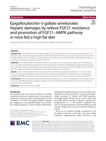
Dig Dis Sci (2014) 59:976–985979RNA Blue kit (TOP-Bio, Prague, Czech Republic). TaqMan Fast Universal PCR Master Mix and pre-designedTaqMan Gene Expression Assay kits were from LifeTechnologies (Carlsbad, CA, USA), and primers and probewere purchased from Generi Biotech (Hradec Králové,Czech Republic). Glyceraldehyde-3-phosphate dehydrogenase was used as reference for normalizing the data.Liver Histology, Incorporation of BromodeoxyuridineAfter the rats had been killed, liver samples were immediately fixed by immersion in 4 % neutral formaldehyde.Paraffin sections were stained with hematoxylin & eosin andchecked for the presence of steatosis and/or inflammation.Furthermore, apoptosis was assessed according to [34], i.e.,the number of apoptotic bodies per visual filed was calculated in hematoxylin & eosin-stained samples. The pathologist evaluating the samples was blinded to the study.The immunohistochemical analysis was made on paraffin sections of the liver tissue as described previously[35]. Briefly, the paraffin sections were incubated withprimary mouse anti-BrdU monoclonal antibody, then withbiotinylated anti-mouse secondary antibody and subsequently with a streptavidin conjugate of peroxidase (Dako,Glostrup, Denmark). Visualization of bound antibody wasperformed using 3,30 -diaminobenzidine-tetrahydrochlorideand hydrogen peroxide. BrdU incorporation was determined by counting of positively stained hepatocyte nucleiin nine representative microscope fields (9100 magnification) using an Olympus IX51 microscope and quantifiedusing a computer-aided image analysis system NIS-Elements AR 2.30 (Nikon, Lewisville, TX, USA).Fig. 1 Body weight of experimental animals at operation (day 1) andat sacrifice (day 2). §, §§, §§§When comparing the same group atsacrifice and at operation. , , When comparing the EGCG50 ? LAP group with the AI ? LAP group; *, **, *** whencompared to laparotomized animals (AI ? PHx vs. AI ? LAP;EGCG20 ? PHx vs. EGCG50 ? LAP; EGCG50 ? PHx vs. EGCG50 ? LAP), p \ 0.05; p \ 0.01; p \ 0.001, respectivelyTable 1 Indices of liver regenerationAI ? PHxEGCG20 ? PHxEGCG50 ? PHxp valueHRR (%)9.5 413.5 68.9 10nsLRR (%)23.3 829.9 918.1 25nsHRR (%) (C - (A - B))/A 9 100 (Murata [26])LRR (%) (C - (A - B))/C 9 100 (Selzner [25])A estimated total liver weight at PHx, B excised liver weight,C weight of regenerated liver at sacrificeStatistical AnalysisResultsfrom further analysis. In group I (AI ? LAP) the bodyweight rose after the operation (p \ 0.05); in all othergroups, there was a non-significant trend to decline in bodyweight after the operation. Body weight at operation didnot differ among groups; however, body weight was higherat sacrifice in the AI ? LAP group when compared to theEGCG50 ? LAP and AI ? PHx groups (p \ 0.001)(Fig. 1).Among hepatectomized groups (III, IV, V), the calculated indices of liver regeneration appeared highest in theEGCG20 ? PHx group; however, due to high variabilitythe differences were not statistically significant (Table 1).Animals, Body and Liver WeightHistological Findings, Incorporation of BrdUDuring our experiment all animals survived until the sacrifice. One animal from the EGCG50 ? PHx group had asurgical complication (hemoperitoneum) and was excludedIn hematoxylin & eosin staining from the caudate lobe,distinct lipid accumulation was visible in the hepatectomized groups (III, IV, V); this is consistent with publishedThe results are expressed as the mean SD. Analyseswere performed using Graph-Pad Prism 4.03 software(Graph Pad Software, San Diego, CA, USA). First, normality was tested by Kolmogorov-Smirnov test. In normaldata, comparisons were made among the groups usingANOVA followed by Tukey-Kramer’s post hoc test. In thecase of non-Gaussian distribution, non-parametric KruskalWallis tests and Dunn’s post hoc test were used. p \0.05was considered statistically significant.123
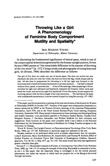
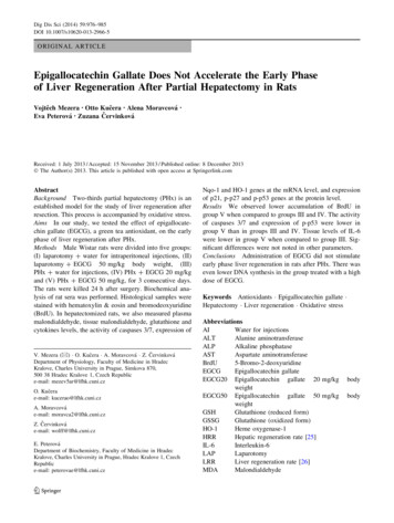
980Dig Dis Sci (2014) 59:976–985Fig. 2 a DNA synthesisestimated by BrdUincorporation. *, **, *** Whencompared to laparotomizedanimals (AI ? PHx vs.AI ? LAP; EGCG20 ? PHxvs. EGCG50 ? LAP;EGCG50 ? PHx vs.EGCG50 ? LAP); ?, ?,?when compared to theAI ? PHx group; 9, 99,999when compared to theEGCG20 ? PHx group,p \ 0.05; p \ 0.01; p \ 0.001,respectively. b Laparotomizedgroups: only sporadic BrdUpositive cells. c Group III(AI ? PHx): numerous BrdUpositive cells. d Group V(EGCG50 ? PHx): numerousBrdU-positive cells, but lessthan in groups III and IV. Notethat the positive nuclei aremostly in periportal areas.e Apoptotic bodies. *, **, ***When compared tolaparotomized animals(AI ? PHx vs. AI ? LAP;EGCG20 ? PHx vs.EGCG50 ? LAP;EGCG50 ? PHx vs.EGCG50 ? LAP)data [36]. In addition, there was vacuolation of the cytoplasm in peripherally located hepatocytes in the hepatectomized groups and sporadically occurring pyknotic nuclei.In the center of the lobe, sparse hepatocytes with granulareosinophilic cytoplasm occurred. We did not observe signsof pronounced inflammatory infiltration in any of the rats.The number of apoptotic bodies was significantly higher inthe hepatectomized groups (III, IV, V) compared to thelaparotomized groups (I, II). The apoptotic bodies weremostly noticed around the central veins. The laparotomized123groups did not differ from one another in apoptotic bodycount; we did not observe differences among the hepatectomized animals either (Fig. 2e).Immunohistochemical staining for incorporated BrdUshowed only sporadic positive hepatocytes in the laparotomized groups (I, II); there was high positivity in thehepatectomized groups (III, IV, V). We observed no difference between groups III and IV; however, the count ofBrdU-positive cells in the EGCG50 ? PHx group waslower when compared to the AI ? PHx and
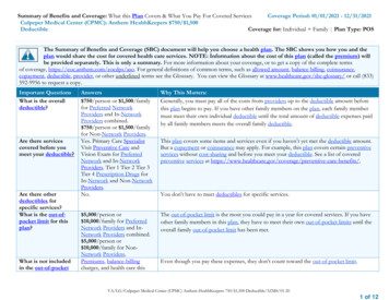
Dig Dis Sci (2014) 59:976–985981Table 2 Serum biochemistryAI ? LAPEGCG 50 ? LAPAI ? PHxEGCG 20 ? PHxEGCG 50 ? PHxp valueGlycemia (mmol/l)7.01 0.6110.49 0.28 7.97 0.578.9 0.69*8.71 0.75*\0.001ALT (lkat/l)1.01 0.570.64 0.064.08 4.762.7 1.38**5.44 4.95**\0.01AST (lkat/l)3.2 1.991.89 0.377.05 6.346.33 2.57**9.92 8.20**\0.016.8 1.29***6.61 1.39***\0.001ALP (lkat/l)2.4 0.452.57 0.327.08 1.11***Bilirubin total (lmol/l)2.0 0.03.0 0.04.0 1.55*3.5 0.555.6 2.19\0.05Bilirubin conj. (lmol/l)1.83 0.412.0 0.02.6 0.892.17 0.414.2 2.28*\0.05 , , When comparing the EGCG50 ? LAP with the AI ? LAP group*, **, *** When compared to laparotomized animals (AI ? PHx vs. AI ? LAP; EGCG20 ? PHx vs. EGCG50 ? LAP; EGCG50 ? PHx vs.EGCG50 ? LAP), p \ 0.05; p \ 0.01; p \ 0.001, respectivelyEGCG20 ? PHx groups (p \ 0.001 for both comparisons)(Fig. 2a–d). BrdU positivity and thus DNA synthesis werenoticed mostly in the periportal areas (Fig. 2c–d).Serum Biochemical CharacteristicsSerum glycemia was higher in group II (EGCG50 ? LAP)when compared to group I (AI ? LAP). There was a trendtowards hyperglycemia even at a dose of 20 mg/kg whencomparing group IV (EGCG20 ? PHx) with group III(AI ? PHx). In addition, we observed lower glycemia inthe hepatectomized groups treated with EGCG whencompared to the EGCG50 ? LAP group.Alanine aminotransferase and aspartate aminotransferaseactivity was higher in hepatectomized animals, but the statistical significance was present only when comparingEGCG20 ? PHx and EGCG50 ? PHx with EGCG50 ? LAP. Furthermore, the ALT and AST activity wassomewhat lower in the EGCG20 ? PHx than in the AI ? PHxgroup, but this difference did not reach statistical significance.Alkaline phosphatase activity was higher in all hepatectomizedgroups when compared to the laparotomized groups.Total bilirubin was higher in the EGCG50 ? LAP groupthan in the AI ? LAP group, and in the hepatectomizedthan in the laparotomized groups; however, a significantdifference was observed only when comparing theAI ? PHx with the AI ? LAP groups. Conjugated bilirubin was higher in the hepatectomized than in the laparotomized groups, reaching significance only whencomparing the EGCG50 ? PHx with the EGCG50 ? LAPgroup. Both total and conjugated bilirubin reached theirhighest values in the EGCG50 ? PHx group; however, thedifferences among hepatectomized groups were not statistically significant (Table 2).Tissue Cytokines TNF-a, IL-6, and TGF-b: Markersof Oxidative StressWe observed an insignificant trend of lower TNF-a inhepatectomized groups treated with EGCG when comparedFig. 3 Tissue levels of IL-6. ?, ?, ?When compared to theAI ? PHx group, p \ 0.05; p \ 0.01; p \ 0.001, respectivelyto the AI ? PHx group. A similar trend was observed withIL-6, and its expression was significantly lower in theEGCG50 ? PHx group when compared to the AI ? PHxgroup (Fig. 3). We did not observe differences in theexpression of liver TGF-b1.A similar non-significant trend of lower tissue MDA,GSH and GSSG content as well as lower plasmatic MDAwas observed in the EGCG20 ? PHx group when compared to the AI ? PHx and EGCG50 ? PHx groups;however, none of these reached the level of statisticalsignificance (Table 3).Nqo-1 and HO-1 Gene Expression at the mRNA LevelThere were no significant differences among hepatectomized groups in mRNA expression of Nqo-1 and HO-1.Because of the small amount of measured samples inexperimental groups (n 3), we observed only an insignificant trend of lower HO-1 expression inEGCG20 ? PHx and higher HO-1 expression in the123
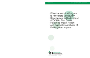
982Dig Dis Sci (2014) 59:976–985Table 3 Cytokines TNF-a, IL-6 and TGF-b: markers of oxidative stressAI ? PHxEGCG 20 ? PHxEGCG 50 ? PHxp valuePlasmatic MDA (lmol/l)1.20 0.181.07 0.181.24 0.14nsTissue MDA (nmol/mg protein)0.35 0.200.27 0.070.47 0.13nsTissue GSH (lmol/l)810 195717 164781 144nsTissue GSSG (lmol/l)238 169188 140241 164nsTissue TNF-a (pg/ml)803 103771 69752 126nsTissue IL-6 (pg/ml)785 122686 111660 99?\0.058,024 4,7756,340 4,0537,647 4,651Tissue TGF-b1 (pg/ml)?, ?, ?nsWhen comparing the EGCG50 ? PHx with the AI ? PHx group, p \ 0.05; p \ 0.01; p \ 0.001, respectivelyDiscussionFig. 4 Activity of executive caspases 3/7; incubation for 1 and 2 h,respectively. ?, ?, ?When compared to the AI ? PHx group; 9,99, 999when compared to the EGCG20 ? PHx group, p \ 0.05;p \ 0.01; p \ 0.001, respectivelyEGCG50 ? PHx group when compared to the AI ? PHxgroup (not shown).Activity of Caspases 3/7After 1 h incubation, lower activity in the EGCG50 ? PHxgroup was observed when compared to the AI ? PHx andEGCG20 ? PHx groups (p \ 0.01 for both comparisons).After 2 h incubation, the EGCG50 ? PHx group also hadlower activity compared to both the AI ? PHx (p \ 0.05)and EGCG20 ? PHx (p \ 0.001) groups (Fig. 4).Protein Expression of Cell-Cycle Regulating ProteinsProtein expression of p21 and p-p27 did not differ amonghepatectomized groups. On the other hand, expression ofp-p53 protein appeared higher in group IV(EGCG20 ? PHx) and lower in group V (EGCG50 ? PHx)than in group III (AI ? PHx) (Fig. 5). We were not able todetect Bcl-2 protein.123Despite several studies describing the beneficial effect ofEGCG on the liver in various experimental models, therewas no beneficial effect on liver regeneration after PHx inour experiment. There was even lower DNA synthesis inanimals receiving higher doses of EGCG. Based on ourresults, the use of EGCG in conditions of organ alteration,such as regenerating liver, cannot be recommended. Theeffect of EGCG is complex: several hundred EGCG-regulated genes were identified in a study with microarrays[37]. Furthermore, anti-proliferative and -cancer propertiesof EGCG are known [38, 39].Since EGCG was reported to decrease the productionof TNF-a [40] and IL-6 [38], which plays an importantrole in liver regeneration [41, 42], we wondered if theinhibiting effect of EGCG on DNA synthesis was mediated in this way. There was only a non-significant trend oflower TNF-a levels in EGCG-treated hepatectomizedanimals when compared to hepatectomized animalsreceiving water for injections. A possible explanation isthat TNF-a is involved in regulation of even earlierevents in liver regeneration [24]. Similarly, the levels ofIL-6 were lower in the EGCG50 ? PHx group comparedto the AI ? PHx group.Furthermore, EGCG was repeatedly reported to activatecaspases and increase apoptosis [43–46]. Surprisingly, wefound rather lower activity of executive caspases 3/7 inliver homogenates from group V (EGCG50 ? PHx) thanfrom animals receiving a lower dose (EGCG20 ? PHx) orvehicle only (AI ? PHx). An explanation could be thedifferent effects of EGCG on tumor and normal cells:EGCG was shown to protect hepatocytes from apoptosiscaused by toxic injury [47, 48]. Direct inhibition of caspase3 by EGCG was also described [49]. We were not able todetect the Bcl-2 protein using Western blot. However, weare not the first workplace to fail to do so: Tzung et al. [50]reported undetectable levels of Bcl-2 mRNA and protein at24 h after PHx. Our results of apoptotic body count are alsoin accord with [50], since the authors showed a peak of
Dig Dis Sci (2014) 59:976–985983Fig. 5 Western blot bands ofp-p53 protein with beta-actin asa loading control (p21 andp-p27 are not shown as nodifferences were observed)high proapoptotic Bad and rather lower expression ofantiapoptotic Bcl-x at 24 h after PHx.Other antiproliferative mechanisms of EGCG arechanges in cell-cycle regulating proteins, especiallyinduction of cyclin-dependent kinase inhibitors p21 andp27 [43, 51, 52], and increased expression and serinephosphorylation of p53 [45, 53]. However, we were notable to prove a difference in p21 and p27 expression amonghepatectomized groups. Expressions of phosphorylated p53appeared higher in the EGCG20 ? PHx group and surprisingly lower in hepatectomized animals receiving ahigher dose of EGCG (EGCG50 ? PHx) when comparedto the group receiving vehicle (AI ? PHx).Many more antiproliferative mechanisms of EGCGwere described, changes in protein kinases, growth factorsand transcription factors [38], e.g., inhibition of NF-KB[54].To check whether the Nrf2 pathway had been activated,we measured the expression of HO-1 and Nqo-1 genes,which belong to Nrf2 effectors, at the mRNA level [55,56]. However, due to the small number of samples, wewere not able to prove activation of this pathway.Interestingly, EGCG led to higher glycemia compared tovehicle-treated rats, which is in contrary to other findings[22, 23]. It should be mentioned that orally administeredEGCG decreases glucose absorption from the intestine, butparenterally administered EGCG hinders glucose uptakeinto the tissues [57].Although some antioxidants accelerated liver regeneration after PHx, as could be seen in a study with vitamin Cand E [11], and a study with bicyclol [58], other antioxidants and hepatoprotective agents revealed a rather inhibitory effect on liver regeneration after PHx, namelyresveratrol [59], curcumin [60] and S-adenosylmethionin[61]. Considering the often complex effect of naturalantioxidants on cells, the diversity of the results is notsurprising. Similarly, the immunosuppressant sirolimuswas able to attenuate ischemia–reperfusion injury, butimpaired liver regeneration after PHx [62].Not only our results, but also other studies suggest thatEGCG should be used with caution. EGCG is present in overthe-counter extracts from green tea [63, 64] and even in otherherbal dietary supplements that do not indicate content ofcatechins [65]. These supplements are not without a risk—there were repeatedly reported cases of acute liver injurycaused by green tea extracts [66–68]. Although these injuriescan be partly explained by toxicity of other substances in thesupplements (especially caffeine and/or undetected contaminants), EGCG itself also revealed important effects,particularly interaction with other drugs [69, 70] and a prooxidant effect at high doses [67, 71]. In mice, the dose of120 mg/kg administered intraperitoneally was hepatotoxicand the dose 150 mg/kg lethal for all mice [71]; the dose of1,500 mg/kg via a gastric tube was lethal for 85 % of mice[64]. In rats, the dose of 2,000 mg/kg administered via agastric tube was lethal. Recently, a cholestatic effect ofEGCG was described [72]; higher levels of conjugated bilirubin (even though not significantly) in EGCG-treatedanimals in our study correspond to these findings.In a study with healthy human volunteers, a single doseof 1,600 mg EGCG taken orally was found to be safe andwell tolerated [73]. Potential application of EGCG inclinical studies is complicated by hardly predictable bioavailability [74].In conclusion, administration of EGCG did not enhancethe early phase of liver regeneration after PHx in rats. Thehigher dose even led to lower DNA synthesis. Although thetests carried out have failed to clarify the mechanism ofthis effect, it was probably mediated by some pathwayother than p53.Acknowledgments We would like to thank to Mr. Rajiv Mandalia,MD, for language review. Thanks to Miroslav Podhola, MD, PhD, forhistopathological evaluation. Also thanks to Monika Pospisilova andHana Lastuvkova for their help with PCR, and to Dr. Tomáš Roušarand Dr. René Endlicher for glutathione measurement. This work wassupported by grants from Charles University in Prague, GA UK668512, PRVOUK P37/02 and SVV-2013-266901.Conflict of interestNone.Open Access This article is distributed under the terms of theCreative Commons Attribution Noncommercial License which permits any noncommercial use, distribution, and reproduction in anymedium, provided the original author(s) and the source are credited.References1. Llovet JM, Di Bisceglie AM, Bruix J, et al. Panel of experts inHC
ORIGINAL ARTICLE Epigallocatechin Gallate Does Not Accelerate the Early Phase of Liver Regeneration After Partial Hepatectomy in Rats Vojteˇch Mezera Otto Kucˇera Alena Moravcova Eva Peterova Zuzana Cˇ ervinkova Received: 1 July 2013/Accepted: 15 November 2013/Published online: 8 December 2013