Transcription
HindawiEvidence-Based Complementary and Alternative MedicineVolume 2022, Article ID 5471586, 13 pageshttps://doi.org/10.1155/2022/5471586Research ArticleAn Shen Ding Zhi Ling Ameliorates the Symptoms of AttentionDeficit Hyperactivity Disorder via Modulating Brain-DerivedNeurotrophic Factor-Related Signaling PathwaysLi Yaqun,1,2,3 Yuan Haixia,1,4 Song Yuchen,1 Zhu Mingxin,4 Lu Manqi,5 Tian Yunlong,2Wang Aizhen ,2 and Han Xinmin 1,41First Clinical Medical College, Nanjing University of Chinese Medicine, Nanjing, ChinaThe Affiliate Taizhou Hospital of Nanjing University of Chinese Medicine, Taizhou, China3Jiangsu Key Laboratory of Pediatric Respiratory Disease, Nanjing, China4Jiangsu Province Hospital of Chinese Medicine, Nanjing, China5College of Traditional Chinese Medicine, Gansu University of Chinese Medicine, Lanzhou, Gansu, China2Correspondence should be addressed to Wang Aizhen; tzszyyek@163.com and Han Xinmin; 15195996828@163.comReceived 30 October 2021; Accepted 16 June 2022; Published 21 July 2022Academic Editor: Gauhar RehmanCopyright 2022 Li Yaqun et al. This is an open access article distributed under the Creative Commons Attribution License,which permits unrestricted use, distribution, and reproduction in any medium, provided the original work is properly cited.Attention deficit hyperactivity disorder (ADHD) is a common childhood neurodevelopmental disorder. It may impact the cognitiveand social functions throughout childhood and determine adult outcomes. Dopamine (DA) deficiency theory is the pathogenesis ofADHD that is recognized by most international literature. Existing studies have shown that DA deficiency is caused by the abnormalfunction of the DA transporter and an imbalance in the DA receptor functionality. Recent clinical and experimental studies havefound that the brain-derived neurotrophic factor (BDNF)/tropomyosin receptor kinase B (TrkB) signaling pathway acts a pivotal partin DA vesicle circulation and ADHD pathogenesis. An Shen Ding Zhi Ling (ASDZL) is a traditional Chinese medicine (TCM)prescription, which was widely prescribed to treat ADHD in Jiangsu, China, but its therapeutic mechanism is unclear. Therefore, weconstructed a spontaneously hypertensive rat (SHR) model to explain its mechanism. SHRs were randomly assigned to four groups:SHR model group (vehicle), methylphenidate hydrochloride group (MPH), ASDZL group, and 7,8-dihydroxyflavone group (7,8DHF). At the same time, the above groups were given continuous medication for four weeks. The results show that ASDZL, MPH,and 7,8-DHF group could significantly improve the spatial memory of SHRs in the Morris water maze tests. ASDZL increased thelevels of BDNF, TrkB, p75 neurotrophin receptor (p75), C-Jun N-terminal kinases 1 (JNK1), and nuclear factor kappa B (NF-κB) inthe prefrontal cortex (PFC) and hippocampus synaptosome of SHRs. The results of this study suggest that ASDZL can relieve thesymptoms of ADHD in SHRs by regulating the balance between the BDNF/TrkB signaling pathway (promoting vesicle circulation)and the BDNF/P75/JNK1/NF-κB signaling pathway (inhibiting vesicle circulation) within the PFC and hippocampus synaptosome toincrease the DA concentration in the synaptic cleft. The BDNF/TrkB signal pathway within the PFC and hippocampus synaptosomewas activated by 7,8-DHF to increase DA concentration in the synaptic cleft. Whether 7,8-DHF can activate or inhibit the BDNF/P75signaling pathway remains unclear.1. IntroductionAttention deficit hyperactivity disorder (ADHD), as acommon mental disorder in children, currently affects 5%of children around the world. Its symptoms are mainlyinattention and hyperactivity-impulsivity [1]. Multivariate meta-regression analyses have shown that the pooledprevalence estimate of ADHD in China was 6.3% [2].ADHD is a prevalent disorder, with the impairing condition beginning in early childhood and continuing intoadolescence and adulthood. ADHD is frequentlycomorbid with other psychiatric disorders that can createsubstantial burdens for patients, their families, andcommunities [3].
2The pathogenesis of ADHD remains unclear. SinceSwanson et al. proposed the dopamine (DA) deficiencytheory, which has been widely recognized and studied by themajority of the international academic community [4]. TheDA system has become the focus of recent studies on theneurobiochemical pathogenesis of ADHD [5]. DA is animportant modulator of learning and motivation [6] thatperforms a significant role in the pathogenesis of ADHD.The literature reports have demonstrated that DA deficiencyis caused by the abnormal function of the dopaminetransporter and an imbalance in the DA receptor function:these findings have formed a basic consensus in the academic community [7]. However, these conclusions do notfully explain the etiology and the pathogenesis of ADHD.Numerous researchers have found that the BDNF/TrkBsignaling pathway performs a crucial role in DA vesiclecirculation and ADHD pathogenesis [8, 9]. Methylphenidate(MPH) is the first-line drug for the treatment of ADHD thatcan significantly improve the low activation state of theBDNF/TrkB signaling pathway [10].Pro- and mature-BDNF activate two distinct receptors:p75 neurotrophin receptor (p75NTR) and TrkB [11]; therefore, the release of BDNF can also activate the P75 neurotrophic factor receptor, thereby activating JNK1 and NFκB, forming the opposite inhibition of the vesicle circulationpathway, inhibiting the sensitization of the AC/cAMP/PKApathway, and affecting transmitter release [12–14].Recently, there has been a significant increase in studiesabout the treatment of ADHD with traditional Chinesemedicine (TCM). In the clinical syndrome definition, wepropose that the positive pathogenesis of the disease ismostly “heart and liver fire, phlegm fire internal disturbance,” and “clearing the heart and calming the liver,eliminating phlegm and resuscitation, soothing the mindand calming the mind” as the treatment. ASDZL as anempirical formula of TCM has achieved positive clinicaloutcomes [15, 16]. Studies have shown that 7,8-DHF is apotent TrkB agonist, which can mimic BDNF and performsan important neuroprotective role in the dopaminergicneurons of mice [17].In this study, our purpose was to explore whetherASDZL affects the function of the SNARE complex byregulating the BDNF/TrkB signaling pathway or downregulating the expression levels of the main factors (P75,JNK1, and NF-κB) that inhibit the activation of relatedpathways and play a regulatory role in the initial process ofvesicle circulation in the prefrontal cortex (PFC), corpusstriatum, and hippocampus of ADHD model SHRs. Inaddition, we also assessed the capacity of the TrkB agonist7,8-DHF in improving the core symptoms of ADHD afteractivating TrkB and the BDNF/P75 signal pathway.2. Materials and Methods2.1. Processing Method of ASDZL Decoction. ASDZL iscomposed of 12 Chinese herbs (Cu Chai Hu, Huang Qin,Lian Qiao, Yu Jin, Jue Ming Zi, Tian Zhu Huang, Gou Teng,Sheng Di Huang, Shi Chang Pu, Dang Gui, Yi Zhi Ren, andYuan Zhi) [18]. All herbs were provided and authenticatedEvidence-Based Complementary and Alternative Medicineby Professor Jianguo Chao. Twelve kinds of Chinese herbswere mixed with distilled water in a ratio of 1:8 and decoctedtwice for 2 hours each time [19]. Then the above decoctingsolution was further concentrated into 1.37 g/mL, andUHPLC was used to analyze the components of the aqueousextract of ASDZL [18].2.2. Animal Experiments. Twelve WKY rats (3–4 weeks ofage) and 48 SHRs (3–4 weeks of age) were purchased fromBeijing Vital River Laboratory Animal Technology Co. Ltd.(Beijing, China, SCXK (Jing) 2016-0006) and maintained ona 12 h light/dark cycle with a temperature of 23 2 C andrelative humidity of 50%–60% in the experimental animalcenter of Nanjing University of Chinese Medicine. Animalexperiments were performed in accordance with the National Institute of Health Guidelines for Laboratory Animalsand were approved by the Animal Ethics Committee ofNanjing University of Chinese Medicine (No. 202010A039).The weight changes in rats of five groups are shown inTable 1.2.3. Drug Administration. MPH is a commonly used drug inthe clinical treatment of ADHD [20]. 7,8-DHF is a BDNFmimetic that acts as a TrkB receptor agonist [21, 22] and canbe used to treat ADHD. MPH and 7,8-DHF were used aspositive control drugs in this study. MPH was obtained fromJanssen Cilag Manufacturing LLC (0AE008) and was mixedin 0.5% CMC-Na to a concentration of 0.1 mg/mL. 7,8-DHFwas obtained from APExBIO (USA, B5484); it was firstdiluted with DMSO to 25 mg/mL and then diluted with PBSto a final drug concentration of 1 mg/mL. WKY rats wereused as a normal control group, and the SHRs were randomly grouped into the following groups (n 12): SHRgroup (model), MPH group (2 mg/kg/d), ASDZL group(27.4 g/kg/d), and 7,8-DHF group (5 mg/kg/d). As shown inTable 1, there was no significant difference in body weightamong all groups. The WKY and SHR groups were orallyadministrated with 0.5% CMC-Na, whereas the MPH andASDZL groups were given the corresponding drugs twice aday for four weeks. The 7,8-DHF group was intraperitoneallyinjected once a day. Dosages were calculated based on theaverage surface area of the bodies of humans and rats. Ratswere weighed daily before administration, with gavage of1.0 mL/100 g each time and injection of 0.5 mL/100 g eachtime according to their body weight. After the last behavioraltest, the abdominal aorta of the rats was taken and sacrificed,and the plasma and brain tissues were stored at 80 C.2.4. Preparation of Synaptosomes. The Percoll density gradient solution was prepared based on the concentration ratiowithin 30 min before the experiment. Fresh brain tissue wastaken after the rats were killed. Impurities such as blood wereremoved with a large amount of freezing point homogenization buffer and blotted on filter paper before the tissuewas weighed. The tissue was cut into a grinding tubeaccording to the requirement of 1:9 (w/v). After centrifugation, the supernatant was taken and further diluted to
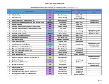
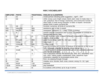
Evidence-Based Complementary and Alternative Medicine3Table 1: Weekly weight changes in rats of five groups.GroupWKYSHRMPHASDZL7,8-DHFDay 0 (g)68.2 5.687.0 5.5100.9 10.899.2 10.6100.3 7.4Day 7 (g)103.8 6.7118.7 7.8139.4 10.7130.4 14.9137.6 8.910–12 mL with freezing point homogenization buffer andstored on ice. The supernatant protein content was measuredand diluted to 5–7 mg/mL. After dilution, 2 mL of supernatant per tube was spread evenly and slowly on a 3%gradient belt and centrifuged at 31876 g for 5 min at 4 C;then the third and fourth layers of the gradient zone wereremoved, diluted with precooled sucrose/EDTA buffer, andcentrifuged. Finally, the synaptosome was collected andfixed for 2 h in 2.5% glutaraldehyde for experimental use[23].2.5. Behavioral Tests. After four weeks of treatment, theopen field test (OFT) and Morris water maze (MWM) testwere performed in each group [24, 25].2.5.1. OFT. The OFT is the most classic evaluation standardfor the ADHD disease model [26]. The open-field arena wasmade of black-colored iron, whose total area(100 100 40 cm) was spaced into 16 squares with sides of25 cm. The movement track of each rat was recorded everyfive minutes from 8:00 to 12:00, mainly including movingdistance (m), time spent in the central area (s), and rearingtimes. The scent marks of each rat’s urine and feces wereremoved with 75% ethanol between tests.2.5.2. MWM Test. The MWM test has been generally used toevaluate the learning and memory abilities of rats. The rats ineach group were placed in MWM equipment with a watertemperature of (24 2 C) for testing, and the spatial acquisition trials, annulus visits, and time spent in the targetquadrant (s) were collected.2.6. Immunofluorescence. The tissues (PFC, corpus striatum,and hippocampus) of each group were sampled, fixed with4% paraformaldehyde, then embedded in paraffin, andfurther cut into 3–4 μm sections. The expressions of BDNF,P75, and NF-κB in brain sections were measured by immunofluorescence. DAPI was used to restain the nuclei forimmunofluorescence staining and microscopic observation.The average fluorescent intensity was analyzed by theImage-J software [25].2.7. Western Blotting. The same amount of protein wasextracted from each tissue and further separated by SDSPAGE and transferred to the PVDF membrane. Anti-BDNF(1:1,000), TrkB (1:1,000), P75 (1:1,000), JNK1 (1:1,000), NFκB (1:1,000), and β-actin (1:2,000) were successively addedDay 14 (g)135.8 8.9158.5 11.9183.9 13.3175.4 17.7181.1 7.5Day 21 (g)190.4 17.0197.9 13.1222.1 12.1213.2 19.7220.9 9.8Day 28 (g)210.7 11.3215.0 14.6232.0 11.7224.7 19.7231.7 12.7and incubated overnight at 4 C. After incubation withsecondary antibody, protein bands were quantitatively analyzed using ChemiDoc MP Imaging System HE ImageLab software. 2.8. Polymerase Chain Reaction (PCR). Total RNA wasisolated from the PFC, corpus striatum, and hippocampustissues, and its purity and concentration were determined.All primers were designed and synthesized by ShanghaiShenggong Biological Engineering Co. Ltd. (Table 2). Fivemicroliter SYBR Green master mix, 0.2 μL reverse primers,and 1.0 μL of cDNA (1:3 dilution) were mixed together toreact. The dissociation curve of the reaction solution wasmeasured to further evaluate the specificity of the amplifiedproducts. The melting curve and amplification plots ofBDNF, TrkB, P75, JNK1, NF-κB, and GAPDH in the PFC,corpus striatum, and hippocampus samples were recordedfor all rats.2.9. Statistical Analysis. All data were processed byGraphPad Prism 8.0 software.3. Results3.1. Effect of ASDZL on the Behavior of SHRs in the OFT.ASDZL and 7,8-DHF had no significant effect on bodyweight and developmental indexes in rats, as shown inTable 1. The results showed a significantly increased indistance and speed of movement among WKY and SHRs.After four weeks of treatment, the distance and speed ofmovement of MPH, ASDZL, and 7,8-DHF groups weresignificantly decreased than that of the SHR group(Figures 1(a) and 1(b)). The representative pathways of allgroups are shown in Figure 1(c).3.2. Effect of ASDZL on the Behavior of SHRs in the MWM Test.The time for locating the escape platform in MPH, ASDZL,and 7,8-DHF groups were less than the SHR group(Figure 1(d)). Compared with the WKY group, the SHRgroup played more visits to the annulus and spent more timein the target quadrant, but the difference was not statisticallysignificant (Figure 1(e)). The number of visits to the annulusby MPH, ASDZL, and 7,8-DHF groups were much greaterthan that of the SHR group (Figure 1(e)), whereas the timespent by the MPH, ASDZL, and 7,8-DHF groups was lessthan that in the SHR group, with no significant difference(Figure 1(f )). The representative swimming patterns areshown in Figure 1(g).
4Evidence-Based Complementary and Alternative MedicineTable 2: Gene sequences of primers.GenesPrimer length -κBGene 5*30moving speed ASDZLMPHSHRWKY0SHRmoving distance HSHRWKY0MPH20**SHR**annulus visitsEscape latency (s)4076543210Time spent in the targetquadrant(c)60(f )MPHASDZL7,8-DHF(g)Figure 1: OFT and MWM test: (a) distance of movement (m) in OFT, (b) time spent (s) in OFT, (c) representative trajectories in OFT, (d)escape latencies in MWM test, (e) number of annulus visits in MWM test, (f ) time spent (s) in the target quadrant in MWM test, and (g)representative trajectories in MWM test. n 8 for each group. # p 0.05, SHR vs. WKY group. p 0.05, compared to the SHR group.
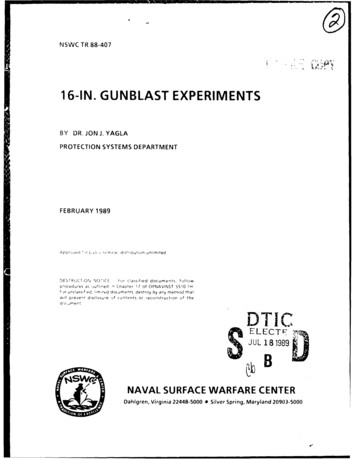
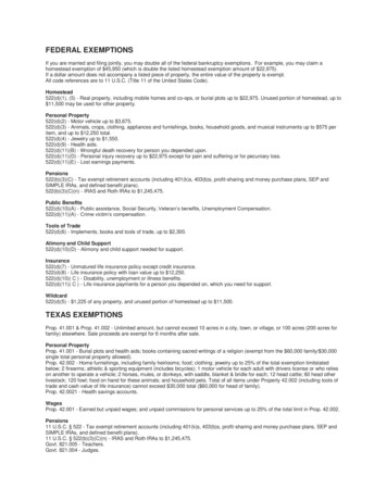
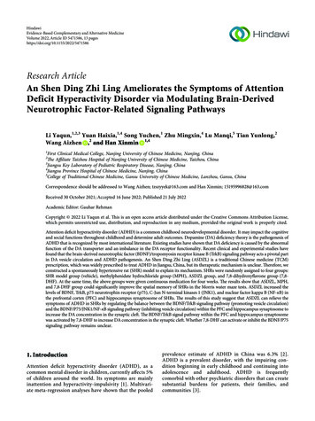
Evidence-Based Complementary and Alternative Medicine3.3. Effect of ASDZL on BDNF, P75, NF-κB in the PFC, CorpusStriatum, and Hippocampus (Immunofluorescence).Higher average fluorescence intensities of BDNF(Figures 2(a), 2(b), and 2(d)), P75 (Figures 2(e), 2(f ), and2(h)), and NF-κB (Figures 2(i), 2(j), and 2(l)) were showed inthe PFC and hippocampus of the MPH, ASDZL, and 7,8DHF groups compared to the SHR group. There was nosignificant discrepancy in fluorescence intensity of P75 inthe corpus striatum between the ASDZL and SHR groups(Figures 2(e) and 2(g)) or NF-κB among the MPH, 7,8-DHF,and SHR groups (Figures 2(i) and 2(k)). The MPH, ASDZL,and 7,8-DHF groups showed that the fluorescent intensity ofBDNF was increasing significantly (Figures 2(a) and 2(c)).3.4. Effect of ASDZL on the BDNF/TrkB Signaling Pathway inthe PFC, Corpus Striatum, and Hippocampus (WesternBlotting). To further observe whether ASDZL affects BDNF/TrkB signaling pathway, the PFC, corpus striatum, andhippocampus proteins were measured. Compare with theWKY group, the levels of BDNF and TrkB were remarkablylower in the SHR group. Following four weeks of treatment,the levels of BDNF and TrkB were significantly increased inthe MPH, ASDZL, 7,8-DHF, and SHR groups, except for thelevel of TrkB in the corpus striatum of the MPH group(Figures 3(a)–3(i)).3.5. Effect of ASDZL on the BDNF/P75/JNK1/NF-κB Pathwayin the PFC, Corpus Striatum, and Hippocampus (WesternBlotting). The total levels of relevant proteins were measured to reveal the effect of ASDZL on the BDNF/P75/JNK1/NF-κB pathway in the PFC, corpus striatum, and hippocampus. The levels of P75, JNK1, and NF-κB were conspicuously lower in the SHR group than in the WKY group.The level of P75 showed a conspicuously increase in the PFCand hippocampus of the MPH and ASDZL groups, whilethere was no conspicuous divergence in the corpus striatumof the MPH, ASDZL, 7,8-DHF, and SHR groups. ASDZLconspicuously increased the level of JNK1 in the PFC, corpusstriatum, and hippocampus, whereas MPH only conspicuously increased the level of JNK1 in the PFC. 7,8-DHFaggrandized the level of JNK1 in the hippocampus. The NFκB/β-actin ratios of the PFC, corpus striatum, and hippocampus were significantly increased in the MPH, ASDZL,7,8-DHF, and SHR groups, except for the level of NF-κBlevel in the corpus striatum of the MPH group (Figure 3(a)and 3(j)–3(r)).3.6. Effect of ASDZL on the BDNF/TrkB Pathway in the PFC,Corpus Striatum, and Hippocampus (qPCR). To determinethe effect of ASDZL on the BDNF/TrkB signaling pathway,we analyzed the relative expression of BDNF and TrkBmRNA in the synaptosomes of the PFC, corpus striatum,and hippocampus. The levels of BDNF and TrkB in the SHRgroup were conspicuously lower than those of the WKYgroup. After treatment, MPH and ASDZL conspicuouslyincreased the expression of BDNF and TrkB in the PFC,corpus striatum, and hippocampus. However, the 7,8-DHF5group displayed no conspicuously divergence in the BDNFof the corpus striatum compared to the SHR group(Figures 4(a)–4(f )).3.7. Effect of ASDZL on the BDNF/P75/JNK1/NF-κB Pathwayin the PFC, Corpus Striatum, and Hippocampus. To understand the effect of ASDZL on the BDNF/P75/JNK1/NFκB pathway in the PFC, corpus striatum, and hippocampus,we also measured the relative expression levels of mRNA.The relative expression levels of P75 and NF-κB wereconspicuously lower in the SHR group than in the WKYgroup. However, the relative expression levels of JNK1 inthe PFC and hippocampus of the SHR group were oppositeto the expression trend in the corpus striatum. The MPHand ASDZL groups conspicuously increased the levels ofP75, JNK1, and NF-κB in the PFC, corpus striatum, andhippocampus; however, there was no conspicuous divergence in the levels of P75 and NF-κB in the corpus striatumbetween the 7,8-DHF and SHR groups. MPH, ASDZL, and7,8-DHF conspicuously decreased the expression of JNK1in the corpus striatum compared with the SHR group(Figures 4(g)–4(o)).4. DiscussionADHD is characterized by the heterogeneity of attentiondeficiency, impulsiveness, hyperactivity, variable, and frequent comorbidities [27]. Although the evidence is lacking,it is generally recommended to use MPH, a central stimulant, to relieve ADHD in children [28]. The most commonadverse events of methylphenidate are headache, loss ofappetite, and insomnia [29]. Parents are often concernedabout the side effects of these stimulants and tend to consider the use of complementary and alternative drugs, suchas TCM to relieve the core symptoms of ADHD [30, 31].ASDZL is a prescription of TCM created by Professor HanXinmin and prescribed to relieve ADHD. Clinical researcheshave demonstrated that ASDZL could relieve symptoms ofADHD, especially the hyperactive-impulsive type [32]. Inthis study, we evaluated the role of ASDZL and 7,8-DHF onthe behavioral performance and synaptic vesicle dopaminerelease mechanism in ADHD, which provided new synapticlevel views for treating ADHD.Synaptosomes can mimic various aspects of synapticfunctions, which are continually used as objects in neurobiology studies [33, 34]. The release of neurotransmitters ismaintained through endocytosis and refilling of presynapticlocal synaptic vesicles [35]. A previous study found thatASDZL significantly upregulated the expression of tyrosinehydroxylase and dopa decarboxylase, which are related toDA synthesis in the presynaptic membrane of SHR rats andincreased the expression levels of synaptic-associated protein 25 and syntaxin-1A involved in the formation ofSNARE complexes and vesicle-associated membrane proteins, among other protein and mRNA, thereby improvingthe core symptoms of ADHD [36]. The main active ingredients of ASDZL (Baicalin) can significantly increase theexpression level of BDNF. Therefore, ASDZL has a special
Evidence-Based Complementary and Alternative Medicine60**7,8-DHFCorpus Striatum*ASDZLPFCBDNF8040200MPHAverage us s StriatumP75807,8-DHFAverage **MPH40Average fluorescenceintensityBDNFSHRAverage 7,8-DHF(f )7,8-DHFASDZLMPH0Corpus Striatum*40200Hippocampus(g)(h)Figure 2: ge fluorescenceintensity80SHRAverage fluorescenceintensity(e)P75
Evidence-Based Complementary and Alternative Medicine7**40*207,8-DHF0ASDZLCorpus Striatum60MPHPFCNF-κB80SHRAverage DZL7,8-DHF(j)200SHR7,8-DHFASDZLMPH0*Corpus Striatum7,8-DHF20**40ASDZL*40NF-κB60MPHAverage fluorescenceintensityNF-κB60SHRAverage fluorescenceintensity(i)Hippocampus(k)(l)Figure 2: Effect of ASDZL on BDNF, P75, NF-κB in PFC, corpus striatum, and hippocampus (immunofluorescence): (a) representativeimages of BDNF-immunopositive microglia (red) with DAPI (blue); average fluorescence intensity of BDNF in PFC (b), in corpus striatum(c), and in hippocampus (d). (e) Representative images of P75-immunopositive microglia (red) with DAPI (blue); average fluorescenceintensity of P75 in PFC (f ), in corpus striatum (g), and in hippocampus (h). (i) Representative images of NF-κB-immunopositive microglia(red) with DAPI (blue); average fluorescence intensity of NF-κB in PFC (j), in corpus striatum (k), and in hippocampus (l). n 3 for eachgroup. p 0.05, compared to the SHR group.PFCCorpus StriatumHippocampusPFC(e)Figure 3: Continued.*Hippocampus(f )7,8-DHF0.0ASDZL7,8-DHFASDZLMPHCorpus ASDZL92KDaMPHTrkBSHR28KDaWKYBDNF
FASDZLWKYCorpus /β-actinratioP75/β-actinratio1.0(k)**1.5Corpus s kB/β-actinratioEvidence-Based Complementary and Alternative pus s(r)Figure 3: Effect of ASDZL on BDNF, TrkB, P75, JNK1, and NF-κB in the PFC, corpus striatum, and hippocampus (western blotting): (a)–(c)western blot analysis of BDNF, TrkB, P75, JNK1, NF-κB, and β-actin; (d)–(f ) relative levels of BDNF; (g)–(i) relative levels of TrkB; (j)–(l)relative levels of P75; (m)–(o) relative levels of JNK1; and (p)–(r) relative levels of NF-κB. n 3 for each group. # p 0.05, SHR vs. WKYgroup. p 0.05, compared to the SHR group. The scale bar corresponds to 1.0 μm.affinity for the DA system and potential regulatory effect onthe DA synaptic vesicle cycle [37–40]. Synaptic-associatedprotein 25 and vesicle-associated membrane proteins are thekey proteins constituting the SNARE complex and cooperatewith syntaxin-1A and other proteins to participate in the keylinks of vesicle circulation such as DA vesicle anchoring,membrane transport, membrane fusion, and vesicle exocytosis in the form of SNARE complexes, as well as affect thebrain. The level of internal DA regulates the learning andmemory abilities of individuals [41–43]. A recent studyshowed that ASDZL regulated the activity of synaptosomeATPase and LDH enzymes in model rats and increased thecontent of AC, cAMP, and PKA in synapses, which suggested that ASDZL may perform a therapeutic role by reducing the inhibitory function of D2RS on the AC/cAMP/PKA signaling pathway [44].Substantial evidence indicates that PFC can regulateattention and working memory by modulating memoryrelated activity within the PFC [45–47]. The striatum isrichly innervated by midbrain DA neurons, which regulate arange of cellular and synaptic functions that control goaldirected behavior and habits [48, 49]. The hippocampus
KY0.5SHRWKY1.0WKYmRNA relativeexpressionJNK1**1.57,8-DHF*mRNA relativeexpression#SHRmRNA SDZL*NF-κB1.5WKY7,8-DHFASDZLMPHSHR0.0WKYmRNA relativeexpression*#0.5**#(k)**0.0Corpus .50.5Corpus StriatumSHR*2.01.51.00.50.0WKY1.0*mRNA relativeexpression*WKYmRNA smRNA relativeexpression*1.0*#(f )P751.57,8-DHFASDZLMPHSHRmRNA relativeexpression*0.0WKYmRNA us StriatumP75*0.0SHR*PFC1.0*mRNA RNA relativeexpression*0.5#(b)TrkBWKYmRNA relativeexpression(a)1.01.0Corpus StriatumPFC1.5mRNA mRNA relativeexpressionBDNF1.5WKYmRNA relativeexpressionEvidence-Based Complementary and Alternative MedicineCorpus StriatumHippocampus(n)(o)Figure 4: Effect of ASDZL on BDNF, TrkB, P75, JNK1, and NF-κB in the PFC, corpus striatum, and hippocampus (qPCR): (a)–(c) relativeexpression of BDNF mRNA levels; (d)–(f ) relative expression of TrkB mRNA levels; (g)–(i) relative expression of P75 mRNA levels; (j)–(l)relative expression of JNK1 mRNA levels; and (m)–(o) relative expression of NF-κB mRNA levels. n 3 for each group. # p 0.05, SHR vs.WKY group. p 0.05, compared to the SHR group.
10serves a key function in memory, navigation, and cognition[50]. Studies have shown that the three brain regions (PFC,corpus striatum, and hippocampus) are closely related to theonset of ADHD [51–53].The DA system in the brain is a large and complex neuralnetwork. Further study will be required to determine thecauses of defects in DA formation. The BDNF/TrkB signaling pathway might participate in regulating the DAsynaptic vesicle circulation by affecting the protein receptorcomplex, which is closely related to ADHD pathogenesis.In this study, we intended to examine the impacts ofdrugs on the BDNF-related pathways that influence therelease of DA from synaptic vesicles in three regions of thebrain and to determine whether drugs affect differently indifferent brain regions. Because of the higher levels of BDNFin brain tissue, BDNF and its receptor TrkB are widelydistributed in the brain cortex, striatum, hippocampus, andother DA neurons. BDNF is involved in DA neuron synapsegrowth, transmitter release, cell survival, membrane transport, and synapse formation [54]. TrkB is a transmembraneprotein with a high-affinity receptor for BDNF [55, 56],which can mediate most of its biological functions. Thedecreased expression of BDNF can cause significant changesin DA neuron synapse morphology and dysfunction, leadingto a decrease in DA content in the brain. This impact may berelated to the low activation of the BDNF/TrkB signalingpathway, which inhibits the function of the SNARE complexand affects presynaptic membrane DA vesicle circulationand transmitter release [57, 58]. The phospholipase Cc wasrecruited and activated by phosphorylation of Tyr816 residue for promoting neuronal survival and implicating neuriteoutgrowth and synaptic plasticity [59]. Numerous literaturehave demonstrated that BDNF
10-12mL with freezing point homogenization buffer and storedonice.esupernatantproteincontentwasmeasured and diluted to 5-7mg/mL. After dilution, 2mL of super-