Transcription
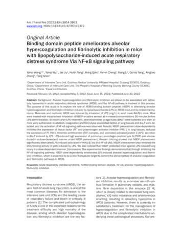
Am J Transl Res 2022;14(6):3854-3863www.ajtr.org /ISSN:1943-8141/AJTR0142372Original ArticleBinding domain peptide ameliorates alveolarhypercoagulation and fibrinolytic inhibition in micewith lipopolysaccharide-induced acute respiratorydistress syndrome Via NF-κB signaling pathwayYahui Wang1,2*, Yanqi Wu1*, Bo Liu1, Huilin Yang1, Hong Qian1, Yumei Cheng1, Xiang Li1, Guixia Yang1, XinghaoZheng1, Feng Shen1Department of Intensive Care Unit, Guizhou Medical University Affiliated Hospital, Guiyang 550001, Guizhou,China; 2Department of Intensive Care Unit, The People’s Hospital of Weining County, Weining County 553100,Guizhou, China. *Equal contributors.1Received February 15, 2022; Accepted May 7, 2022; Epub June 15, 2022; Published June 30, 2022Abstract: Background: Alveolar hypercoagulation and fibrinolytic inhibition are shown to be associated with refractory hypoxemia in acute respiratory distress syndrome (ARDS), and the NF-κB pathway is involved in this process.The purpose of this study is to explore the role of NEMO-binding domain peptide (NBDP) in alleviating alveolarhypercoagulation and fibrinolytic inhibition induced by lipopolysaccharide (LPS) in ARDS mice and its related mechanisms. Materials and methods: ARDS was induced by inhalation of LPS (mg/L) in adult male BALB/c mice. Micewere treated with intratracheal inhalation of NBDP or saline aerosol at increased concentrations 30 minutes beforeLPS administration. Six hours after LPS treatment, bronchoalveolar lavage fluids (BALF) were collected and then allmice were euthanized. In addition, coagulation and fibrinolysis associated factors in lung tissues and BALF were detected, and the activation of NF-κB signaling pathway was observed. Results: NBDP pretreatment dose-dependentlyinhibited the expression of tissue factor (TF) and plasminogen activator inhibitor (PAI) 1 in lung tissues, reducedthe secretions of TF, PAI-1, thrombin-antithrombin (TAT) complex, and promoted activated protein C (APC) secretionin BALF induced by LPS. LPS-induced high expression of pulmonary procollagen peptide type lll (PIIIP) was also reduced in a dose-dependent manner under NBDP pretreatment. Western blotting showed that NBDP pretreatmentsignificantly attenuated LPS-induced activation of IKKα/β, Iκα and NF-κB p65. NBDP pretreatment also inhibited theDNA binding activity of p65 induced by LPS. We also noticed that NBDP protected mice against LPS-induced lunginjury in a dose-dependent manner. Conclusions: The experimental findings demonstrate that through inhibiting theNF-κB signaling pathway, NBDP dose-dependently ameliorates LPS-induced alveolar hypercoagulation and fibrinolytic inhibition, which is expected to be a new therapeutic target to correct the abnormalities of alveolar coagulationand fibrinolytic pathways in ARDS.Keywords: Acute respiratory distress syndrome, NEMO-binding domain peptide, NF-κB, alveolar hypercoagulation,fibrinolytic inhibitionIntroductionRespiratory distress syndrome (ARDS), the severe form of acute lung injury (ALI), is one of themost common diseases for admission to theintensive care unit (ICU) and the leading causeof respiratory failure and death in critically illpatients [1]. The complicated pathophysiologyof ARDS is one of the important reasons for thetreatment difficulty and high mortality of thedisease, among which alveolar hypercoagulation and fibrinolytic inhibition are the key fac-tors [2]. Alveolar hypercoagulation and fibrinolysis inhibition results in extensive microthrombus formation in pulmonary vessels, and massive fibrin deposition in the airspace [3, 4],which is closely related to decreased lung compliance, V/Q ratio imbalance and arteriovenousshunting, resulting in refractory hypoxemia inARDS patients. However, there is currently nosatisfactory treatment for the management ofhypercoagulation and fibrinolytic inhibition inARDS due to the complicated mechanisms underlying these pathological processes. Our pre-
The role of NEMO-binding domain peptide in acute respiratory distress syndromevious studies [5, 6] and other published data[7] have confirmed that the nuclear factorkappa B (NF-κB) signaling pathway plays a pivotal role in the pathogenesis of alveolar hypercoagulation and fibrinolytic inhibition.NF-κB is mainly involved in the regulation ofmany important physiopathologic processes,such as immunity, inflammation, tumorigenesisand stress responses [8, 9]. Activation of NF-κBresults in its translocation from the cytoplasmto the nucleus. Under normal conditions, NF-κBis sequestered in the cytoplasm, where it bindsto its inhibitor protein members, including IκBα,IκBβ and IκBγ. The IκB kinase (IKK) complex isrequired for the activation of NF-κB and consists of three subunits, namely IKKα, IKKβ andIKKγ, of which IKKγ is also known as NEMO.NEMO itself does not have a catalytic domain,but plays a key role in biology as a part ofthe IKK complex [10, 11]. The NH2-terminus ofNEMO binds to a hexapeptide sequence LeuAsp-Trp-Ser-Trp-Leu at the COOH terminus ofIKKα and IKKβ, called the NEMO-binding domain (NBD) [12], which is the basic structurefor the crosstalk among IKKα, IKKβ and NEMO,maintaining the biological activity of the IKKcomplex [13]. Although there are many waysto inhibit NF-κB, such as NF-κB/IKKβ geneknockout or knockdown, NF-κB-specific inhibition, blocking P65 translocation from cytoplasm into the nucleus or preventing P65from binding to its specific DNA sequence (κBsequence) etc., these methods also inevitablyinhibit some basal biological activities of NFκB [14, 15]. A small molecular NBD peptide(NBDP), however, has been shown not only toselectively inhibit the NF-κB-mediated targetgene transcription through targeting the crosstalk between IKK and NEMO [16], but alsomaintain the important basal activities of NFκB [17]. In addition, previous studies havedemonstrated that NBDP effectively inhibitedNF-κB pathway activation [18-21]. Based onthese findings, we speculate that NBDP cancorrect alveolar coagulation and fibrinolysis abnormalities via the NF-κB signaling pathway inARDS. Thus, the innovation of our study is toinvestigate the underlying mechanism of NBDPin ARDs. In our study, it is demonstrated thatNBDP dose-dependently attenuated lipopolysaccharide (LPS)-induced alveolar hypercoagulation and fibrinolytic inhibition by inactivatingthe NF-κB signaling pathway in mice, and NBDPis expected to be a new significant target in thetreatment of ARDS.3855Materials and methodsExperimental animalsBALB/c male mice, aged 6-8 weeks and weighing 20-25 g, were purchased from the AnimalCenter of Guizhou Medical University. All micewere fed a normal standard diet in a controlledenvironment (temperature 22 1 C) with a 12hour light/dark cycle and controlled humidity.The mice were given 7 days to acclimatize tothe environment prior to the experiment. Thestudy was approved by the Animal EthicsCommittee of Guizhou Medical University andwas conducted in accordance with guidelinesof the Chinese Laboratory Animal ManagementRegulations.Animal model establishmentA mouse model of ARDS was established byaerosol inhalation of LPS. Seventy-two micewere randomly divided into the following sixgroups: Control, Model, N-NBD, L-NBD, M-NBDand H-NBD, with 12 in each group. The mice inModel, N-NBD, L-NBD, M-NBD and H-NBD received 50 μl of LPS (4 mg/ml, Sigma-Aldrich)while those in the control group received thesame volume of saline inhalation. Thirty minutes before LPS administration, mice in L-NBD,M-NBD and H-NBD groups inhaled 50 μl ofNBDP (MERCK) with the concentration of 120μg/ml, 240 μg/ml and 360 μg/ml, respectively.The N-NBD group served as a negative controland received a non-functional NBDP analogue(50 μl, MERCK) at a concentration of 240 μg/ml. Six hours after LPS or saline inhalation, allmice were euthanized via cervical dislocationand exsanguination under anesthesia with pentobarbital sodium (50 mg/kg). Bronchoalveolarlavage fluid (BALF) samples were collected forthe detection of coagulation-related factors.Left lung tissues of mice were collected for histopathological and immunohistochemical analysis while right lungs were immediately frozenin liquid nitrogen and stored at -80 C forenzyme-linked immunosorbent assay (ELISA)and western blot (WB) analysis.Real-time quantitative PCR (RT-qPCR)RT-qPCR was performed to detect tissue factor(TF) and plasminogen activator inhibitor-1 (PAI1) gene expression. The concentration of totalRNA was detected by using a NanoDrop-2000spectrophotometer (NanoDrop Technologies,Am J Transl Res 2022;14(6):3854-3863
The role of NEMO-binding domain peptide in acute respiratory distress syndromeTable 1. Gene sequences of TF, PAI-1GeneTFPAI-1β-actinSequences5-AGA CGG AGA CCA ACT TGT GAT-35-CTG CTG AAT TAC TGG CTG TCC-35-CTG CAA AAG GTC AGG ATC GAG-35-CAT CAC TTG GCC CAT GAA GAG-35-CAC CCG CGA GTA CAA CCT TC-35-CCA ATA CCC ACC ATC ACA CC-3Germany) and the A260/A280 ratio of theextracted RNA was controlled between 1.8 to2.0. Primers were designed based on the TFand PAI-1 gene sequences supplied by the NCBIgene database. The primers were synthesized by Guangzhou Aiji Biotechnology Co., Ltd.(Table 1). PCR amplification was performedusing cDNA as the template. The reaction system was set as follows: SYBR Green Mix 10 μl,forward primer and reverse primer 0.8 μl each,cDNA template 0.8 μl, and ddH2O 7.6 μl into asystem containing 20 μl reagents. The dissolution and amplification curves of genes wererecorded following gene amplification. The relative expression levels of target genes were calculated using the 2-ΔΔCt method.Western blotCytoplasmic proteins were extracted using theCell Solute Extraction Kit according to the manufacturer’s instructions (Solebao TechnologyCo., Ltd, Beijing, China). Briefly, the concentration of protein was measured using a bicinchoninic acid (BCA) assay kit (Thermo Scientific,Waltham, MA). An equal amount of protein fromeach sample was resolved in 12% Tris-glycinesodium dodecyl sulfate (SDS) polyacrylamidegel. The protein bands were blotted onto anitrocellulose membrane. After incubating for2 hours in blocking solution, the membranewas incubated with p-P65 (ab76302, Abcam,UK), p-IKKα/β (ab194528, Abcam, UK), p-IκBα(#2859S, Cell Signaling Technology, USA), PAI-1(ab222754, Abcam, UK), and TF (ab228968,Abcam, UK) antibodies (all diluted at 1:1000)for 24 hours. Then the secondary antibody horseradish peroxidase-conjugated goat anti-rabbit immunoglobulin (1:2000 dilution, #7074S,Cell Signaling Technology, USA) was added andincubated for 2 hours at 37 C. The target protein bands were visualized using an enhancedchemiluminescence detection system (Millipore, MA, USA). Relative band densities werequantified by Image J software.3856ELISA assayThe collected BALF was centrifuged at 4500rpm for 10 minutes at 4 C, and the resultingsupernatant was collected and stored at -80 Cfor testing. TF, PAI-1 and activated protein C(APC) levels were determined using ELISA kits(Huamei Bio-company, Wuhan, China) according to the manufacturer’s instructions.HistopathologyThe lung tissues were fixed with 4% paraformaldehyde for 24 hours, followed by dehydration,paraffin-embedding and slicing (4 μm). Thenthe slices were stained with hematoxylin-eosin(H&E). The scores of lung injury were blindlyevaluated by pathologists, as described previously [22]. Each histological change was scored(lung injury score, LIS) from 0 to 3 according tothe lesion range, including alveolar wall thickening, edema, inflammatory cell infiltration, hemorrhage and cellulose deposition (0: normal;1: injury 25% of the field; 2: injury within 25%50% of the field; 3: injury 50% of the field).NF-κB p65 DNA binding activityThe DNA binding activity of NF-κB p65 in theright upper lung tissues was detected usingthe Universal EZ-TFA Transcription Factor Chemiluminescence Kit (Millipore, Germany) according to the manufacturer’s instructions.The nuclear extract of lung tissues was addedto a plate containing biotinylated oligonucleotide which had NF-κB binding site. After incubating for 1 hour at room temperature, theplate was washed and incubated with rabbitanti-NF-κB p65 (1:1000 dilution) for 1 hour.After washing the plate, an anti-rabbit horseradish peroxidase-conjugated antibody (1:500dilution) was added and incubated for 30 minutes. This was followed by the addition of thechemiluminescent substrate solution for 5minutes of incubation. The sample OD valuewas read by a microplate reader at 1-s integration time.Immunohistochemistry (IHC)After dehydration of paraffin sections in ethanol series, the antigens were retrieved using acitrate buffer. The nonspecific binding site wasblocked by 3% BSA. The sections were incubated with rabbit anti-mouse P65 (1:600, #6956S,Cell Signaling Technology, USA) and type III colAm J Transl Res 2022;14(6):3854-3863
The role of NEMO-binding domain peptide in acute respiratory distress syndromeFigure 1. Pathological changes in the lung tissues. A: Lung sections were stained with hematoxylin and eosin 6hours after LPS administration. LPS destroyed the lung tissues, resulting in hemorrhage ( ), inflammatory cellinfiltration ( ), alveolar wall thickening ( ) and so on, which were all attenuated by NBDP pretreatment; B: NBDPpretreatment significantly alleviated these pathological changes; C: NBDP pretreatment significantly alleviated pulmonary edema. Data were expressed as mean SD (n 6). *P 0.05 compared with Control. #P 0.05 compared withModel. &P 0.05 compared with N-NBD. %P 0.05 compared with L-NBD.lagen (1:200, ab7778, Abcam, UK) antibodiesat 4 C overnight, followed by washing and incubating with corresponding HRP-labeled secondary antibodies (1:1000, ab6721, Abcam, UK)for 1 hour at room temperature. The expressionlevels of antigens were visualized using peroxidase activity developed by DAB staining solution and observed at a magnification of 400X.Statistical analysisStatistical analyses were performed with SPSS.Data was expressed as mean SD. Statistical3857differences were determined by one-way analysis of variance (ANOVA) and the StudentNewman-Keuls (SNK) method, with the significance level set as P 0.05.ResultsNBDP improved pulmonary pathologicalchanges induced by LPS inhalation in miceH&E staining and histopathological analysiswere performed to assess the pathologicalchanges of pulmonary tissues and the degreeAm J Transl Res 2022;14(6):3854-3863
The role of NEMO-binding domain peptide in acute respiratory distress syndromeFigure 2. Changes of TF and PAI-1 expression in pulmonary tissue 6 hours after LPS inhalation with or without NBDPpretreatment. A: RT-qPCR was performed to detect TF mRNA expression in lung tissues; B: RT-qPCR was performedto detect PAI-1 mRNA expression in lung tissues; C: Western blotting was performed to detect TF protein expressionin lung tissues; D: Western blotting was performed to detect PAI-1 protein expression in lung tissues. Each bar represents the mean SD of 6 mice. *P 0.05 compared with Control. #P 0.05 compared with Model. &P 0.05 comparedwith N-NBD. %P 0.05 compared with L-NBD. P 0.05 compared with M-NBD.of lung injury. It was found that LPS inducedexcessive edema, obvious inflammatory cell infiltration, alveolar collapse, alveolar wall thickening, and severe hemorrhage, which were alldose-dependently inhibited by NBDP. The LPSinduced high W/D ratio and LIS were also significantly reduced by NBDP treatment (Figure1).NBDP attenuated mRNA and protein levels ofTF and PAI-1 in LPS-induced ARDS miceIn order to evaluate the coagulation and fibrinolytic status of LPS-induced lung tissues, TF andPAI-1 were measured by RT-qPCR and WB. Theresults showed that LPS stimulated high mRNAand protein expression of TF and PAI-1 in lungtissues, which were effectively attenuated byNBDP pretreatment (Figure 2).3858NBDP inhibited LPS-induced secretions of TF,PAI-1, and thrombin-antithrombin (TAT) in lungtissue and promoted APC productionThe levels of TF, PAI-1, TAT and APC in BALFwere determined by ELISA to evaluate thedegree of alveolar hypercoagulation and fibrinolytic inhibition. LPS stimulation for 6 hoursresulted in obvious increases in TF, PAI-1 andTAT levels and a decrease in APC level in BALF,all of which were reversed by NBDP in a dosedependent manner (Figure 3).NBDP alleviated LPS-induced PIIIP depositionin mouse lung tissueThe levels of PIIIP in mouse lung tissue wasdetected using immunohistochemistry to evaluate the effect of LPS on fibrinolystic inhibi-Am J Transl Res 2022;14(6):3854-3863
The role of NEMO-binding domain peptide in acute respiratory distress syndromeFigure 3. NBDP significantly inhibited TF, PAI-1, and TAT, and promoted APC secretions in LPS-induced lung tissues.A: ELISA detection of the changes of TF following LPS induction with or without NBDP. B: ELISA detection of thechanges of PAI-1 following LPS induction with or without NBDP; C: ELISA detection of the changes of TAT followingLPS induction with or without NBDP; D: ELISA detection of the changes of APC secretions following LPS inductionwith or without NBDP; Values were presented as mean SD. *P 0.05 compared with Control. #P 0.05 comparedwith Model. &P 0.05 compared with N-NBD. %P 0.05 compared with L-NBD. P 0.05 compared with M-NBD.tion. The results demonstrated that LPS induced a large amount of PIIIP in pulmonary tissues.After pretreatment with NBD, LPS-induced PIIIPdeposition was significantly reduced in a dosedependent manner (Figure 4).NBDP inhibited LPS-induced NF-κB activationTo assess the effects of NBDP on NF-κB signaling pathway, WT was performed to detectthe phosphorylation of IKKα/β, IκBα and P65after LPS stimulation. Significant increaseswere observed in IKKα/β (p-IKKα/β), p-IκBαand p-P65 phosphorylation levels in LPS-induced lung tissues, which were weakened byNBDP pretreatment (Figure 5).NBDP decreased P65 DNA binding activity initiated by LPS stimulationNF-κB p65 DNA binding activity is related toP65 translocation from cytoplasm to nucleus.3859Our data showed that NF-κB p65 DNA bindingactivity was significantly increased after LPSstimulation, which was dose-dependently decreased by NBDP (Figure 6).DiscussionIn this study, an ARDS mouse model simulatingthe pathogenesis of ARDS was successfullyconstructed [22]. Meanwhile, we found thatNBDP effectively ameliorated LPS-induced hypercoagulation and fibrinolysis inhibition inlung tissues and the airspace, which was consistent with the research of Huang et al. [23].These findings suggested that NBD has potential as a therapeutic target in the treatmentof acute inflammatory diseases, including ALI,ARDS, and infectious diseases.TF is a potent procoagulant that initiates theextrinsic coagulation cascade mainly throughinteracting with Factor VII in the presence ofAm J Transl Res 2022;14(6):3854-3863
The role of NEMO-binding domain peptide in acute respiratory distress syndromeFigure 4. Expression of PIIIP in lung tissues in LPS-induced lung injury mice. A: Expression of PIIIP in lungtissues in different groups of mice; B: PIIIP IHC score;PIIIP reflected the level of fibrosis in lung tissues andit was highly expressed in Model and N-NBD groups(Red arrow presented positive staining). Each barrepresents the mean SD of PIIIP IHC score in eachgroup. *P 0.05 compared with Control. #P 0.05compared with Model. &P 0.05 compared with NNBD. %P 0.05 compared with L-NBD.calcium, resulting in activation of Factor X [24,25]. PAI-1 is a major physiological inhibitor ofthe fibrinolytic system that can also regulatethrombosis [26]. PAI-1 binds to and inhibitstissue and urokinase-type plasminogen activators (tPA and uPA), thereby reducing plasminproduction and fibrin clot lysis [27]. The results of our study showed that the mRNA andprotein expression of both TF and PAI-1 werehighly expressed in pulmonary tissue underLPS stimulation, indicating the presence ofprocoagulation and fibrinolytic defects in lungtissues [28].TAT is a complex of thrombin and antithrombinthat directly reflects the generation of thrombin, with an increase in TAT suggesting a stateof procoagulant activity [29]. APC is a proteinsynthesized by the liver and exerts anticoagulant activity by hydrolyzing blood coagulationfactors Va and VIIIa [30]. Our experimental datashowed that in BALF, the concentrations of TF,PAI-1, and TAT were all significantly increased,3860while APC concentration was statistically decreased, indicating hypercoagulation and fibrinolytic inhibition in the airspace in LPS-inducedARDS [31].PIIIP is mainly synthesized and secreted byfibroblasts and transformed in myofibroblasts.In addition, PIIIP is the main component ofthe extracellular matrix (ECM), and the excessive accumulation of PIIIP suggests pulmonaryfibrous deposition [32]. High expression of pulmonary PIIIP under LPS treatment in our studyindicated an increase in fibrous tissue in thelung due to fibrinolytic inhibition.NBDP is a protein peptide that has been shown to inhibit the activation of the classicalNF-κB signaling pathway by interfering with theNEMO-IKKα/IKKβ interaction [33]. Our datademonstrated that NBDP pretreatment significantly inhibited LPS-induced NF-κB pathwayactivation, manifested as decreased levels ofp-IKKα/β, p-Iκα and p-P65, and decreased P65Am J Transl Res 2022;14(6):3854-3863
The role of NEMO-binding domain peptide in acute respiratory distress syndromeFigure 5. NBDP inhibited LPS-induced activation of NF-κB signal pathway. A: Western blotting results of p-IKKα/β;B: Western blotting results of IκBα; C: Western blotting results of p-IκBα; D: Western blotting results of p-P65. Thequantitative data were presented as mean SD of 6 mice. *P 0.05 compared with Control. #P 0.05 compared withModel. &P 0.05 compared with N-NBD. %P 0.05 compared with L-NBD.NDA binding activity. In addition, NBDP effectively suppressed TF and PAI-1 expression inpulmonary tissue, and reduced TF, PAI-1 andTAT secretions in BALF while promoting APCproduction in BALF. Therefore, NBDP can correct alveolar hypercoagulation and fibrinolyticinhibition induced by LPS via inactivating theNF-κB pathway. Interestingly, we found that thehigher the dose of NBDP, the more obvious theeffects of NBDP on coagulation and fibrinolysisassociated factors and NF-κB inactivation, indicating a dose-dependent manner.Figure 6. NBDP decreased the enhanced p65 DNAbinding activity induced by LPS inhalation. DNAbinding activity of NF-κB p65 was examined by aTransAM p65 transcription factor ELISA kit. Each barrepresents the mean SD of 6 mice. *P 0.05 compared with Control. #P 0.05 compared with Model.&P 0.05 compared with N-NBD. %P 0.05 comparedwith L-NBD. P 0.05 compared with M-NBD.3861Unlike other inhibitors or methods, such asgene knockdown, knockout or specific inhibitor, NBDP has been shown to selectively inhibitNF-κB-mediated target gene transcription whilemaintaining the important basal activities ofNF-κB [16, 17], allowing it to be a new potentialtherapeutic target in ARDS treatment.Am J Transl Res 2022;14(6):3854-3863
The role of NEMO-binding domain peptide in acute respiratory distress syndromeIn this study, a negative control group with nonfunctional NBDP analogue (50 μl, MERCK) wasset up to eliminate the effect of NBDP itselfon the experimental results. Overall, however,there are still some limitations to be addressed. First, the lack of arterial blood gas analysismade the diagnosis of ARDS insufficient. Second, although we pretreated LPS inducedmice with different doses of NBDP, the optimaldose to weaken hypercoagulability and fibrinolysis inhibition and the optimal timing ofadministration remain to be clarified. Nevertheless, the findings of our study showed thatNBDP dose-dependently ameliorates LPS-induced alveolar hypercoagulation and fibrinolytic inhibition through inhibiting the NF-κB signaling pathway, suggesting that NBDP is apromising therapeutic choice for the treatmentof ARDS and deserves further exploration.ConclusionsNBDP dose-dependently ameliorates alveolarhypercoagulation and fibrinolysis inhibition viathe NF-κB signaling pathway, which is expected to be a new effective therapeutic target forARDS.AcknowledgementsWe thank Mr. Yi FANG and Professor Xu LIU,who greatly assisted our experiments. Thisstudy was supported by a grant from Guizhou Science and Technology Plan Project([2019]1261), and also was supported by theNational Natural Science Foundation of China[82160365].Disclosure of conflict of interestNone.AbbreviationsNBDP, NEMO-Binding Domain Peptide; ARDS,Acute respiratory distress syndrome; ALI, Acutelung injury; BALF, Bronchoalveolar lavage fluids;TF, Tissue factor; PAI-1, Plasminogen activatorinhibitor 1; TAT, Thrombin-antithrombin complex; APC, Activated protein C; PIIIP, Procollagenpeptide type III; WB, Western blotting; IHC,Immunohistochemistry; tPA, Tissue plasmiogen activator; uPA, Urokinase-type plasminogen activators.3862Address correspondence to: Feng Shen, Department of Intensive Care Unit, Guizhou MedicalUniversity Affiliated Hospital, No. 28, Guiyi Street,Yunyan District, Guiyang 550001, Guizhou, China.Tel: 86-135-1199-9117; E-mail: gyyxydrshen@hotmail.comReferences[1]Bellani G, Laffey JG, Pham T, Fan E, BrochardL, Esteban A, Gattinoni L, Van Haren F, LarssonA and McAuley DF. Epidemiology, patterns ofcare, and mortality for patients with acute respiratory distress syndrome in intensive careunits in 50 countries. JAMA 2016; 315: 788800.[2] de Luis Cabezón N, Sánchez Castro I, Bengoetxea Uriarte UX, Rodrigo Casanova MP,García Peña JM and Aguilera Celorrio L. Acuterespiratory distress syndrome: a review of theBerlin definition. Rev Esp Anestesiol Reanim2014; 61: 319-327.[3] Mokra D and Kosutova P. Biomarkers in acutelung injury. Respir Physiol Neurobiol 2015;209: 52-58.[4] Ware LB and Matthay MA. The acute respiratory distress syndrome. N Engl J Med 2000;342: 1334-1349.[5] Liu B, Wu Y, Wang Y, Cheng Y, Yao L, Liu Y, QianH, Yang H and Shen F. NF-κB p65 Knock-downinhibits TF, PAI-1 and promotes activated protein C production in lipopolysaccharide-stimulated alveolar epithelial cells type II. Exp LungRes 2018; 44: 241-251.[6] Liu B, Wang Y, Wu Y, Cheng Y, Qian H, Yang Hand Shen F. IKKβ regulates the expression ofcoagulation and fibrinolysis factors throughthe NF‑κB canonical pathway in LPS‑stimulatedalveolar epithelial cells type II. Exp Ther Med2019; 18: 2859-2866.[7] Ding R, Zhao D, Li X, Liu B and Ma X. Rhokinase inhibitor treatment prevents pulmonaryinflammation and coagulation in lipopolysaccharide-induced lung injury. Thromb Res 2017;150: 59-64.[8] Bonizzi G and Karin M. The two NF-κB activation pathways and their role in innate andadaptive immunity. Trends Immunol 2004; 25:280-288.[9] Hayden MS and Ghosh S. Signaling to NF-κB.Genes Dev 2004; 18: 2195-2224.[10] Zheng C, Yin Q and Wu H. Structural studies ofNF-κB signaling. Cell Res 2011; 21: 183-195.[11] Israël A. The IKK complex, a central regulatorof NF-κB activation. Cold Spring Harb PerspectBiol 2010; 2: a000158.[12] May MJ, Marienfeld RB and Ghosh S. Characterization of the IκB-kinase NEMO binding domain. J Biol Chem 2002; 277: 45992-46000.Am J Transl Res 2022;14(6):3854-3863
The role of NEMO-binding domain peptide in acute respiratory distress syndrome[13] Kensche T, Tokunaga F, Ikeda F, Goto E, Iwai Kand Dikic I. Analysis of nuclear factor-κB (NFκB) essential modulator (NEMO) binding to linear and lysine-linked ubiquitin chains and itsrole in the activation of NF-κB. J Biol Chem2012; 287: 23626-23634.[14] Ward C, Schlingmann B, Stecenko AA, GuidotDM and Koval M. NF-κB inhibitors impair lungepithelial tight junctions in the absence of inflammation. Tissue Barriers 2015; 3: e982424.[15] Zhang Q, Lenardo MJ and Baltimore D. 30years of NF-κB: a blossoming of relevance tohuman pathobiology. Cell 2017; 168: 37-57.[16] May MJ, D’Acquisto F, Madge LA, Glockner J,Pober JS and Ghosh S. Selective inhibition ofNF-κB activation by a peptide that blocks theinteraction of NEMO with the IκB kinase complex. Science 2000; 289: 1550-1554.[17] Khaja K and Robbins P. Comparison of functional protein transduction domains using theNEMO binding domain peptide. Pharmaceuticals 2010; 3: 110-124.[18] venningsson A, Falk E, Celius EG, Fuchs S,Schreiber K, Berkö S, Sun J and Penner IK;Tynergy Trial Investigators. Natalizumab treatment reduces fatigue in multiple sclerosis.Results from the TYNERGY trial; a study in thereal life setting. PLoS One 2013; 8: e58643.[19] Huang J, Li L, Yuan W, Zheng L, Guo Zand Huang W. NEMO-binding domain peptideattenuates lipopolysaccharide-induced acutelung injury by inhibiting the NF-κB signalingpathway. Mediators Inflamm 2016; 2016:7349603.[20] Wu Y, Wang Y, Liu B, Cheng Y, Qian H, YangH, Li X, Yang G, Zheng X and Shen F. SN50 attenuates alveolar hypercoagulation and fibrinolysis inhibition in acute respiratory distresssyndrome mice through inhibiting NF-κB p65translocation. Respir Res 2020; 21: 1-10.[21] Zhuang Z, Li H, Lee H, Aguilar M, Gocho T, Ju H,Iida T, Ling J, Fu J and Wu M. NEMO peptideinhibits the growth of pancreatic ductal adenocarcinoma by blocking NF-κB activation.Cancer Lett 2017; 411: 44-56.[22] Zilberman-Rudenko J, Shawver LM, WesselAW, Luo Y, Pelletier M, Tsai WL, Lee Y, VonortasS, Cheng L and Ashwell JD. Recruitment ofA20 by the C-terminal domain of NEMO suppresses NF-κB activation and autoinflammatory disease. Proc Natl Acad Sci U S A 2016; 113:1612-1617.[23] Schuster DP. ARDS: clinical lessons from theoleic acid model of acute lung injury. Am JRespir Crit Care Med 1994; 149: 245-260.3863[24] Bastarache JA, Wang L, Geiser T, Wang Z,Albertine KH, Matthay MA and Ware LB. Thealveolar epithelium can initiate the extrinsiccoagulation cascade through expression of tissue factor. Thorax 2007; 62: 608-616.[25] Ahmad S, Ahmad A, Rancourt RC, Neeves KB,Loader JE, Hendry-Hofer T, Di Paola J, ReynoldsSD and White CW. Tissue factor signals airwayepithelial basal cell survival via coagulationand protease-activated receptor isoforms 1
of procoagulant activity [29]. APC is a protein synthesized by the liver and exerts anticoagu-lant activity by hydrolyzing blood coagulation factors Va and VIIIa [30]. Our experimental data showed that in BALF, the concentrations of TF, PAI-1, and TAT were all significantly increased, while APC concentration was statistically de-