Transcription
Pan et al. Journal of Inflammation (2017) 14:7DOI 10.1186/s12950-017-0154-7RESEARCHOpen AccessUlinastatin ameliorates tissue damage ofsevere acute pancreatitis throughmodulating regulatory T cellsYu Pan1, Haizong Fang1, Fengchun Lu1, Minggui Pan2, Fei Chen1, Ping Xiong3, Yi Yao1 and Heguang Huang1*AbstractBackground: Ulinastatin or urinary trypsin inhibitor (UTI) has been shown to ameliorate the inflammatory responseinduced by experimental severe acute pancreatitis (SAP) and hence reduce the mortality, however the mechanismof its action remains incompletely understood. We have investigated the effect of ulinastatin on regulatory T-cells(Tregs) in an established rat model of SAP.Methods: We established a rat SAP model by injecting 5% Na-taurocholate into the pancreatic duct and treatedthe SAP rats with ulinastatin with different dose level (5000, 10000, 30000 U/kg) through intraperitoneal injection at0, 6 and 12 h.Results: We showed that the tissue damage of pancreas and the mortality of the SAP rats were significantlyreduced by ulinastatin. We also showed that in the SAP rats the frequencies of CD4 T cells and Tregs, as well asthe expressions of TGF-β1, CTLA-4, and Foxp3 were decreased in the SAP animals while IL-1β, IL-10 and TNF-α weresignificantly increased. Treatment with ulinastatin up-regulated the proportion of Tregs in CD4 T cells and theexpression of IL-10, Foxp3 and CTLA-4 in the SAP rats in a dose dependence fashion, while down-regulating thelevels of L-1β and TNF-α, myeloperoxidase (MPO) activity.Conclusions: Our findings suggest that ulinastatin alleviates inflammatory response and tissue damage in SAP ratsby increasing the proportion of Tregs. Our study provides a new mechanism for the beneficial effect of ulinastatinin SAP rat model.Keywords: Severe acute pancreatitis, Inflammatory responses, Ulinastatin, Regulatory T cellsBackgroundAcute pancreatitis (AP) is a common abdominal disordercharacterized by systemic inflammation as well as singleorgan or multi-organ failure associated with high mortality[1, 2]. The pro-inflammatory cytokines such as IL-1β, IL10, TNFα have been shown to play an important role in thepathogenesis of AP causing tissue damage and organdysfunction. In the course of AP, multi-organ damagecaused in the early stage associated with systemic inflammatory response, followed by pancreatic necrosis in the latestage are the two most important events responsible for themortality [3]. The intervention in the early stage by* Correspondence: heguanghuang2@163.com1General Surgery Department, Fujian Medical University Union Hospital,No.29 Xinquan Road, Fuzhou 350001, People’s Republic of ChinaFull list of author information is available at the end of the articletargeting inflammatory response may be an effective treatment strategy in the treatment of AP [4].Urinary trypsin inhibitor (ulinastatin, UTI), a proteaseinhibitor that can be obtained from the urine of healthypeople [5], was reported to have strong inhibitory activity on the pancreatic enzymes and anti-inflammatoryeffect on AP [6]. Maciejewski et al. [7] showed thatulinastatin administration increased the survival rate inexperimental animals of AP. Furthermore, previousstudies [6, 8] have shown that ulinastatin can inhibit theexpression of tumor necrosis factor-α (TNF-α), andinterleukin-1β (IL-1β), and increase the levels of IL-2and IL-10. The studies on the mechanism of the antiinflammatory effect of ulinastatin have mainly beenfocused on the roles of these cytokines. The Author(s). 2017 Open Access This article is distributed under the terms of the Creative Commons Attribution 4.0International License (http://creativecommons.org/licenses/by/4.0/), which permits unrestricted use, distribution, andreproduction in any medium, provided you give appropriate credit to the original author(s) and the source, provide a link tothe Creative Commons license, and indicate if changes were made. The Creative Commons Public Domain Dedication o/1.0/) applies to the data made available in this article, unless otherwise stated.
Pan et al. Journal of Inflammation (2017) 14:7Regulatory T cells (Tregs), a critical immune cell lineage,develops and matures in the thymus to regulate immuneresponse and maintain the immune homeostasis. Conventionally, Tregs have been characterized by high expressionof CD25 in CD4 T cells [9]. Similarly, they express cytotoxic T-lymphocyte antigen 4 (CTLA-4) at a high level[10]. The fork-head/winged helix transcription factorp3(Foxp3) is a key nuclear transcription factor to thedevelopment and function of CD4 Tregs [11, 12]. Inaddition to regulating immune system, there is growingevidence that Treg cells play crucial roles in controllingprogressive inflammation of many diseases [13–15].Recently, Zheng et al. [16] demonstrated that increasing the percentage of CD4 CD25 Tregs in peripheralblood can reduce the pancreatic inflammation and mortality in a mouse model of severe AP (SAP). Anotherstudy indicated that ulinastatin can enhance immunological function and reduce the injury in SAP ratsthrough inhibiting the apoptosis of CD4 T cells [17].Hao et al. [18] showed that UTI can attenuate inflammatory response of patients undergoing cardiopulmonarybypass by inducing the expansion of CD4 CD25 Tregs.These studies suggested that CD4 CD25 Tregs mayplay important roles in preventing the inflammatoryresponse of AP.In the current study, we investigated the effect and therelated mechanism of ulinastatin on CD4 CD25 Tregsin an established rat model of SAP. We studied theeffects of ulinastatin on the dynamic changes of CD4 CD25 Tregs, CTLA-4, certain nuclear transcriptionfactor, inflammatory response, and the pathologicalstructure in the SAP rats.MethodsPage 2 of 9weight, sc). With experience, animal survival for 24 hfollowing infusion reached almost 95%. The controlgroups underwent laparotomy and were subjected toinsertion of a cannula into the biliopancreatic duct, butinfused nothing.Experimental protocolsTotal of 100 rats weighing 200 to 250 g were used in thestudy. Fifty rats were divided into 5 groups according tothe animal model and treatment: control group, SAPgroup, SAP with ulinastatin (5000 U/kg; Techpool Biochemical Pharmaceutical Corporation, Guangzhou,China), SAP with ulinastatin (10000 U/kg), SAP with ulinastatin (30000 U/kg). In ulinastatin treatment groups,SAP-induced animals were administered with 1 ml ulinastatin through intraperitoneal injection at 0 h, 6 h and12 h, and control group and SAP group were injectedwith 1 mL normal saline instead. The 0 indicates themoment when abdominal incision was closed. All ratswere sacrificed at 24 h, by intraperitoneal injection ofChloral hydrate that induces lethal anesthesia. Some peripheral blood samples were collected to procure mononuclear cells and perform Flow Cytometry. The otherblood samples were obtained from all previously mentioned rats for RNAs extraction and the assessment ofserum amylase, lipase, and serum cytokines. Longitudinal dissected parts of the pancreas were removed andfrozen in liquid nitrogen to prevent degradation for tissue MPO activity detection; other parts were fixed in 4%formaldehyde solution and then embedded in paraffinfor histologic analysis. The other 50 rats, also dividedinto 5 groups as above, were used to evaluate the effectof ulinastatin on the mortality rate after SAP operation,respectively.AnimalsMale Sprague–Dawley (SD) rats, weighing 200–250 g,were obtained from the Fujian Medical UniversityLaboratory Animal Center (Fuzhou, China) and housedin rooms with 12-h light–dark cycle for at least oneweek. Food and tap water were provided ad libitum. Allexperimental protocols were approved by the EthicalCommittee for Animal Research of Fujian MedicalUniversity.Preparation of severe AP animal modelSevere AP was induced in male SD rats by retrogradeinjection of 5% Na-taurocholate (1 ml/kg body weight,Inalco S.p.A., Milano, Italy) into the pancreatic duct ofthe rats in accordance with the method of George Perides et al. [19]. Anesthesia was performed with intraperitoneal injection of 10% Chloral hydrate (3 ml/kg bodyweight; Bio Basic, Markham, ON, Canada). Afterwards,the abdominal incision was closed with sutures, and theanimals received normal saline (40 mL/kg by bodySerum amylase analysis and ELISABlood samples were centrifuged at 3000 g for 10 min at4 C to separate the serum. The serum amylase andlipase level were measured with Olympus AV2700 automated clinical biochemistry analysis equipment (Olympus, Tokyo, Japan), according to the manufacturer’sinstructions. Serum levels of interleukin 1β (IL-1β),tumor necrosis factor α (TNF-α), IL-10, and TGF-β1were measured with a rat enzyme-linked immunosorbent assay (ELISA) kit (CUSABIO, Wuhan, China) inaccordance with the manufacturer’s instructions.Pancreatic MPO assay and histological analysisThe pancreatic MPO assay was performed on frozen tissue using colorimetry assay kits (Jiancheng BioengineeringInstitute, Nanjing, China), according to the manufacturer’sinstructions. Four-micrometer sections were stained withhematoxylin and eosin (H&E) to observe the morphological changes under the light microscope. The degree of
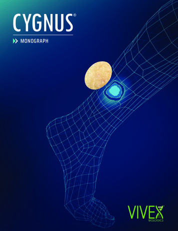
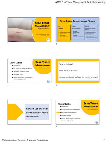
Pan et al. Journal of Inflammation (2017) 14:7development of pancreatic lesions was evaluatedaccording to point Spormann scale, as previouslydescribed [20]. Histological analysis was conducted incontrol group, SAP group, and SAP treated with ulinastatin group (30000U/kg).Cell isolation from the peripheral bloodThe mononuclear cells were isolated from the peripheralblood by Ficoll-Hypaque gradient centrifugation (Solarbio Biotech, Beijing, China), washed twice with PBS, andkept on ice until labeling.Page 3 of 9Statistical analysisQuantitative data were expressed as the mean standarddeviation (SD) and analyzed with a one-way ANOVA.Fisher least significant difference was used to evaluatesignificant differences between the groups. The survivalrate was calculated as percentage of survivors at thedescribed certain time point relative to the total numberof rats that received a given treatment. The survival ratedata were analyzed using the log-rank test. P values lessthan 0.05 were considered statistically significant. Allstatistical analyses were performed using SPSS statisticalsoftware version 19.ResultsFlow cytometry analysisThe expression markers on T cells from the peripheralblood were determined by flow cytometry after stainingwith anti-rat specific Abs conjugated with PE, FITC, orAPC. The rat Abs including anti-CD4, anti-CD25, andanti-FoxP3 that were purchased from eBioscience (SanDiego, CA, USA). To determine the proportion of CD4 T cells in lymphocytes and CD25 expression on the surface of CD4 T cells, the cells were stained with CD25PE mAb and CD4-FITC mAb for 30 min in darkness.Concomitantly, for the detection of intranuclear Foxp3,the cells were reacted with 1 mL of freshly prepared fixation/permeabilization working solution for 2 h at 4 C.After washing the cells with the permeabilization buffertwice, the cells were stained with anti-rat Foxp3-APCantibody for 30 min in the dark. After washing, the cellswere performed on a BD Accuri C6 flow cytometer (BDBiosciences) using the FlowJo Software (Ashland, Kentucky, USA).Real-time polymerase chain reactionTotal RNAs were extracted from peripheral blood withTRIzol reagent (Takara, Dalian, China) followed by reversetranscription into complementary DNA (cDNA) according to the manufacturer’s instructions (Thermo Scientific,Waltham, MA, USA). Real-time quantitative polymerasechain reaction (PCR) was performed with QuantiTectSYBR Green PCR Kit (Applied Biosystems, Foster City,CA, USA) in ABI PRISM 7500 PCR instrument (AppliedBiosystems) according to the manufacturer’s instructions.Glyceraldehyde-3-phosphate dehydrogenase (GAPDH)served as an internal reference. The primers were as follow: CTLA-4, 5′-TGCGGCAGACAAATGACCA-3′ and5′-CAAAGTATGGCGGTGGGTA-3′; Foxp3, 5′-TTCTCAAGCACTGCCAAGC-3′ and 5′-GTCTCCGCACAGCAAACAA-3′; GAPDH, 5′-CTGAGTATGTCGTGGAGTCTAC-3′ and 5′-AGTCTTCTGAGTGGCAGTGATG-3′. The relative gene expression level was calculated bythe 2-ΔΔCt method.Ulinastatin significantly improves survival in SAP ratmodelAll rats in the sham group survived, whereas the ratsthat received SAP operation all died within 72 h (P 0.01, Fig. 1). Intraperitoneal injection of ulinastatin(5000 U/kg) after SAP operation reduced the mortalityrate (P 0.05, Fig. 1). Furthermore, treatment with ulinastatin at higher dose (10000 and 30000 U/kg) significantly protected the rats from SAP (P 0.05, Fig. 1).Effect of ulinastatin on SAP-induced enzyme productionand MPOWe assessed the severity of AP by measuring amylaseand lipase levels, the known biomarkers of SAP. Neutrophils are known to contribute to the activation oftrypsinogen to cause further damage in SAP. Therefore we next examined neutrophil infiltration bymeasuring MPO in pancreas homogenates. The serumamylase and lipase levels were increased significantlyin the SAP group compared to the control group,indicating that SAP rat model was induced successfully (P 0.01, Fig. 2a and b). Treatment withFig. 1 Ulinastatin treatment improves survival in SAP rats. Ulinastatinwas administered immediately when SAP operation achieved andadministered again in 6 h later and again in 12 h later throughintraperitoneal injection. Data are shown as percent of rats surviving(n 10). *P 0.05 when compared with the SAP group
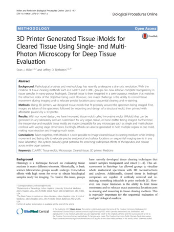
Pan et al. Journal of Inflammation (2017) 14:7Page 4 of 9Fig. 2 Effects of ulinastatin on SAP-induced enzyme production and MPO. a Serum levels of amylase. b Serum levels of lipase. c MPO activity.Data were expressed as means SD. n 10 for each group. **P 0.05 when compared with the control group; *P 0.05 when compared with theSAP group; #P 0.05 when compared with the SAP Ulinastatin (5000U/kg) group; ##P 0.05 when compared with the SAP Ulinastatin(10000U/kg) groupulinastatin significantly reduced the levels of amylaseand lipase in the SAP-induced rats (P 0.01) compared to the untreated animals. In addition, this effectwas dose dependent as ulinastatin at 30000U/kgshowed pronounced effect than the 5000U/kg dose(Fig. 2a and b). Furthermore, a similar result was obtained in pancreatic MPO activities (Fig. 2c).Ulinastatin reduced SAP-induced tissue damage of pancreasHistologically, the pancreatic sections from the controlgroup showed normal architecture (Fig. 3a). In contrast,the pancreatic sections from the SAP animals showedapparent destruction of normal architecture, tissueedema, necrosis of acinar cells, and infiltration of inflammatory cells, with the median point Spormann scale ofFig. 3 Histopathology of pancreas. a The pancreatic parenchyma showed typical normal architecture in control group, white arrow indicatenormal acinar cell. b The pancreatic section showed significant destruction of structure in SAP group. c After ulinastatin (3000U/kg) treatment, thepancreatic damage was less severe than that in the SAP group. Single black arrows indicate glands damage and acinar cells necrosis, doubleblack arrows indicate tissue oedema, treble black arrows indicate inflammatory cells infiltration. d Pancreas histopathologic score were evaluatedaccording to point Spormann scale, and the results were expressed as means SE. n 10 for each group. **P 0.05 when compared with thecontrol group; *P 0.05 when compared with the SAP group. Magnification, 200
Pan et al. Journal of Inflammation (2017) 14:716 (P 0.01, Fig. 3b and d). After treatment with ulinastatin (3000U/kg), the tissue damage was significantlyreduced with the median point Spormann scale of 11 (P 0.05, Fig. 3c and d).Effect of ulinastatin on cytokines productionThe injury of acinar cells in acute pancreatitis isfollowed by a pro-inflammatory cascade leading to pancreatic necrosis and systemic inflammatory responsesyndrome both in human and in experimental animalmodels. TNF-α and IL-1β are pro-inflammatory cytokines that are known for their ability to induce systemicinflammation. In contrast to the control group, theexpression of both cytokines in SAP was significantlyelevated compared to the normal animals (Fig. 4a andb). Treatment with ulinastatin reduced the level of TNFα and IL-1β in a dose-dependent way (P 0.05). TGF-β1and IL-10 are Treg-related cytokines characterized asimmunosuppressive or anti-inflammatory mediators. IL10 level was increased in the SAP animals compared tothe controls while TGF-β1 showed decrease instead (P 0.05). Ulinastatin treatment (10000 and 30000 U/kg) ofSAP rats led to a significant increase in the serumPage 5 of 9concentration of IL-10, but no change in serum TGF-β1level (Fig. 4c and d).Ulinastatin does not alter the proportion of CD4 Tlymphocytes in the SAP ratsWe explored the effect of ulinastatin on the peripheralblood CD4 T cells in SAP rats by flow cytometry(Fig. 5). The SAP animals showed a lower percentage ofCD4 T lymphocytes compared to the controls (P 0.05). There was no significant difference in the proportion of CD4 T lymphocytes between SAP animals andSAP animals treated with ulinastatin.Ulinastatin enhances the percentage of Treg in CD4 Tcells in the SAP ratsWe next studied the change in Tregs of the SAP animalstreated with ulinastatin. The proportion of CD4 CD25 Tcells was decreased significantly in the SAP group, compared to the control group (P 0.05, Fig. 6a and b). Incontrast, ulinastatin (10000 and 30000 U/kg) effectivelyprevented the decline of CD4 CD25 T cells (P 0.05,Fig. 6a and b). Tregs were also defined as CD4 CD25 FoxP3 T cells. We studied the effect of ulinastatin onFig. 4 Effects of ulinastatin on serum cytokines in the SAP rats. a Serum levels of IL-1β. b Serum levels of TNF-α. c Serum levels of IL-10. d Serumlevels of TGF-β1. Data were expressed as means SD. n 10 for each group. **P 0.05 when compared with the control group; *P 0.05 whencompared with the SAP group; #P 0.05 when compared with the SAP Ulinastatin (5000U/kg) group; ##P 0.05 when compared with the SAP Ulinastatin (10000U/kg) group
Pan et al. Journal of Inflammation (2017) 14:7Page 6 of 9Fig. 5 The effect of ulinastatin on circulating CD4 T cells in SAP rats. R1 region represents CD4 T cell in lymphocytes. The frequencies (%) ofCD4 T Cells were measured by using flow cytometry. Results were shown as means SD. n 10 for each group. **P 0.05 when compared withthe control groupFig. 6 The effect of ulinastatin on Treg cells in SAP rats. a R2 region represents CD4 CD25 T cells from lymphocytes. b The frequencies (%) ofCD4 CD25 T cells. c R3 region represents CD4 CD25 FoxP3 T cells from CD4 T cells. d The frequencies (%) of CD4 CD25 FoxP3 T cells.Results were measured by using flow cytometry and shown as means SD. n 10 for each group. **P 0.05 when compared with the controlgroup; *P 0.05 when compared with the SAP group; #P 0.05 when compared with the SAP Ulinastatin (5000U/kg) group; ##P 0.05 whencompared with the SAP Ulinastatin (10000U/kg) group
Pan et al. Journal of Inflammation (2017) 14:7the proportion of CD4 CD25 FoxP3 T cells in CD4 Tcells, and obtained similar results (Fig. 6c and d).Effect of ulinastatin on Foxp3 and CTLA-4 geneexpressionTo explore the potential influence of ulinastatin on immunoregulatory molecules in Tregs, the expression ofFoxp3 and CTLA-4 was determined with RT-PCR usinglymphocytes isolated from the peripheral blood. Compared to the control group, the messenger RNA (mRNA)levels of Foxp3 and CTLA-4 in SAP group were significantly decreased (P 0.05, Fig. 7). Moreover, treatmentwith ulinastatin further up-regulated the expression ofFoxp3 and CTLA-4 in SAP rats in a dose dependencefashion, and at the 30000U/kg dose level the effect wasthe most pronounced (P 0.05).DiscussionThe techniques of inducing animal model of SAPinclude pancreatic under-capsule injection or biliopancreatic duct retrograde injection of sodium taurocholate,intraperitoneal injection of caerulein alone or injectionof caerulein combined with lipopolysaccharide, ethioninediet and hypercalcemia derivation [21, 22]. The technique of Aho et al. [23] by retrogradely injecting sodiumtaurocholate into biliopancreatic duct through duodenalpapilla puncture could more reliably induce the SAPanimal model. It could reflect the patho-physiologicmechanism of acute biliary pancreatitis, a common category of clinical SAP. However, the technique causes ahigh damage rate of biliopancreatic duct and results inbile leakage. To overcome this problem, George Perideset al. [19] changed the way of injection by creating asmall hole in the duodenal wall opposite to the papilla,followed by puncturing a cannula to complete the injection. In our study, a stable and reliable animal modelPage 7 of 9was induced successfully by the modified technique ofGeorge Perides. Levels of serum amylase and lipase inSAP group were consistently elevated over the controlgroup. Furthermore, the pathologic changes such as largenecrosis area, edema and infiltration of inflammatory cellswere observed in pancreatic tissue of the SAP rats.Inflammatory cytokines including IL-1, IL-2, IL-4, IL10 and TNF-α, are released early in the course of SAPleading to the systemic inflammatory response whichrepresents the core problem of SAP. In the recent years,evidence has accumulated that immune reactions andimmune cells like CD4 T cells and Tregs play importantroles in the AP pathogenesis [24, 25]. Hence, inhibitingpro-inflammatory mediators [26, 27] and regulating immune reactions [25] are important considerations in thetherapy of SAP. It has been confirmed that ulinastatinprevents the inflammatory response induced by SAP[28], and evidence also indicates that ulinastatin canregulate the immunological function through these special immune cells [18, 29]. In the present study, we demonstrated that in SAP rat model, pro-inflammatorycytokines TNF-α and IL-1β and anti-inflammatory cytokine IL-10 all were increased significantly, while TGF-βwas decreased. Treatment of ulinastatin led to decreasedlevels of TNF-α, IL-1β, and increased level of IL-10, thusattenuated the acute inflammatory response and improved the survival rate in the SAP rats. We also foundthat CD4 T cells and Tregs were significantly decreased.The mRNA levels of CTLA-4 and Foxp3 were decreasedin the SAP animals. These results indicate that not onlyinflammatory response, but also immune dysfunctionplay important roles in SAP, resulting in high mortalityrate of SAP.Tregs are pivotal to the maintenance of immunesystem, and key to a measured inflammatory response.Treg-related cytokines, IL-10 and TGF-β are importantFig. 7 The effect of ulinastatin on mRNA expression of CTLA-4 and Foxp3 in SAP rats. a mRNA expression of CTLA-4. b mRNA expression ofFoxp3. Results were expressed as means SD. n 10 for each group. **P 0.05 when compared with the control group; *P 0.05 when comparedwith the SAP group; #P 0.05 when compared with the SAP Ulinastatin (5000U/kg) group; ##P 0.05 when compared with the SAP Ulinastatin(10000U/kg) group
Pan et al. Journal of Inflammation (2017) 14:7regulators of inflammation. During the inflammatoryprocess, IL-10 plays the role of an anti-inflammatorycytokine, and its anti-inflammatory effect in turnregulates the function of Tregs. Some studies havedemonstrated that IL-1 can affect the function of Tregs[30, 31]. Tregs express IL-1 receptor (IL-1R) and canactivate p38/JNK signaling in response to IL-1 [32]. LikeIL-1, TNF-α is a pleiotropic cytokine promoting inflammatory mediator cascade reaction and also impairingTreg stability and function. For instance, several studieshave revealed that Tregs were functionally abnormal inpatients with rheumatoid arthritis (RA), and TNF-α wasthe key mediator of this abnormal immune regulation[33, 34]. We found in our study that, in SAP-inducedrats treated with ulinastatin, the percentage of Tregsincreased significantly, in addition the expression ofCTLA-4 and Foxp3 were increased, in a dosedependence manner. Ulinastatin did not affect TGF-β1level or the proportion of CD4 T cells. Treatment withulinastatin lessened the pancreatic tissue injury and theMPO activity, likely by enhancing the anti-inflammatoryfunction of Tregs with the increased proportion of CD4 T cells. The mechanism of ulinastatin inducing theexpansion of Tregs may be through up-regulating IL-10level and down-regulating TNF-α and IL-1β.However, at present, it is still not clear whether thechanges of these inflammatory cytokines are the primarymechanism of ulinastatin or indirect consequence of thereduced inflammatory injury after treatment with ulinastatin. In addition, the mechanism is not yet clear ifulinastatin up-regulates Tregs directly or indirectly. Alsosome studies showed that TNF-α can enhance thesuppressive activity of Tregs [35]. The interplay betweenTregs and inflammatory cytokines is complex and intimately connected and remains to be further studied.ConclusionsIn conclusion, our study showed ulinastatin alleviatesthe pancreatic injury through reducing the level ofMPO, pro-inflammatory cytokines, serum amylase andlipase. Our study also showed that ulinastatin upregulated the percentage of Tregs and their antiinflammatory function through regulating inflammatorycytokines and increasing the level of Foxp3 and CTLA-4expression, providing evidence for a new mechanism responsible for the effect of ulinastatin in SAP.AbbreviationsMPO: Myeloperoxidase; SAP: Severe acute pancreatitis; IL-1β: Interleukin-1β;TNF-α: Tumor necrosis factor-α; IL-10: Interleukin-10; TGF-β1: Transforminggrowth factor-β1; Foxp3: Forkhead/winged helix transcription factor p3;CTLA-4: Cytotoxic T-lymphocyte antigen 4; mRNA: Messenger RNAAcknowledgementsThe authors thank Chengguang He for his technical support.Page 8 of 9FundingThis work was supported by the Foundation of Techpool Research (No.UF201318).Availability of data and materialsPlease contact author for data requests.Authors’ contributionsYP, HH and FL conceived and designed the experiments; YP, HF and MPdrafted and revised the manuscript; YP, HF, FC,YY and PX carried outexperiments. YP, HF and PX performed the statistical analysis. All authorsread and approved the final manuscript.Competing interestsThe authors declare that they have no competing interests.Consent for publicationNot applicable in this section.Ethics approvalAll experimental protocols were approved by the Ethical Committee forAnimal Research of Fujian Medical University.Publisher’s NoteSpringer Nature remains neutral with regard to jurisdictional claims inpublished maps and institutional affiliations.Author details1General Surgery Department, Fujian Medical University Union Hospital,No.29 Xinquan Road, Fuzhou 350001, People’s Republic of China.2Department of Oncology and Hematology, Kaiser Permanente MedicalCenter, 710 Lawrence Expressway, Santa Clara, CA 95051, USA. 3Fuzhou,People’s Republic of China.Received: 19 December 2016 Accepted: 14 March 2017References1. Lankisch PG, Apte M, Banks PA. Acute pancreatitis. Lancet. 2015;386:81–92.2. Mayerle J, Hlouschek V, Lerch MM. Current management of acutepancreatitis. J Gastrointest Surg. 2005;9:440–52.3. Minkov GA, Halacheva KS, Yovtchev YP, Gulubova MV. Pathophysiologicalmechanisms of acute pancreatitis define inflammatory markers of clinicalprognosis. Pancreas. 2015;44:713–7.4. Sendler M, Dummer A, Weiss UF, Kr BG, Wartmann T, Scharffetter KK, VanNR, Malla RS, Aghdassi A, Halangk W. Tumour necrosis factor α secretioninduces protease activation and acinar cell necrosis in acute experimentalpancreatitis in mice. Gut. 2013. doi:10.1136/gutjnl-2011-300771.5. Kobayashi H, Hirashima Y, Sun GW, Fujie M, Shibata K, Tamotsu S, Miura K,Sugino D, Tanaka Y, Kondo S. Identification and characterization of the cellassociated binding protein for urinary trypsin inhibitor. Biochimic Et BiophysActa. 1998;1383:253–68.6. Liu R, Qi H, Wang J, Wang Y, Cui L, Wen Y, Yin C. Ulinastatin activates therenin-angiotensin system to ameliorate the pathophysiology of severeacute pancreatitis. J Gastroenterol Hepatol. 2014;29:1328–37.7. Maciejewski R, Burdan F, Burski K, Madej B, Ziemiakowicz R, Dąbrowski A,Wallner G. Selected biochemical parameters and ultrastructural picture ofpancreas due to Ulinastatin treatment of experimental acute pancreatitis.Exp Toxicol Pathol. 2005;56:305–11.8. Ma T, Kang C, Shao H, Qi Q, Hu W. Protective Effects of Ulinastatin onProliferation and Cytokine Release of Splenocytes from Rats with SevereAcute Pancreatitis. Eur Surg Res. 2006;38:445–50.9. Venet F, Chung C-S, Kherouf H, Geeraert A, Malcus C, Poitevin F, Bohé J,Lepape A, Ayala A, Monneret G. Increased circulating regulatory T cells(CD4 CD25 CD127 ) contribute to lymphocyte anergy in septic shockpatients. Intensive Care Med. 2008;35:678–86.10. Sakaguchi S, Sakaguchi N. Regulatory T Cells in Immunologic Self-Toleranceand Autoimmune Disease. Int Rev Immunol. 2009;24:211–26.11. Chauhan SK, Saban DR, Lee HK, Dana R. Levels of Foxp3 in regulatory T cellsreflect their functional status in transplantation. J Immunol.2009;182:148–53.
Pan et al. Journal of Inflammation (2017) 14:712. Lin J, Lu Y, Silva HM, Trzeciak A, Choi Y, Schwab SR, Dustin ML, Lafaille JJ.Increased generation of Foxp3 regulatory T cells by manipulating antigenpresentation in the thymus. Nat Commun. 2016. doi:10.1038/ncomms10562.13. Morita H, Arae K, Unno H, Miyauchi K, Toyama S. An Interleukin-33-MastCell-Interleukin-2 Axis Suppresses Papain-Induced Allergic Inflammation byPromoting Regulatory T Cell Numbers. Immunity. 2015;43:175–86.14. Villalta SA, Rosenthal W, Martinez L, Kaur A, Sparwasser T, Tidball JG,Margeta M, Spencer MJ, Bluestone JA. Regulatory T cells suppress muscleinflammation and injury in muscular dystrophy. Sci Transl Med. 2014;6(258):258ra142. doi:10.11
CA, USA) in ABI PRISM 7500 PCR instrument (Applied Biosystems) according to the manufacturer's instructions. Glyceraldehyde-3-phosphate dehydrogenase (GAPDH) served as an internal reference. The primers were as fol-low: CTLA-4, 5′-TGCGGCAGACAAATGACCA-3′ and 5′-CAAAGTATGGCGGTGGGTA-3′;Foxp3,5′-TTCT CAAGCACTGCCAAGC-3′ and 5 .