Transcription
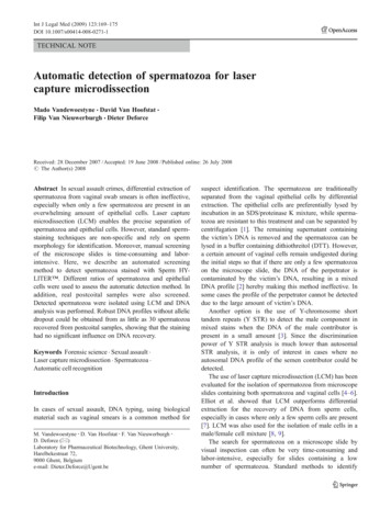
Int J Legal Med (2009) 123:169–175DOI 10.1007/s00414-008-0271-1TECHNICAL NOTEAutomatic detection of spermatozoa for lasercapture microdissectionMado Vandewoestyne & David Van Hoofstat &Filip Van Nieuwerburgh & Dieter DeforceReceived: 28 December 2007 / Accepted: 19 June 2008 / Published online: 26 July 2008# The Author(s) 2008Abstract In sexual assault crimes, differential extraction ofspermatozoa from vaginal swab smears is often ineffective,especially when only a few spermatozoa are present in anoverwhelming amount of epithelial cells. Laser capturemicrodissection (LCM) enables the precise separation ofspermatozoa and epithelial cells. However, standard spermstaining techniques are non-specific and rely on spermmorphology for identification. Moreover, manual screeningof the microscope slides is time-consuming and laborintensive. Here, we describe an automated screeningmethod to detect spermatozoa stained with Sperm HYLITER . Different ratios of spermatozoa and epithelialcells were used to assess the automatic detection method. Inaddition, real postcoital samples were also screened.Detected spermatozoa were isolated using LCM and DNAanalysis was performed. Robust DNA profiles without allelicdropout could be obtained from as little as 30 spermatozoarecovered from postcoital samples, showing that the staininghad no significant influence on DNA recovery.Keywords Forensic science . Sexual assault .Laser capture microdissection . Spermatozoa .Automatic cell recognitionIntroductionIn cases of sexual assault, DNA typing, using biologicalmaterial such as vaginal smears is a common method forM. Vandewoestyne : D. Van Hoofstat : F. Van Nieuwerburgh :D. Deforce (*)Laboratory for Pharmaceutical Biotechnology, Ghent University,Harelbekestraat 72,9000 Ghent, Belgiume-mail: Dieter.Deforce@Ugent.besuspect identification. The spermatozoa are traditionallyseparated from the vaginal epithelial cells by differentialextraction. The epithelial cells are preferentially lysed byincubation in an SDS/proteinase K mixture, while spermatozoa are resistant to this treatment and can be separated bycentrifugation [1]. The remaining supernatant containingthe victim’s DNA is removed and the spermatozoa can belysed in a buffer containing dithiothreitol (DTT). However,a certain amount of vaginal cells remain undigested duringthe initial steps so that if there are only a few spermatozoaon the microscope slide, the DNA of the perpetrator iscontaminated by the victim’s DNA, resulting in a mixedDNA profile [2] hereby making this method ineffective. Insome cases the profile of the perpetrator cannot be detecteddue to the large amount of victim’s DNA.Another option is the use of Y-chromosome shorttandem repeats (Y STR) to detect the male component inmixed stains when the DNA of the male contributor ispresent in a small amount [3]. Since the discriminationpower of Y STR analysis is much lower than autosomalSTR analysis, it is only of interest in cases where noautosomal DNA profile of the semen contributor could bedetected.The use of laser capture microdissection (LCM) has beenevaluated for the isolation of spermatozoa from microscopeslides containing both spermatozoa and vaginal cells [4–6].Elliot et al. showed that LCM outperforms differentialextraction for the recovery of DNA from sperm cells,especially in cases where only a few sperm cells are present[7]. LCM was also used for the isolation of male cells in amale/female cell mixture [8, 9].The search for spermatozoa on a microscope slide byvisual inspection can often be very time-consuming andlabor-intensive, especially for slides containing a lownumber of spermatozoa. Standard methods to identify
170spermatozoa are based on non-specific staining techniquessuch as hematoxylin/eosin (HE) and nuclear fast red/picroindigocarmine (CTS or “Christmas tree stain”) [10],which rely on sperm morphology for identification [4] andcannot be used for automated sperm detection. MoreoverHE staining reduces the yield of DNA [4, 5, 11] and CTSstaining results in DNA damage, probably due to the picricacid component [4].Sperm HY-LITER (Independent Forensics, Hillside,IL, USA) is a fluorescent kit for the detection of humanspermatozoa which does not rely on morphological characteristics or non-specific staining for identification. All relevantinformation concerning this kit can be found at http://www.spermhy-liter.com/. The spermatozoa are detected using anAlexa Fluor 488 derivatized mouse monoclonal antibodyagainst proteins contained in the human sperm heads. Inaddition, a 4′,6-diamidino-2-phenylindole staining is used todetect all nuclei present on the slide. Processed slides can beviewed at low magnification, greatly increasing the speed ofsperm identification.The PALM MicroBeam system (P.A.L.M. MicrolaserTechnologies, Bernried, Germany), allows scanning ofmicroscope slides [5]. The system can be supplementedwith image analyzing software modules allowing automated specimen identification and image processing [12] e.g.the AxioVision Commander (Carl Zeiss, Hallbergmoos,Germany). This image analyzing software can be optimizedto automatically detect cells with specific features. In thecurrent report we have optimized for the automaticdetection of spermatozoa, stained with Sperm HY-LITER (Independent Forensics). The detected spermatozoa canthen be collected by a defined laser pulse using the PALMlaser pressure catapulting (LPC) function. This contact-freecell collection is an ideal approach to avoid samplecontamination in forensic cases.In the present study, an automated scanning method wasdeveloped to identify sperm heads, stained with Sperm HYLITER (Independent Forensics), on smear preparationsfrom postcoital vaginal swabs. LPC was used to catapult theauto-detected spermatozoa and DNA analysis was performed.After optimization of the DNA extraction protocol, the effectof the Sperm HY-LITER staining on downstream analysisof laser pressure catapulted spermatozoa was examined andthe ability to obtain DNA profiles from spermatozoa catapulted from stained postcoital vaginal smears was assessed.Materials and methodsSample preparationVaginal epithelial cells from healthy volunteers werecollected onto sterile cotton swabs. These swabs wereInt J Legal Med (2009) 123:169–175agitated in phosphate buffered saline (PBS, Gibco, Paisly,UK). After cell counting, PBS was added to obtain anepithelial cell working solution of 2 106 cells/ml. Semensamples were obtained from healthy volunteers. Semenworking solutions of 2 106 spermatozoa/ml were preparedby diluting the liquid semen samples in PBS. Mixtures wereprepared by combining semen working solution withepithelial cell working solution in the following ratios: 1:2,1:10, and 1:50. Of each mixture 30 μl was used to makesmear preparations on routine glass object slides (Dakosilanized slides, Glostrup, Denmark). In addition, pure semensmears and postcoital vaginal swab smears were made.The preparations were all dried overnight at roomtemperature and stained using the Sperm HY-LITER kit(Independent Forensics) according to manufacturer’s suggested protocol with slight modifications: the preparationswere fixed for 2 min using two drops of a 70% ethanolsolution (absolute ethanol, Merck BV, Schiphol-Rijk, TheNetherlands) in pure water (MilliQ, Millipore, Billerica,MA, USA) in stead of the Sperm HY-LITER fixationsolution. After the last washing step in the protocol, theslides were washed with 2 ml of pure water (MilliQ,Millipore) to remove adherent salts from the Sperm HYLITER wash buffer and the stained slides were visualizedimmediately without mounting.Fluorescence scanningThe scanning stage was controlled by the PALM RoboSoftware version 4 (P.A.L.M. Microlaser Technologies). Imageacquisition was carried out with the AxioVision multichannelfluorescence module (Carl Zeiss) and the AxioCam MRmcamera (Carl Zeiss).The cell nuclei, including epithelial and sperm cell nuclei,were visualized using Zeiss filter set no. 49 (G 365 nm, FT495, BP 445/50). The spermatozoa were visualized usingZeiss filter set no. 38 (BP 470/40, FT 495, BP 525/50). Theslides were scanned at 20 magnification using a Carl Zeisslong distance Plan-Neofluar objective. From every slide,100 images were acquired using the scanning mode. Theacquired images were displayed as an overview image inthe PALM Navigator window and the individual imageswere stored as tiff-files.Segmentation and maskingFor automated detection of spermatozoa, the image processing AxioVision Commander module (Carl Zeiss) wasused. All steps of processing, analysis, and evaluation werestored in an AxioVision Commander Script, which could berun automatically on the stored images.In a first step the sperm heads, detected in the greenfluorescence channel, were discriminated from the back-
Int J Legal Med (2009) 123:169–175171ground using an interactive threshold. The resulting imagewas a binary image where the background was black andthe detected regions were white. In a following step alldetected regions below 3 μm or above 7 μm were removedfrom the binary image to eliminate artefacts smaller orbigger than a sperm head (approximately 5 μm).Then the nuclei detected in the blue fluorescence channelwere distinguished from the background using an interactivethreshold. Afterwards artefacts (regions with an area above70 μm2) were also eliminated from the resulting binaryimage. Following this procedure all nuclei still touchingeach other were separated using a watershed algorithm, tomake sure every nucleus was counted separately. Thisalgorithm splits the image into disjoint regions containingonly one nucleus and is based on the topology of the image.In a next step the spermatozoa were identified by maskingthe binary image of the green channel and the binary imageof the blue channel, as shown in Fig. 1. Only regions of thebinary image of the nuclei which were overlaid by at leastone pixel of the binary image of the sperm heads were keptin the resulting image. In this way, remaining artefacts withthe same dimensions as a sperm head but without a nucleuswere removed from the resulting image.The coordinates of the detected spermatozoa weretransferred automatically to an element list in the PALMRoboSoftware and after visual verification of the detectedspermatozoa a catapulting point was set on the sperm head.Fig. 1 Principle of the AxioVision Commander Script. a Pseudocolored original image of the sperm heads, b pseudo-colored originalimage of the nuclei, c overlay of both pseudo-colored images, d binaryimage of the sperm heads after segmentation, e binary image of thenuclei after segmentation, f final masked image outlining the detectedsperm headsLaser pressure catapultingThe detected spermatozoa were collected by laser pressurecatapulting using a pulsed nitrogen UV-A laser (wavelength355 nm). The high energy generated by the focused laserlight was used to catapult the detected spermatozoa into thecap of a standard 0.2 ml microfuge tube (Westburg,Leusden, The Netherlands) containing 18 μl of PicoPureDNA extraction buffer (PicoPure DNA extraction kit,Arcturus, Mountain View, CA, USA) supplemented with2 μl of 1 M DTT (MP Biomedicals, Solon, OH, USA). Thiscontact-free method avoids contamination of the sample.After the catapulting process, the recovery of thespermatozoa could be verified microscopically in the collection caps. In addition, as a verification of the catapultingprocess, fluorescent images were acquired before and afterLPC, as shown in Fig. 2.DNA extractionDNA was extracted from the catapulted spermatozoa, usingthe PicoPure DNA extraction kit (Arcturus). The samples
172Int J Legal Med (2009) 123:169–175The amplified fragments were then separated andanalyzed by capillary electrophoresis using an ABI 3100Genetic Analyzer (Applied Biosystems).ResultsAutomated detection of spermatozoaAs the Sperm HY-LITER kit (Independent Forensics) hasbeen designed for microscopic screening of mounted slides,a few modifications of the manufacturer’s suggestedprotocol had to be carried out to make it compatible withFig. 2 Laser pressure catapulting of detected spermatozoa. Same fieldof view: a fluorescent image before laser pressure catapulting, bfluorescent image after laser pressure catapultingwere incubated at 65 C overnight, centrifuged briefly andheated to 95 C for 30 min to inactivate proteinase K. If thepolymerase chain reaction (PCR) could not be performedimmediately, the samples were stored at 20 C.Amplification and detectionFor assessing the profile recovery after LPC from puresemen samples, a multiplex of four STR loci (CD4, TH01,D21S11, and SE33) was used as described earlier [13] withslight modifications. In short, 1.3 units of hotstar Taq DNApolymerase (Qiagen, Huntsville, AL, USA) was used andthe samples were amplified on an Applied BiosystemsGeneAmp 9700 60 Well thermal cycler (Foster City, CA,USA). Amplification parameters were: preincubation at95 C for 15 min, followed by 33 cycles of denaturation for60 s at 94 C, annealing for 60 s at 58 C and extension for80 s at 72 C. This was followed by a final elongation stepof 5 min at 72 C. At the end of the PCR reaction thetemperature was kept at 4 C.For the amplification of the DNA extracted from thespermatozoa isolated by LPC from the postcoital samples,the AmpFℓ STR Profiler Plus kit from AppliedBiosystems was used according to the manufacturer'sinstructions, except that Hotstar Taq DNA polymerase wasused instead of Taq Gold. The total number of cycles was 33.Fig. 3 Automatic detection of spermatozoa by the AxioVisionCommander Script. Same field of view: a bright field image,containing six spermatozoa, vaginal epithelial cells and Lactobacillusorganisms, b fluorescent image, c resulting binary image, outlining thesix sperm heads
Int J Legal Med (2009) 123:169–175173Fig. 4 Automatic scanning of the microscope slide and result of theAxioVision Commander Script. a Overview image of the 100 imagesacquired from the postcoital vaginal smear. b Zoomed in image (100 zoom): vaginal epithelial cells are outlined in light blue, sperm headsare outlined in yellowLPC. An additional washing step with pure water wasadded to the protocol, to remove salt crystals originatingfrom the Sperm HY-LITER wash buffer. This wasnecessary because the salt crystals were highly autofluorescent in the green fluorescence channel and interfered with thedetection of the stained sperm heads. After this additionalwashing step, the slides could be visualized unmounted,which is necessary for LPC.Figure 3 shows that spermatozoa stained with SpermHY-LITER kit are more easily detectable than unstainedspermatozoa. The AxioVision Commander Script generatesa binary image that outlines the sperm heads and transfersthe coordinates into a PALM RoboSoftware element list forLPC.To determine whether the AxioVision CommanderScript could be used to detect sperm heads in mixtures ofspermatozoa and vaginal epithelial cells, different ratios ofthese mixtures were used to make smears on microscopeslides. To control the efficiency on a real sample, a smear,made from a vaginal swab taken 3-h postcoitus, was alsoscanned. For every slide, 100 images were acquired asshown in Fig. 4. The detected ratios of spermatozoa andnuclei are represented in Table 1.The postcoital vaginal swab smear was re-examinedmanually and only 2.8% false positives were found. Boththe low amount of false positives and the good correlationbetween the ratio used to make the smears and the detectedratio, show the very high reliability of the developedAxioVision Commander Script.The detected spermatozoa were catapulted by LPC in toa standard microfuge tube. Images acquired before and aftercatapulting clearly show that the catapulting processremoves the sperm head from the glass slide.Profile recovery after laser pressure catapultingThree DNA isolation methods were evaluated (data notshown): an alkaline extraction method [14], DNA IQ System (Promega Corporation, Madison, WI, USA) andPicoPure DNA extraction kit (Arcturus) and the best resultswere obtained with the latter. After optimization, this DNAextraction method was used for further assessment ofprofile recovery after LPC.Different amounts of spermatozoa (200, 100, 75, 50, 40,30, 20, and 10) were collected by LPC from the pure spermsmears to assess the effect of the Sperm HY-LITER staining on the recovery of DNA. A full DNA profile of thefour assessed loci (CD4, TH01, D21S11, and SE33) couldbe recovered from every sample. This proves that theSperm HY-LITER staining has no negative influence onthe DNA quality of the spermatozoa.In addition, profile recovery from spermatozoa isolatedby LPC from postcoital samples was analyzed using theAmpFℓ STR Profiler Plus kit from Applied Biosystems.Table 1 Automatically detected spermatozoa and nuclei in 100 acquired images per slideRatio of spermatozoa/nucleiused to make the smears1:21:101:50Postcoital sampleNumber of spermheads detectedTotal number of nuclei (spermatozoaand epithelial cells) detectedDetected ratio 7841:2.61:7.31:42.71:4.97
174Table 2 Profile recovery fromspermatozoa after LPC frompostcoital samplesInt J Legal Med (2009) 123:169–175Number of isolatedspermatozoaNumber of expectedmale allelesNumber of detectedmale allelesNumber of contaminatingfemale 0202030305050Different amounts of spermatozoa (two times 50, 30, 20,and 10 spermatozoa) were collected. The results arepresented in Table 2. The two samples where 10 spermatozoa were collected and one sample where 20 spermatozoaFig. 5 DNA profile recoveryafter LPC. Profile derived froma postcoital sample aftercatapulting 50 spermatozoawere collected show allelic drop out. This is in agreementwith the theoretical expected probability of full allelicrepresentation from this number of haploid cells [15]. FullDNA profiles were recovered when 30 or more spermatozoa
Int J Legal Med (2009) 123:169–175were collected. Figure 5 shows a full DNA profile obtainedafter catapulting 50 spermatozoa from a postcoital sample. Inone of the samples a few extra alleles from the female donorwere recovered. This mixture may be due to the adherence offemale DNA from lysed epithelial cells to spermatozoa [7,16]. Despite the presence of female DNA, the male DNAprofile was easily interpreted.DiscussionThe results demonstrate that the AxioVision CommanderScript has sufficient sensitivity to identify sperm headsstained with Sperm HY-LITER (Independent Forensics),present in an overwhelming background of epithelial cells.The sperm heads and the cell nuclei are easily detectableand the background staining is negligible. The nucleusstaining provides additional confirmation that the AlexaFluor 488 signal observed is related to a DNA-containingsperm head. The coordinates of the single cells assessed asspermatozoa are transferred to an element list of the PALMRoboSoftware. Before laser catapulting, the detected cellscan be verified easily through bright field microscopy basedon sperm cell morphology.The scanning, analysis and identification of spermatozoacan be performed on normal microscopic slides, which areused with the Belgian sexual assault sets, allowing its use inexisting preparations from sexual assault cases. Processedslides can be scanned at low magnification, greatlyincreasing the speed of sperm identification.For forensic DNA profiling, it is important that thestaining reagents used have no adverse effects on therecovery of DNA from spermatozoa on postcoital vaginalswabs. As robust DNA profiles without allelic drop outcould be obtained from as little as 30 spermatozoa recoveredfrom postcoital samples after LPC, it can be concluded thatthe Sperm HY-LITER staining has no influence ondownstream DNA analysis, in contrast to traditional nonspecific sperm-staining methods as HE and CTS.In one of the postcoital samples, female DNA waspresent in the profile after LPC. This may be due toadherence of female DNA to the spermatozoa [7, 16].Despite the presence of female DNA in one of the samplesafter LPC, the male DNA profile was interpreted easily.Nevertheless, further work will concentrate on generatingpure male DNA profiles from this kind of samples.As the whole procedure of staining, scanning, andcatapulting can be performed in half a day, it can beconcluded that, used in combination with the automaticdetection of spermatozoa by the AxioVision CommanderScript, the LCM method is a fast, sensitive and non-contactprocedure for DNA profiling in cases of sexual assault.175Acknowledgements The Research was funded by a PhD grant fromthe Institute for the Promotion of Innovation through Science andTechnology in Flanders (IWT-Vlaanderen) to Mado Vandewoestyne.The authors are grateful to Wouter Christiaens (Zeiss, Belgium) andRenate Burgemeister & colleagues (P.A.L.M. Microlaser, Germany) fortheir help in the development of the AxioVision Commander Script.Disclaimer The authors declare that they have no competingfinancial, personal nor professional interests.Open Access This article is distributed under the terms of theCreative Commons Attribution Noncommercial License which permits any noncommercial use, distribution, and reproduction in anymedium, provided the original author(s) and source are credited.References1. Gill P, Jeffreys AJ, Werrett DJ (1985) Forensic application ofDNA ‘fingerprints’. Nature 318:577–5792. Wiegand P, Schurenkamp M, Schutte U (1992) DNA extractionfrom mixtures of body fluid using mild preferential lysis. Int JLegal Med 104:359–3603. Cerri N, Ricci U, Sani I, Verzeletti A, De Ferrari F (2003) Mixedstains from sexual assault cases: autosomal or Y-chromosomeshort tandem repeats? Croat Med J 44:289–2924. Sanders CT, Sanchez N, Ballantyne J, Peterson DA (2006) Lasermicrodissection separation of pure spermatozoa from epithelialcells for short tandem repeat analysis. J Forensic Sci 51:748–7575. Seidl S (2005) Contact-free isolation of sperm and epithelial cellsby laser microdissection and pressure catapulting. Forensic SciMed Pathol 1:153–1586. Di Martino D, Giuffre G, Staiti N, Simone A, Le Donne M,Saravo L (2004) Single sperm cell isolation by laser microdissection.Forensic Sci Int 146:S151–S1537. Elliott K, Hill DS, Lambert C, Burroughes TR, Gill P (2003) Useof laser microdissection greatly improves the recovery of DNAfrom sperm on microscope slides. Forensic Sci Int 137:28–368. Anslinger K, Mack B, Bayer B, Rolf B, Eisenmenger W (2005)Digoxigenin labelling and laser capture microdissection of malecells. Int J Legal Med 119:374–3779. Anslinger K, Bayer B, Mack B, Eisenmenger W (2007) Sexspecific fluorescent labelling of cells for laser microdissection andDNA profiling. Int J Legal Med 121:54–5610. Allery JP, Telmon N, Mieusset R, Blanc A, Rouge D (2001)Cytological detection of spermatozoa: comparison of threestaining methods. J Forensic Sci 46:349–35111. Ehrig T, Abdulkadir SA, Dintzis SM, Milbrandt J, Watson MA(2001) Quantitative amplification of genomic DNA from histological tissue sections after staining with nuclear dyes and lasercapture microdissection. J Mol Diagn 3:22–2512. Niyaz Y, Sägmüller B (2005) Non-contact laser microdissectionand pressure catapulting: automation via object-oriented imageprocessing. Med Laser Appl 20:223–23213. Van Hoofstat DE, Deforce DL, Millecamps RE et al (1998) Populationgenetic study of four short tandem repeat loci in the Belgian population,using capillary electrophoresis. Electrophoresis 19:719–72214. Rudbeck L, Dissing J (1998) Rapid, simple alkaline extraction ofhuman genomic DNA from whole blood, buccal epithelial cells,semen and forensic stains for PCR. Biotechniques 25:588–590 59215. Lucy D, Curran JM, Pirie AA, Gill P (2007) The probability ofachieving full allelic representation for LCN-STR profiling ofhaploid cells. Sci Justice 47:168–17116. Spadafora C (1998) Sperm cells and foreign DNA: a controversialrelation. Bioessays 20:955–964
DNA extraction DNAwas extracted from the catapulted spermatozoa, using the PicoPure DNA extraction kit (Arcturus). The samples Fig. 1 Principle of the AxioVision Commander Script. a Pseudo-colored original image of the sperm heads, b pseudo-colored original image of the nuclei, c overlay of both pseudo-colored images, d binary