Transcription
Introduction toFlow Cytometry:A Learning Guide11-11032-03 rev. ADecember 2002
Introduction to Flow Cytometry: A Learning GuideCopyright 2002 Becton, Dickinson and Company. All rights reserved. No part of this publication may be reproduced,transmitted, transcribed, stored in retrieval systems, or translated into any language or computer language, in anyform or by any means: electronic, mechanical, magnetic, optical, chemical, manual, or otherwise, without the priorwritten permission of BD Biosciences, 2350 Qume Drive, San Jose, CA 95131, United States of America.DisclaimerBD Biosciences reserves the right to change its products and services at any time to incorporate the latesttechnological developments. This guide is subject to change without notice. BD Biosciences welcomes customerinput on corrections and suggestions for improvement.Although this guide has been prepared with every precaution to ensure accuracy, BD Biosciences assumes noliability for any errors or omissions, nor for any damages resulting from the application or use of this information.TrademarksApple, the Apple logo, Mac, Macintosh, and Power Macintosh are trademarks of Apple Computer, Inc., registeredin the U.S. and other countries. Finder is a trademark of Apple Computer, Inc.Modfit LT and QuantiCALC are trademarks of Verity Software House, Inc.BD Attractors, BD CellQuest, BD FACS, BD FACSCalibur, BD FACSDiVa, BD FACScan, BD FACSort,BD FACSVantage, BD FACSVantage SE, BD MultiSET, are trademarks of Becton, Dickinson and Company.
Table of ContentsPreface . . . . . . . . . . . . . . . . . . . . . . . . . . . . . . . . . . . . . . . . . . . . . . . . . . . . . . . . . . vFor More Information . . . . . . . . . . . . . . . . . . . . . . . . . . . . . . . . . . . . . . . . . . . vChapter 1 Overview . . . . . . . . . . . . . . . . . . . . . . . . . . . . . . . . . . . . . . . . . . . . . . . . 1Review Questions: Overview . . . . . . . . . . . . . . . . . . . . . . . . . . . . . . . . . . . . 3Chapter 2 Fluidics . . . . . . . . . . . . . . . . . . . . . . . . . . . . . . . . . . . . . . . . . . . . . . . . . . 5Review Questions: Fluidics . . . . . . . . . . . . . . . . . . . . . . . . . . . . . . . . . . . . . 8Chapter 3 Generation of Scatter and Fluorescence . . . . . . . . . . . . . . . . . . . . . . . . . 93.1 Light Scatter . . . . . . . . . . . . . . . . . . . . . . . . . . . . . . . . . . . . . . . . . . . . . . . . . 10Review Questions: Light Scatter . . . . . . . . . . . . . . . . . . . . . . . . . . . . . . . . 123.2 Fluorescence . . . . . . . . . . . . . . . . . . . . . . . . . . . . . . . . . . . . . . . . . . . . . . . . . 13Review Questions: Fluorescence . . . . . . . . . . . . . . . . . . . . . . . . . . . . . . . . 15Chapter 4 Optical System . . . . . . . . . . . . . . . . . . . . . . . . . . . . . . . . . . . . . . . . . . . 174.1 Optical Bench . . . . . . . . . . . . . . . . . . . . . . . . . . . . . . . . . . . . . . . . . . . . . . . 17Review Questions: Optical Bench . . . . . . . . . . . . . . . . . . . . . . . . . . . . . . . . 194.2 Optical Filters . . . . . . . . . . . . . . . . . . . . . . . . . . . . . . . . . . . . . . . . . . . . . . . 20Review Questions: Optical Filters . . . . . . . . . . . . . . . . . . . . . . . . . . . . . . . . . 214.3 Signal Detection . . . . . . . . . . . . . . . . . . . . . . . . . . . . . . . . . . . . . . . . . . . . . . 22Review Questions: Signal Detection . . . . . . . . . . . . . . . . . . . . . . . . . . . . . . . 244.4 Threshold . . . . . . . . . . . . . . . . . . . . . . . . . . . . . . . . . . . . . . . . . . . . . . . . . . 25Review Questions: Threshold . . . . . . . . . . . . . . . . . . . . . . . . . . . . . . . . . . . . 25Chapter 5 Data Analysis . . . . . . . . . . . . . . . . . . . . . . . . . . . . . . . . . . . . . . . . . . . . 275.1 Data Collection and Display . . . . . . . . . . . . . . . . . . . . . . . . . . . . . . . . . . . . 27Review Questions: Data Collection and Display . . . . . . . . . . . . . . . . . . . . . 285.2 Gating . . . . . . . . . . . . . . . . . . . . . . . . . . . . . . . . . . . . . . . . . . . . . . . . . . . . . 29Review Questions: Gating . . . . . . . . . . . . . . . . . . . . . . . . . . . . . . . . . . . . . . 295.3 Data Analysis for Subsetting Applications . . . . . . . . . . . . . . . . . . . . . . . . . 305.4 Data Analysis for Other Applications . . . . . . . . . . . . . . . . . . . . . . . . . . . . 34Review Questions: Data Analysis . . . . . . . . . . . . . . . . . . . . . . . . . . . . . . . . . 36Introduction to Flow Cytometry: A Learning Guideiii
Chapter 6 Sorting . . . . . . . . . . . . . . . . . . . . . . . . . . . . . . . . . . . . . . . . . . . . . . . . . 396.1 Sorting . . . . . . . . . . . . . . . . . . . . . . . . . . . . . . . . . . . . . . . . . . . . . . . . . . . 39Review Questions: Sorting . . . . . . . . . . . . . . . . . . . . . . . . . . . . . . . . . . . . 41Chapter 7 Lasers and Alignment . . . . . . . . . . . . . . . . . . . . . . . . . . . . . . . . . . . . . . 437.1 How Lasers Work . . . . . . . . . . . . . . . . . . . . . . . . . . . . . . . . . . . . . . . . . . . . . 437.2 Laser Alignment . . . . . . . . . . . . . . . . . . . . . . . . . . . . . . . . . . . . . . . . . . . . 44Review Questions: Lasers and Alignment . . . . . . . . . . . . . . . . . . . . . . . . . . . 45Answer Key . . . . . . . . . . . . . . . . . . . . . . . . . . . . . . . . . . . . . . . . . . . . . . . . . . . . . 47Review Questions: Overview on page 3 . . . . . . . . . . . . . . . . . . . . . . . . . . . . 47Review Questions: Fluidics on page 8 . . . . . . . . . . . . . . . . . . . . . . . . . . . . . . 48Review Questions: Light Scatter on page 12 . . . . . . . . . . . . . . . . . . . . . . . . . 48Review Questions: Fluorescence on page 15 . . . . . . . . . . . . . . . . . . . . . . . . . 49Review Questions: Optical Bench on page 19 . . . . . . . . . . . . . . . . . . . . . . 49Review Questions: Optical Filters on page 21 . . . . . . . . . . . . . . . . . . . . . . . . 49Review Questions: Signal Detection on page 24 . . . . . . . . . . . . . . . . . . . . . . 50Review Questions: Threshold on page 25 . . . . . . . . . . . . . . . . . . . . . . . . . . . 50Review Questions: Data Collection and Display on page 28 . . . . . . . . . . . . 50Review Questions: Gating on page 29 . . . . . . . . . . . . . . . . . . . . . . . . . . . . . 50Review Questions: Data Analysis on page 36 . . . . . . . . . . . . . . . . . . . . . . . . 51Review Questions: Sorting on page 41 . . . . . . . . . . . . . . . . . . . . . . . . . . . . . 51Review Questions: Lasers and Alignment on page 45 . . . . . . . . . . . . . . . . . . 52ivIntroduction to Flow Cytometry: A Learning Guide
PrefaceLearning to operate a flow cytometer is best achieved by using the instrument.However, understanding the principles underlying this technology greatly facilitatesthe process.This document contains basic information on flow cytometry. Differences betweenflow cell–based benchtop cytometers (BD FACScan , BD FACSort ,BD FACSCalibur , and BD LSR cytometers) and stream-in-air cytometers(BD FACS Vantage and BD FACSVantage SE cytometers) are described inrelevant sections. Reading this material and answering the questions at the end of eachsection will enhance your hands-on training experience during Operator Training atBD Biosciences.This assignment will take approximately 2.5 hours to complete. Please review itbefore you attend the training session. An answer key is provided.If you have any questions or problems in the US, call 1.877.232.8995, prompts 2, 2.In Europe or Canada, contact your local application specialist.For More InformationFor more information on general flow cytometry, review the following: Givan AL. Flow Cytometry: First Principles. New York, NY: Wiley-Liss; 2001(ISBN 0-471-38224-8). Melamed MR. Flow Cytometry and Sorting. New York, NY: Wiley-Liss; 1990(ISBN 0-471-56235-1). Shapiro H. Practical Flow Cytometry. 3rd ed. New York, NY: Alan R. Liss; 1995(ISBN 0-471-30376-3).v
Introduction to Flow Cytometry: A Learning Guidevi
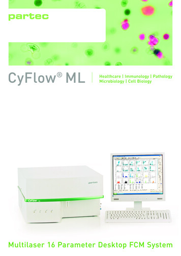
1OverviewFlow cytometry is a technology that simultaneously measures and then analyzesmultiple physical characteristics of single particles, usually cells, as they flow in a fluidstream through a beam of light. The properties measured include a particle’s relativesize, relative granularity or internal complexity, and relative fluorescence intensity.These characteristics are determined using an optical-to-electronic coupling systemthat records how the cell or particle scatters incident laser light and emits fluorescence.A flow cytometer is made up of three main systems: fluidics, optics, and electronics. The fluidics system transports particles in a stream to the laser beam forinterrogation. The optics system consists of lasers to illuminate the particles in the sample streamand optical filters to direct the resulting light signals to the appropriate detectors. The electronics system converts the detected light signals into electronic signalsthat can be processed by the computer. For some instruments equipped with asorting feature, the electronics system is also capable of initiating sorting decisionsto charge and deflect particles.In the flow cytometer, particles are carried to the laser intercept in a fluid stream. Anysuspended particle or cell from 0.2–150 micrometers in size is suitable for analysis.Cells from solid tissue must be desegregated before analysis. The portion of the fluidstream where particles are located is called the sample core. When particles passthrough the laser intercept, they scatter laser light. Any fluorescent molecules present1
Introduction to Flow Cytometry: A Learning Guideon the particle fluoresce. The scattered and fluorescent light is collected byappropriately positioned lenses. A combination of beam splitters and filters steers thescattered and fluorescent light to the appropriate detectors. The detectors produceelectronic signals proportional to the optical signals striking them.List mode data are collected on each particle or event. The characteristics orparameters of each event are based on its light scattering and fluorescent properties.The data are collected and stored in the computer. This data can be analyzed toprovide information about subpopulations within the sample (Figure1-1).sample corelaserdata displayselectronic pulsesFigure1-1 Scattered and emitted light signals converted to electronic pulses2
Chapter : OverviewReview Questions: Overview1 What properties of a cell or particle can be measured by a flow cytometer?2 What light source is used in most flow cytometers?3 What are the three main systems in a flow cytometer?4 What type of biological sample is best suited for flow cytometric analysis?5 What is the name given to the portion of the fluid stream where the cells arelocated?6 When cells labeled with fluorescent molecules pass through the focused laserbeam, what two types of light signals are generated?7 Light emitted from a particle is collected by8 The electronic signal produced by the detectors is proportional to:9 All flow cytometric measurements can be made simultaneously on a single cell.TF10 Particles must be in single-cell suspension before flow cytometric analysis.TF3
Introduction to Flow Cytometry: A Learning Guide4
2FluidicsThe purpose of the fluidics system is to transport particles in a fluid stream to the laserbeam for interrogation. For optimal illumination, the stream transporting theparticles should be positioned in the center of the laser beam. In addition, only onecell or particle should move through the laser beam at a given moment.To accomplish this, the sample is injected into a stream of sheath fluid within the flowchamber. The flow chamber in a benchtop cytometer is called a flow cell and the flowchamber in a stream-in-air cytometer is called a nozzle tip. The design of the flowchamber causes the sample core to be focused in the center of the sheath fluid wherethe laser beam will then interact with the particles.Based on principles relating to laminar flow, the sample core remains separate butcoaxial within the sheath fluid. The flow of sheath fluid accelerates the particles andrestricts them to the center of the sample core. This process is known ashydrodynamic focusing. For an illustration of hydrodynamic focusing in each type offlow cell, see Figure 2-1 and Figure 2-2.5
Introduction to Flow Cytometry: A Learning Guidelow samplepressure(12 µL/min)sheathfluidlaser beamsamplehigh samplepressure(60 µL/min)sheathfluidsheathfluidlaser beamsamplesheathfluidFigure 2-1 Hydrodynamic focusing of the sample core through a flow cellsamplesheathsheathsheathlasersheathlaserlow differential pressurehigh differential pressureFigure 2-2 Hydrodynamic focusing of the sample core through a nozzle tipThe sample pressure and the sheath fluid pressure are different from each other. Thesample pressure is always greater than the sheath fluid pressure. The sample pressureregulator controls the sample flow rate by changing the sample pressure relative to thesheath pressure. 6In BD benchtop cytometers, the sample stream is pressurized upward through anoptically clear region of the flow cell or cuvette; particles pass through the laserbeam while they are still within this flow cell (Figure 2-1). Most benchtopcytometers have fixed sample pressure settings (LO, MED, and HI). The
Chapter : Fluidicsbenchtop BD LSR cytometer also has a “fine” adjustment knob for intermediatesettings. In stream-in-air cytometers, the sample stream passes through a small orifice in anozzle tip before being intersected by the light beam in the open air (Figure 2-2).Sample pressure settings can be adjusted within a dynamic range.Increasing the sample pressure increases the flow rate by increasing the width of thesample core. This, in turn, allows more cells to enter the stream within a givenmoment. With a wid
Flow cytometry is a technology that simultaneously measures and then analyzes multiple physical characteristics of single particles, usually cells, as they flow in a fluid stream through a beam of light. The properties measured include a particle’s relative size, relative granularity or internal complexity, and relative fluorescence intensity. These characteristics are determined using an .
