Transcription
Published 2003 Wiley-Liss, Inc.†Cytometry Part A 54A:48 –55 (2003)Analysis of Violet-Excited Fluorochromes by FlowCytometry Using a Violet Laser DiodeWilliam G. Telford,1* Teresa S. Hawley,2 and Robert G. Hawley21Experimental Transplantation and Immunology Branch, Center for Cancer Research, National Cancer Institute,National Institutes of Health, Bethesda, Maryland2Holland Laboratory, American Red Cross, Rockville, MarylandReceived 9 October 2002; Revision Received 22 January 2003; Accepted 13 February 2003Background: Low power violet laser diodes (VLDs) havebeen evaluated as potential replacements for water-cooledargon-ion and krypton-ion ultraviolet and violet lasers forDNA content analysis using the Hoechst dyes and 4,6diamidino-2-phenylindole (Shapiro HMN, Perlmutter NG:Cytometry 44:133–136, 2001). In this study, we used aVLD to excite a variety of violet-excited fluorescent molecules important in biomedical analysis, including thefluorochromes Cascade Blue and Pacific Blue, the expressible fluorescent protein cyan fluorescent protein (CFP),and the fluorogenic alkaline phosphatase (AP) substrate2-(5 -chloro-2 -phosphoryloxyphenyl)-6-chloro-4-(3H)quinazoline (ELF-97; for endogenous AP detection and cellsurface labeling with AP-conjugated antibodies).Methods: Comparisons were made between VLD excitation and a krypton-ion laser emitting at 407 nm (both athigher power levels and with the beam attenuated atlevels approximating the VLD) on the same FACSVantageSE stream-in-air flow cytometer. We evaluated a PowerTechnology 408-nm VLD (30 mW) equipped with circularization optics (18 mW maximum output, set to 15 mW)Flow cytometry and its allied technologies are dependenton lasers as an excitation source for the ever expandinggroup of fluorogenic molecules available for biological andbiomedical analyses. Technologic development in flow cytometry has largely paralleled the development of laser technology. Water-cooled gas lasers have been a traditionalchoice for fluorochrome excitation in flow cytometry foralmost 30 years; they possess extremely low noise levels,stable power levels, and true TEM00 beam configurations.Their high power levels have allowed useful fluorochromedetection on the fluorescence-activated cell sorter, wherethe ability to physically sort cells has required a compromiseof poorer light-collecting optics. Their power levels also haveprovided practical emission levels for a larger group of laseremission lines that possess relatively weak emission energies.For some of these wavelengths (e.g., ultraviolet [UV], nearUV, and violet), they have traditionally represented the onlyand a Coherent I-302C krypton-ion laser emitting at powerlevels ranging from 15 to 75 mW.Results: Cascade Blue, Pacific Blue, and CFP showedcomparable signal-to-noise ratios and levels of sensitivitywith VLD excitation versus the krypton-ion laser at highand VLD-matched power outputs. Multicolor fluorescentprotein analysis with 488-nm excitation of green fluorescent protein and DsRed and VLD excitation of CFP wastherefore feasible and was demonstrated. Similar levels ofexcitation efficiency between krypton-ion and VLDsources also were observed for ELF-97 detection.Conclusions: These evaluations confirmed that VLDsmay be cost- and maintenance-effective replacements forwater-cooled gas lasers for applications requiring violetexcitation in addition to DNA binding dyes. CytometryPart A 54A:48 –55, 2003. Published 2003 Wiley-Liss, Inc.†Key terms: violet laser diode; Cascade Blue; Pacific Blue;cyan fluorescent protein; 2-(5 -chloro-2 ractical option for flow cytometric applications. The plethora of fluorescent probes now available to biomedical investigators for flow cytometry requires the availability of a widevariety of excitation wavelengths. In the UV and near-UVranges, for example, important probes such as the calciumchelator indo-1, the Hoechst and 4,6-diamidino-2-phenylindole (DAPI) DNA binding dyes, and the expressible fluorescent proteins cyan fluorescent protein (CFP) and blue fluorescent protein require UV or near-UV/violet excitation.†This article is a US government work and, as such, is in the publicdomain in the United States of America.*Correspondence to: William G. Telford, Ph.D., National Cancer Institute,Building 10, Room 12C121, 9000 Rockville Pike, Bethesda, MD 20892.E-mail: telfordw@mail.nih.govPublished online in Wiley InterScience (www.interscience.wiley.com).DOI: 10.1002/cyto.a.10046
VIOLET LASER DIODE EXCITATION FOR FLOW CYTOMETRYNevertheless, these lasers possess a number of disadvantages, including great expense in initial purchase andmaintenance, a requirement for large volumes of coolingwater, and considerable size and weight. Air-cooled gaslasers (including argon, helium-cadmium, and heliumneon) have become common on flow cytometers in recent years due to their lower cost, smaller size, and simpler electrical and cooling requirements. Nevertheless,they are currently limited in their useful laser wavelengths. Low power argon-ion lasers usually produce useful laser light only in their most powerful emission lines(488 and 514.5 nm), which do not include the UV orviolet range. Helium-neon lasers with higher power ratings have been developed almost exclusively for 633-nmred emission, with other wavelengths only built withlower power ratings and seeing limited use in flow cytometry. Helium-cadmium lasers emit in the UV (325 nm) andblue (442 nm) ranges but remain large and expensive;they also develop considerable levels of laser “noise” overtime (1).Solid-state lasers, in particular diodes, are becomingmore prevalent in fluorescence analysis instrumentation,including flow cytometers. These lasers are small andinexpensive, have low electrical and cooling requirements, and are increasingly reliable for long-term use (1).Red laser diodes now are used extensively in flow cytometers. Diode-pumped solid-state 532-nm green lasers havebeen incorporated into several small flow cytometers.Recently developed diode-pumped solid-state lasers emitting at 488 nm may one day replace argon-ion lasers inmany instruments. The gallium nitride/indium gallium nitride UV and violet laser diodes (VLDs) recently developedby Nakamura and colleagues (2) have opened up thepossibility of placing small, low-cost, and low–maintenance lasers in flow cytometers that cover the near-UV toviolet excitation spectrum, thereby allowing the use offluorochromes previously excitable only by more expensive gas lasers. Shapiro and colleagues recently incorporated a VLD into a flow cytometer and were able to carryout DNA content analysis with the UV-excited DNAprobes DAPI and Hoechst 34580 with precisions approaching those of gas lasers (3). In this study we evaluated the ability of VLDs to excite a variety of importantviolet-excited fluorescent probes important to biologicaland biomedical research, including the phenotyping fluorochromes Cascade Blue and Pacific Blue, the expressiblefluorescent protein CFP, and the fluorogenic alkaline phosphatase (AP) substrate 2-(5 -chloro-2 -phosphoryloxyphenyl)-6-chloro-4-(3H)-quinazoline (ELF-97). This study and recent commercial efforts to incorporate violet lasers into flowcytometric instrumentation suggest that these laser sourceswill be useful and cost-effective alternatives to traditionalwater-cooled gas lasers.MATERIALS AND METHODSViolet Laser DiodeThese evaluations and comparisons used a single transverse mode VLD with circularization optics and built-in49Peltier temperature control (Power Technology, Inc., Alexander, AR). Beam shape was circular to within 1.2milliradians and was approximately 2 mm in diameter.Maximum laser power was 30 mW, and maximum outputpost-circularization optics was 18 mW. Power level wasset at 15 mW for these experiments and confirmed with alaser power meter (Melles Griot, Carlsbad, CA). The VLDtemperature controller was set to maximize emission at408 nm.Krypton-Ion LaserThese evaluations also used a Coherent (Santa Clara,CA) I-302C krypton-ion water-cooled laser emitting at 407or 413 nm in single-line mode. Power levels were set from15 to 75 mW. Because internal laser power metering inhigher power gas lasers becomes inaccurate at very lowvoltage levels, an external meter (Melles Griot, Carlabad,CA) was used to monitor krypton laser power levels below 50 mW.Instrumentation and Analysis ConditionsAll experiments were performed on a FACSVantage SE(Becton-Dickinson [BD] Biosciences, San Diego, CA) withDiVa digital upgrade and a custom 11-color optical bench.Violet laser excitation was integrated into the cytometerin one of two ways: (i) VLD as the secondary laser on aFACSVantage SE with a Coherent I-90 argon-ion laser providing primary laser excitation and triggering or (ii) aCoherent I-302C krypton-ion laser as the secondary laseron the same FACSVantage SE with the same primary lasersource. The FACSVantage SE used a standard 50-mm focallength laser focusing lens and a 70- m stream-in-air nozzle. Cascade Blue was detected through a 440/10-nm narrow bandpass filter, and Pacific Blue was optimally detected through a 463/50-nm filter sandwiched with a488-nm notch filter; the notch filter was necessary toprevent spillover noise from the 488-nm primary lasersource, particularly at low laser power. Similarly, CFP wasdetected with a 485/22-nm filter also sandwiched with a488-nm notch filter, and green fluorescent protein (GFP)and DsRed were detected through 530/30- and 610/20-nmfilters respectively. ELF-97 alcohol was detected with a535/45-nm filter. Data was acquired with DiVa digitalacquisition software (BD Biosciences) and analyzed withWinMDI 2.8 (Dr. Joseph Trotter, Scripps Institute, and BDBiosciences). Software compensation was carried outwith WinList 4.0 (Verity Software House, Topsham, ME).All comparisons were made from the same samples on thesame day, with the same detector voltage settings, whenpossible (except where noted). In some cases, photomultiplier (PMT) voltage was increased by a fixed amount tobring the lower background autofluorescence generatedby low power krypton-ion or VLD excitation onto the logscale to reduce the error inherent in low-level fluorescence measurement and to allow more accurate calculation of signal-to-noise ratio; this is noted and illustrated inthe figures where appropriate. Median fluorescence intensity values for background and specific labeling and theirratios were used for approximate comparisons between
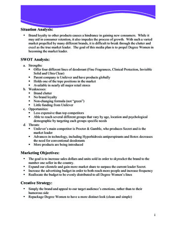
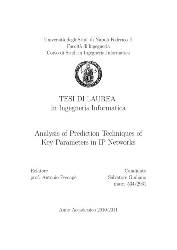
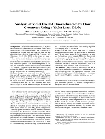
50TELFORD ET AL.excitation sources. Day-to-day variations in laser excitation and emission detection were quality controlled withviolet-excited single-intensity 2- m green-yellow fluorescent beads (Polysciences, Harrington, PA) and Inspeckmultiple-intensity 2.5- m bead cocktails (MolecularProbes, Eugene, OR). InSpeck beads were also used tomonitor PMT detection linearity.Cells and Cell LabelingAll cell lines were obtained from the American TypeCulture Collection (Manassas, VA). Immunophenotypingexperiments with Cascade Blue and Pacific Blue werecarried out with EL4 murine thymoma cells labeled withbiotin-conjugated anti-CD44 or anti-CD90 (Caltag Laboratories, Burlingame, CA) and streptavidin-conjugated Cascade Blue or Pacific Blue (Molecular Probes), followed byfixation in 1% paraformaldehyde and analysis within 24 h.Immunophenotyping with ELF-97 was also carried out inEL4 cells labeled with biotin plus anti-CD44 or anti-CD90and streptavidin-AP (Life Technologies, Gaithersburg,MD). Cells were subsequently washed in phosphate buffered saline and labeled with ELF-97 substrate (MolecularProbes) at 2 mM in phosphate buffered saline containing2% fetal bovine serum, followed by fixation in 1% paraformaldehyde and analysis. For CFP experiments, pECFP,pEGFP-1, and pDsRed1-1 plasmids encoding the ECFP,EGFP, and DsRed fluorescent proteins, respectively, wereobtained from Clontech Laboratories, Inc. (Palo Alto, CA).The MCIN and MGIN retroviral vectors containing theEGFP gene expressed from the retroviral long terminalrepeat from a murine stem cell virus on a bicistronictranscript, which also contained a downstream neomycinphosphotransferase (neo) gene linked via an internal ribosome entry site from the encephalomyocarditis virus, hasbeen described elsewhere (4). The MYIN, MCIN, andMRIN retroviral vectors containing the ECFP, EGFP, andDsRed genes, respectively, were similarly constructed byinserting the respective fluorescent protein genes into thesame murine stem cell virus– based neo-linked internalribosome entry site retroviral vector backbone (5,6). Stable helper-free retroviral producer lines were generatedby transduction of GP E-86 ecotropic packaging cellsfollowed by G418 selection, as described previously (7).RESULTSInstrument ConfigurationThe purpose of this study was to determine whethersome commonly used violet-excited fluorescent molecules could be detected on a flow cytometer equippedwith a VLD and to determine whether this excitation wassimilar to that observed with a more powerful krypton-ionviolet laser source. The prototype VLD used in thesestudies is shown in Figure 1a and was tuned to emit atapproximately 408 nm at 15-mW power output. The laserand instrument configurations used in the study were setup on the same FACSVantage SE equipped with the DiVadigital acquisition system. The VLD was set up in thesecondary position with an argon-ion laser emitting at 488FIG. 1. Violet laser diode (VLD). a: The Power Technology 408-nm,30-mW VLD used in these studies. b: Polyscience 2- m green-yellow (left)and InSpeck 2.5- m blue (right) fluorescent bead standards with Kr407-nm, 75-mW excitation. c, d: These bead standards with Kr 407-nm,15-mW and VLD 408-nm ,15-mW excitations, respectively, at the samephotomultiplier (PMT) voltage setting as that shown in panel b. InSpeckbead fluorescence was detected through a 440/10-nm filter, and Polyscience bead fluorescence was detected through a 535/45-nm filter.Median fluorescent intensity values are shown.nm in the primary position; for comparison, the kryptonion source emitting at 407 nm was set up in the sameposition. The krypton-ion laser was set to a standard
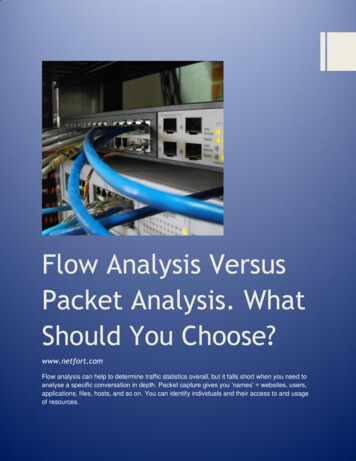
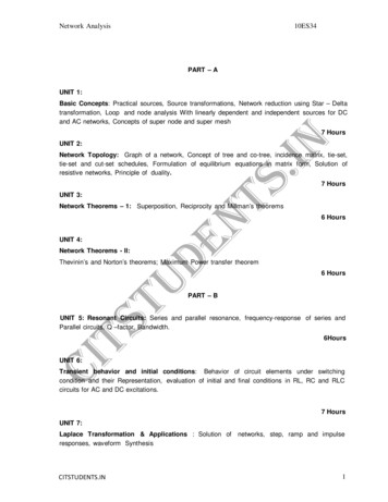
VIOLET LASER DIODE EXCITATION FOR FLOW CYTOMETRY5175-mW power level or at an attenuated 15-mW level tomore closely approximate the VLD power level, withexternal power level confirmation. Unlabeled and fluorochrome-labeled cells were analyzed for median fluorescence intensity, and the ratios were calculated betweenbackground and specific labeling. Although the large errors associated with background fluorescence measurements are a limitation on this method of comparison,calculation of the labeling ratio provides an approximatevalue for the signal-to-noise ratio for rough comparisonsbetween laser sources. In cases in which the low powerlasers produced a very low background value (in comparison to higher power sources), the PMT gain was increased by an indicated amount to decrease this error andprovide a more accurate signal-to-noise calculation.To initially determine the excitation efficiency of bothlaser sources, UV-excited Polyscience green-yellow 2- mbeads and InSpeck bead mixtures (Molecular Probes) containing 2.5- m beads at 1%, 3%, 10%, 30%, and 100%relative intensities were analyzed with all configurations(Fig. 1b– d). Krypton at 407-nm excitation at 75 mW easilydistinguished the 3% InSpeck bead population from unlabeled background, with some loss of sensitivity with reduction to 15 mW. The VLD showed a roughly 10-folddecrease in excitation efficiency and easily distinguishedonly the 10% relative population from background. Analysis of green-yellow beads showed a similar relative decrease in excitation efficiency. Nevertheless, these commercial bead preparations were not optimally excited inthe violet range. Analysis of violet-excited fluorochromesproduced much closer values between excitation sources(below).Cascade Blue and Pacific BlueCascade Blue and Pacific Blue are low-molecular-weightfluorochromes developed by Haugland and colleagues (8)and are optimally excited by violet laser lines (9). Theirexcitation and emission spectra are shown in Figures 2aand 3a. They have proven useful in polychromatic flowcytometry due to their relative brightness (approximatingfluorescein) and their red-shifted excitation maxima, thusavoiding much of the autofluorescence associated withUV-excited fluorochromes such as the coumarins (10 –12).Krypton-ion excitation at 407 or 413 nm is traditionallyused to excite these fluorochromes. This is shown for 3FIG. 2. Violet laser diode (VLD) excitation of Cascade Blue. a: Excitation and emission spectra for Cascade Blue. Excitation spectral traces aredotted, and emission traces are solid. All curves are normalized. b– e: Flowcytometric detection of Cascade Blue. Mouse EL4 cells were labeled withbiotin plus anti-CD44 (left column) or anti-CD90 (right column), followedby streptavidin conjugated to Cascade Blue, and analyzed with (b) the407-nm, 75-mW krypton-ion laser as a secondary source, (c) the 407-nm,15-mW krypton-ion laser as the secondary source, (d) the 408-nm, 15-mWVLD, and (e) the 408-nm, 15-mW VLD with photomultiplier (PMT) amplifier voltage increased by 100 V. All samples were analyzed on the sameinstrument, on the same day, and at the same PMT voltage level (with theexception of e), with a 440/10-nm filter. Specific labeling (solid areas) andbackground autofluorescence (open areas) peaks are shown. Medianfluorescence intensity values are shown for each peak at the top of eachhistogram, with the ratio in boldface.
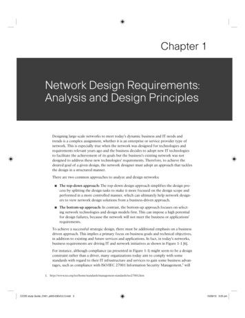
52TELFORD ET AL.Cascade Blue in Figure 2b and 2c, where Cascade BlueCD44- and CD90-labeled EL4 cells were excited with krypton-ion at 407 nm and at laser powers of 75 mW (Fig. 2b)and attenuated to 15 mW (Fig. 2c) at equivalent detectorvoltages. Although the higher laser power level resulted ina higher background signal (and higher raw fluorescencevalue for specific labeling), Cascade Blue showed similarsignal-to-noise ratios and sensitivities at high and lowpower krypton-ion laser excitations. Excitation of CascadeBlue with the VLD produced levels of detection comparable with those of the attenuated krypton-ion laser (Fig. 2dand 2e). Similar results were obtained with Pacific Blue(Fig. 3b– d).Cyan Fluorescent ProteinCFP is an Aequorea victoria– derived fluorescent protein that is important as a single expressible fluorescentmarker, a component of multicolor fluorescent proteinprotocols, and a donor fluorochrome for fluorescenceresonance energy transfer experiments (13,14). The excitation and emission spectra for CFP are shown in Figure4a. CFP is optimally excited by violet to blue, 420- to440-nm, light, a spectral area with little coverage by gasand solid-state lasers typically found on flow cytometers.Most investigators use the violet krypton-ion lines or theblue 457-nm line found on argon-ion lasers or, less commonly, a 442-nm helium-cadmium laser, all of which produce reasonable CFP excitation (14,15). Krypton-ion violet excitation of CFP in shown in Figure 4b and 4c, whereCFP-expressing 3T3 cells were excited at 407 nm at laserpowers of 75 mW (Fig. 4b) and attenuated to 15 mW (Fig.4c). CFP appeared to be excited equally well with higherand lower krypton-ion laser power levels. Once again, theVLD produced acceptable levels of detection sensitivityfor CFP when compared with high power and voltagematched krypton-ion excitations after accounting for reduced autofluorescence background (Fig. 3d and 3e).More importantly, VLD excitation of CFP was compatiblewith other fluorescent proteins in multicolor experiments(15–17). Figure 3e shows the analysis of a “cocktail” of3T3 cells expressing no fluorescent protein, GFP, DsRed,or CFP on a FACSVantage SE equipped with the VLD. TheVLD was able to adequately excite CFP and functioncompatibly with argon-ion 488-nm excitation of GFP andDsRed in a
cyan fluorescent protein; 2-(5 -chloro-2 -phosphoryloxy-phenyl)-6-chloro-4-(3H)-quinazoline Flow cytometry and its allied technologies are dependent on lasers as an excitation source for the ever expanding group of fluorogenic molecules available for biological an