Transcription
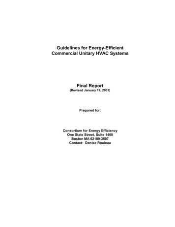
Open Access CaseReportDOI: 10.7759/cureus.8742Bilateral Congenital Nasolacrimal Duct Cyst: ARare Cause of Nasal ObstructionManish Gupta 1 , Habibulla Khan 2 , Monica Gupta 31. Otorhinolaryngology, Maharishi Markandeshwar Institute of Medical Sciences and Research, Ambala, IND 2.Otorhinolaryngology, Maharishi Markandeshwar Institute of Medical Science and Research, Ambala, IND 3. GeneralMedicine, Government Medical College and Hospital, Chandigarh, INDCorresponding author: Monica Gupta, monicamanish2001@gmail.comAbstractNasal obstruction in neonates often results in respiratory discomfort, as neonates are obligate nasalbreathers. Congenital bilateral nasal obstruction is an emergency situation which is generally secondary tochoanal atresia. Rarely bilateral nasolacrimal duct (NLD) cyst causing intranasal swelling may be theunderlying etiology. Neonatal respiratory distress warrants immediate measures to secure the airway andprompt investigations to reach a diagnosis for a definitive management. We describe a case of two-day-oldgirl with intermittent breathing difficulty because of bilateral NLD cysts causing nasal obstruction. Thepatient improved with conservative medical management.Categories: Emergency Medicine, Otolaryngology, PediatricsKeywords: nasolacrimal duct cyst, nasal obstruction, intra-nasal cyst, pediatric airway, dacryocystoceleIntroductionCongenital anomaly of nasolacrimal system is common, but dacryocystocele is an uncommon manifestationespecially when it presents as bilateral intranasal cyst. The dacryocystocele is formed when there isconcomitant proximal (valve of Rosenmuller) and distal (valve of Hasner) membranous obstruction of thenasolacrimal drainage system, thus causing lacrimal sac and/or duct distension [1,2]. The patient maypresent with bluish cystic swelling inferomedial to medial canthus externally or as intranasal inferior meatalmass [3].Usually, the garden variety of dacryocystocele presents with unilateral facial swelling located inferomedialto the medial canthus of the eye. In the case of rare congenital intranasal cysts, dacryocystocele may extendintranasally forming a nasal cyst located in the inferior meatus and present predominantly with respiratorydistress, especially if bilateral. Most patients have epiphora due to functional closure of Rosenmuller valvepresent at the superior end of the tear sac and failure to open Hasner valve at the distal end of thenasolacrimal duct (NLD) [4].Received 06/03/2020Case PresentationReview began 06/12/2020Review ended 06/16/2020Published 06/21/2020 Copyright 2020Gupta et al. This is an open access articledistributed under the terms of theCreative Commons Attribution LicenseCC-BY 4.0., which permits unrestricteduse, distribution, and reproduction in anyA two-day-old girl, born as full-term normal vaginal delivery, was referred to our department forintermittent episodes of difficult breathing as noticed by her mother. She also complained that the child hadnoisy respiration while sleeping and had excessive tearing. The symptoms neither progressed nor relievedon crying or on making the baby prone. There was no history of breath holding, difficulty while suckling, orbluish discoloration of her skin. There were no antepartum or intrapartum complications; the Apgar scorewas 10/10 and the birth weight was adequate.medium, provided the original author andsource are credited.On examination, the baby was lying comfortably beside her mother and her respiratory rate was withinnormal limits (40/min). On closer examination, there was an intermittently occurring audible rush of airthrough the nasal passage during inspiration, more noticeable with the child’s mouth closed. There was mildepiphora and concomitant sticky mucopurulent eye discharge bilaterally. There were no signs of respiratorydistress in the form of grunting, stertor, stridor, alar collapse, sternal or intercostal recession, and theoxygen saturation at room air was 97%. There were no episodes of apnea, cyanosis, or epistaxis. Importantly,there was no rhinorrhea or mucus impaction on nasal inspection. Additionally, there were no signs of anycraniofacial dysmorphism, ear or nasal malformations, or any comorbidities. Cardiac and other systemicexaminations were within normal limits.Examination of the nasal cavity with an otoscope revealed lateral wall bulge inferolateral to inferiorturbinate, almost touching nasal septum, bilaterally. A nasogastric tube could be negotiated through thenares, which ruled out choanal atresia. There was no facial swelling and eye examination byophthalmologist was reported as normal.Non-contrast computed tomogram (NCCT) of the nose and paranasal sinuses revealed hypodense cysticHow to cite this articleGupta M, Khan H, Gupta M (June 21, 2020) Bilateral Congenital Nasolacrimal Duct Cyst: A Rare Cause of Nasal Obstruction. Cureus 12(6):e8742. DOI 10.7759/cureus.8742
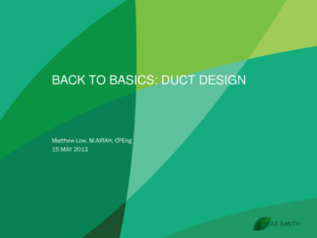
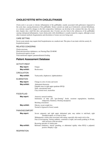
swellings in bilateral nasal cavities. The lesions were located inferior and lateral to inferior turbinates,pushing them medially in apposition with nasal septum (Figures 1, 2).FIGURE 1: Non-contrast computed tomogram coronal section showinghypodense cystic mass in bilateral nasal cavity (yellow arrows),inferolateral to inferior turbinate.2020 Gupta et al. Cureus 12(6): e8742. DOI 10.7759/cureus.87422 of 5
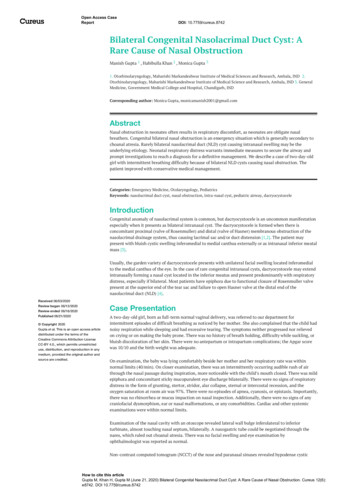
FIGURE 2: Non-contrast computed tomogram axial section showingintranasal cystic masses (black arrows) pushing inferior turbinates (redarrows) medially touching the nasal septum (yellow arrow) andcompletely obstructing bilateral nasal cavity.It was decided to give a conservative trial of warm compresses, nasal decongestants, antibiotic eye drops,and repeated lacrimal sac pressure and manual digital massage medial to medial canthus, towards theinferior direction. The patient responded well and was symptom free on the fifth day. The masses showedsignificant resolution and normal nasal cavity patency was noted on intranasal examination on the 10thday.Discussion2020 Gupta et al. Cureus 12(6): e8742. DOI 10.7759/cureus.87423 of 5
Newborns are obligate nasal breathers until they are at least two months old. Thus, any nasal obstructionmay have serious consequences and lead to respiratory distress. The most common cause of nasalobstruction is inflammatory edema; nevertheless, examination may reveal structural abnormalities,necessitating surgical intervention. The main surgical differential diagnosis is choanal atresia which is acongenital narrowing of the posterior nasal airway by a bony or membranous septum. In bilateral choanalatresia, respiratory distress in the neonate is relieved on crying, as it allows the baby to breathe throughmouth. In laryngomalacia, the stridor worsens with crying, feeding, and lying in the supine position,whereas it improves in the prone position. Other differentials to be considered for congenital nasalobstruction are nasal trauma, such as septal hematomas and septal subluxation, dacryocystocele,meningoencephalocele, dermoid cyst, Thornwald’s cyst, hemangioma, pyriform aperture stenosis, nasalhypoplasia, and glioma [2].Congenital defects in nasolacrimal drainage occur in 20% of newborns, but its severe form, i.e.dacryocystocele, is rarely observed. Its frequency is in two in 1,000 live births [5]. Approximately 90% ofdacryocystoceles are unilateral, thus making bilateral presentation further rare, as in our case [5]. Literaturesuggests a higher incidence of congenital lacrimal pathology in female babies due to a narrower NLDcompared to males [6]. A case report of monozygotic twins, having bilateral congenital dacryocystocele, hadsuggested some genetic basis for its familial predisposition and formation [7]. The exact genetic basis hasnot been elucidated yet due to the rarity of such an association.Imaging with CT and MRI techniques are helpful in distinguishing lesions and identifying intracranialcommunication. CT is quick and easy, and its diagnostic features include triad of lacrimal sac cysticdilatation, NLD dilatation, and intranasal cystic mass that appear homogeneous, well-defined, thin walledwith fluid attenuation [8]. CT is the imaging of choice although MRI has the advantage of characterizing thecontent of the lesion without radiation exposure.Asymptomatic dacryocystocele may be managed conservatively with lacrimal sac massage as most (96%)resolve spontaneously by one year of age [9]. Dacryocystitis, if evident by the presence of pain, fever,purulent discharge, and eye congestion, needs systemic antibiotics. Untreated dacrocystitis may complicateand lead to orbital and facial cellulitis [9]. If lacrimal sac distension is excessive, it may lead to narrowing ofthe lid fissure or corneal astigmatism resulting in anisometropic amblyopia and permanent canthalasymmetry [10].Surgical interventions, such as lacrimal probing by an ophthalmologist and intranasal endoscopic cystmarsupialization by an otorhinolaryngologist, may be required in patients with large external or intranasalcysts who develop apnea or stridor [10]. Mechanical probing through the valve of Rosenmuller and irrigationdecompresses the nasolacrimal drainage system proximally, while endoscopy identifies intranasal cyst andmarsupialization decompresses the system distally [11]. This combined surgical intervention helps inrestoring continuity of nasolacrimal system. In our case, there was no respiratory distress or features ofdacryocystitis as highlighted above; therefore, the child was managed conservatively. Kuboi et al. reportedone case of congenital bilateral dacryocystocele with respiratory difficulty which was managedconservatively with nasal continuous positive airway pressure [4].ConclusionsCongenital nasal anomalies as seen in our patient are uncommon yet potentially life-threatening reasons ofupper airway obstruction in newborn and can result in neonatal respiratory distress. Through this two-dayold newborn baby’s case, we wish to highlight the importance of the broad differential diagnosis andstructural abnormalities that dealing physicians should know so that appropriate referrals are done andtreatment can be promptly initiated. Timely CT allows for optimal bony definition, and it is the diagnosticmodality of choice. Management depends on the severity of distress, type, and location of the anomaly, themain goal being establishment of nasal patency. Fortunately, most infants often improve with conservativemedical management as seen in our case.Additional InformationDisclosuresHuman subjects: Consent was obtained by all participants in this study. Conflicts of interest: Incompliance with the ICMJE uniform disclosure form, all authors declare the following: Payment/servicesinfo: All authors have declared that no financial support was received from any organization for thesubmitted work. Financial relationships: All authors have declared that they have no financialrelationships at present or within the previous three years with any organizations that might have aninterest in the submitted work. Other relationships: All authors have declared that there are no otherrelationships or activities that could appear to have influenced the submitted work.References1.Sevel D: Development and congenital abnormalities of the nasolacrimal apparatus . J Pediatr OphthalmolStrabismus. 1981, 18:13-19.2020 Gupta et al. Cureus 12(6): e8742. DOI 10.7759/cureus.87424 of 5
2.3.4.5.6.7.8.9.10.11.Leonard DS, O'Keefe M, Rowley H, Hughes JP: Neonatal respiratory distress secondary to bilateral intranasaldacryocystocoeles. Int J Pediatr Otorhinolaryngol. 2008, 72:1873-1877. 10.1016/j.ijporl.2008.09.011Kim YH, Lee YJ, Song MJ, Han BH, Lee YH, Lee KS: Dacryocystocele on prenatal ultrasonography: diagnosisand postnatal outcomes. Ultrasonography. 2015, 34:51-57. 10.14366/usg.14037Kuboi T, Okazaki K, Kusaka T, Shimada A: Congenital dacryocystoceles controlled by nCPAP via nasal maskin a neonate. Pediatr Int. 2015, 57:475-477. 10.1111/ped.12516Macewen CJ, Young JDH: Epiphora during the first year of life . Eye. 1991, 5:596-600. 10.1038/eye.1991.103Shekunov J, Griepentrog GJ, Diehl NN, Mohney BG: Prevalence and clinical characteristics of congenitaldacryocystocele. JAAPOS. 2010, 14:417-420. 10.1016/j.jaapos.2010.07.006Barham HP, Wudel JM, Enzenauer RW, Chan KH: Congenital nasolacrimal duct cyst/dacryocystocele: anargument for a genetic basis. Allergy Rhinol. 2012, 3:46-49. 10.2500/ar.2012.3.0024Rand PK, Ball WS Jr, Kulwin DR: Congenital nasolacrimal mucoceles: CT evaluation . Radiology. 1989,173:691-694. 10.1148/radiology.173.3.2813773Shashy RG, Durairaj VD, Holmes JM, Hohberger GG, Thompson DM, Kasperbauer JL: Congenitaldacryocystocele associated with intranasal cysts: diagnosis and management. Laryngoscope. 2003, 113:3740. 10.1097/00005537-200301000-00007Yin T, van der Meer G: Neonatal airway obstruction in bilateral congenital dacryocystocoele: case report andreview of the literature. Int J Pediatr Otorhinolaryngol. 2017, 92:161-164. 10.1016/j.ijporl.2016.11.027Cunningham MJ: Endoscopic management of pediatric nasolacrimal anomalies . Otolaryngol Clin North Am.2006, 39:1059-1074. 10.1016/j.otc.2006.07.0042020 Gupta et al. Cureus 12(6): e8742. DOI 10.7759/cureus.87425 of 5
Usually, the garden variety of dacryocystocele presents with unilateral facial swelling located inferomedial to the medial canthus of the eye. In the case of rare congenital intranasal cysts, dacryocystocele may extend intranasally forming a nasal cyst located in the inferior meatus and present predominantly with respiratory