Transcription
Timm et al. Microbiome(2020) ARCHOpen AccessIsolation and characterization of diversemicrobial representatives from the humanskin microbiomeCollin M. Timm1, Kristin Loomis1, William Stone1, Thomas Mehoke1, Bryan Brensinger1, Matthew Pellicore1,Phillip P.A. Staniczenko2, Curtisha Charles3, Seema Nayak3 and David K. Karig1,4*AbstractBackground: The skin micro-environment varies across the body, but all sites are host to microorganisms that canimpact skin health. Some of these organisms are true commensals which colonize a unique niche on the skin,while open exposure of the skin to the environment also results in the transient presence of diverse microbes withunknown influences on skin health. Culture-based studies of skin microbiota suggest that skin microbes can affectskin properties, immune responses, pathogen growth, and wound healing.Results: In this work, we greatly expanded the diversity of available commensal organisms by collecting 800organisms from 3 body sites of 17 individuals. Our collection includes 30 bacterial genera and 14 fungal genera,with Staphylococcus and Micrococcus as the most prevalent isolates. We characterized a subset of skin isolates forthe utilization of carbon compounds found on the skin surface. We observed that members of the skin microbiotahave the capacity to metabolize amino acids, steroids, lipids, and sugars, as well as compounds originating frompersonal care products.Conclusions: This collection is a resource that will support skin microbiome research with the potential fordiscovery of novel small molecules, development of novel therapeutics, and insight into the metabolicactivities of the skin microbiota. We believe this unique resource will inform skin microbiome management tobenefit skin health.Keywords: Skin microbiome, Isolate collection, Carbon source utilizationBackgroundSkin is a constantly growing and changing barrier tissuethat acts as a first line of defense against many environmental factors. The skin micro-environment variesacross the body, but all sites are host to microorganismsthat can affect the maintenance and disruption of skinhealth. Some of these organisms are true commensalswhich colonize a unique niche on the skin, while open* Correspondence: dkarig@clemson.edu1Research and Exploratory Development Department, Johns HopkinsUniversity Applied Physics Laboratory, Laurel, MD, USA4Department of Bioengineering, Clemson University, Clemson, SC, USAFull list of author information is available at the end of the articleexposure to the environment results in transientcolonization by diverse microbes with an unknown contribution to skin function. Culture-based studies of skinmicrobiota suggest that skin microbes can affect skinproperties [1], immune responses [2, 3], pathogengrowth [4], wound healing [5], and even disease vectorattraction [6].The skin microbiome has been studied by both culturing and sequencing, and Actinobacteria, Firmicutes, Bacteriodetes, and Proteobacteria have been identified asmajor dominating phyla [7]. The proportion of organisms within these groups changes by body site, with The Author(s). 2020 Open Access This article is licensed under a Creative Commons Attribution 4.0 International License,which permits use, sharing, adaptation, distribution and reproduction in any medium or format, as long as you giveappropriate credit to the original author(s) and the source, provide a link to the Creative Commons licence, and indicate ifchanges were made. The images or other third party material in this article are included in the article's Creative Commonslicence, unless indicated otherwise in a credit line to the material. If material is not included in the article's Creative Commonslicence and your intended use is not permitted by statutory regulation or exceeds the permitted use, you will need to obtainpermission directly from the copyright holder. To view a copy of this licence, visit http://creativecommons.org/licenses/by/4.0/.The Creative Commons Public Domain Dedication waiver ) applies to thedata made available in this article, unless otherwise stated in a credit line to the data.
Timm et al. Microbiome(2020) 8:58sebaceous (oily) sites such as the forehead representedby Cutibacterium, Staphylococci, and Corynebacteria.Moist sites such as the antecubital fossa (inner elbow)are dominated by Proteobacteria and Staphylococci, anddry sites such as the forearm or legs being the most diverse but primarily dominated by Corynebacteria, Flavobacteriales, or β-Proteobacteria [8]. A number of factorslikely influence the microbiome composition at differentbody sites. These factors include moisture content,temperature, pH, and the resources available for microorganism metabolism at each site. Sequencing studieshave associated bacterial genes in skin swabs with metabolic pathways for compounds present on the skin suchas sugars, lipids, and iron [9]. Some bacteria have beenfound to co-localize at body locations with specific skinresidual compounds. For example, Cutibacterium isenriched at sites with compounds such as oleic acid andpalmitic acid [10]. Similarly, the use of personal careproducts that introduce additional compounds to theskin surface has been found to alter the microbiomecomposition [11].For decades, culture-based studies were conducted tocharacterize the bacterial flora of the skin, probing differences among body sites, effects of hygiene and behavior, and roles in disease [12]. While revealing keyinsights, these studies were limited by the ability to culture diverse microbes from the skin. DNA sequencinghas since driven far more detailed characterizations. Forexample, in chronic wound studies, molecular testingwas compared to culture-based methods, and resultsshowed that molecular techniques identified 85% of cultured representatives, while culturing only identified 16%of genera detected by sequencing [13]. Nonetheless,while offering powerful sensitivity, sequencing information is also associated with various forms of error andbias. Reagent contamination and imperfect samplingconditions can generate false positives, particularly dueto the low input nature of skin swabs [14, 15]. Inaddition to these error sources, bias can be introducedduring DNA extraction, as efficient disruption of all taxain a diverse sample is challenging. For amplicon sequencing, bias can also result from primer mismatches anddeviations from balanced GC-content [16].Perhaps more importantly, insights gained fromsequencing-based studies are based on correlations,often without definitive identification of the causative relationships between taxa/genes and function. Thus, regardless of the new wealth of information on the spatialecology of the skin microbiome [8], its temporal stability[17], its variation across individuals [18], and its response to disruption [19, 20], culture-based bottom-upstudies will remain important for revealing mechanisticexplanations of results stemming from sequence-based topdown measurements [21]. These mechanistic explanationsPage 2 of 12will be essential for ultimately tapping into the therapeuticpotential of microbiome manipulation.Indeed, cultured isolates have offered tremendousvalue to the study of multiple microbiome systems [12].In the gut microbiome, cultured representatives havecontributed to our understanding of vitamin productionby probiotic organisms [22], microbiome structure including adhesion [23], the chemical modification andmetabolism of drugs [24], and the tracking of microbes[25]. In plant environments, bacterial strains were usedto show synergistic effects of microbiome members incomplex communities [26]. On the skin, cultured microorganisms have been shown to activate TLR2 and stimulate the immune response, leading to increasedresistance to skin pathogens [3]. Cultured isolates fromskin have also been used to study VOC profile modification, which could lead to strategies of skin microbiomemanagement that ultimately affect vector attraction [27].More conclusively, culture-based studies have shownthat S. epidermidis, when cultured and re-applied to itsoriginal human host, can inhibit the growth of S. aureusin a skin microbiome [4].Recognizing the need for a diverse and controlled setof commensal isolates to study the skin microbiome, wecollected samples from 17 diverse, healthy research participants. Organisms were collected from a dry site, theforearm (AM), a moist site, the antecubital fossa (AF),and a sebaceous site, the forehead (FM), from each individual by swabbing and aerobic isolation on blood agar.For four of the 17 participants, additional growth conditions were used, including anaerobic cultures, potatodextrose agar for fungi [28], low nutrient agar to captureslow-growing organisms [29], and spore treatments. Theisolates described here have been cataloged and storedfor future research, and include accompanying surveydata on participant history and skin functional data fromisolation sites. To showcase the utility of this collectionand gain insight into the functional capabilities of skinmicrobiome constituents, we characterized a subset ofisolates for their ability to metabolize skin surface compounds. While we found some phylogenetic patterns inmetabolic abilities, we also observed significant variations across taxa within the same genus, thus drawingattention to the need for analyzing functional activity atthe species or strain strain-level. This characterizationeffort exemplifies one of many possible applications ofthe isolates collection to gain information and insightsinto the forces shaping skin microbial communities.ResultsIsolation conditions and countsWe isolated microbial strains from the forehead, forearm, and antecubital fossa (inner elbow) to represent sebaceous, dry, and moist sites from 17 healthy volunteers.
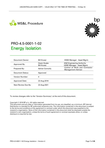
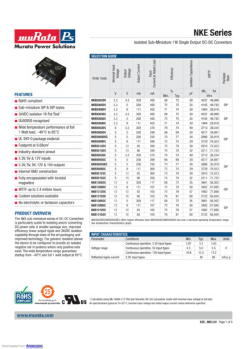
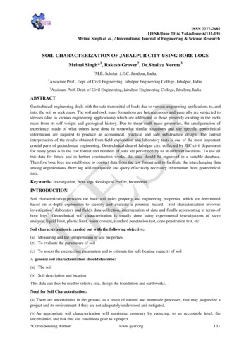
Timm et al. Microbiome(2020) 8:58Volunteers ranged in age from 22 to 45 with 5 malesand 12 females. Metadata collected includes ethnicity,country of birth, travel patterns, diet, and site-specificmeasures including transepidermal water loss and sebumcontent (Fig. 1a). Using 12 unique growth conditions, wecollected 842 strains. Colonies were picked based onphenotypic diversity (representative plate shown in Fig.1b) with 310 strains from the forehead, 250 from theforearm, and 282 from the antecubital fossa (Fig. 1c).From 4 of the 17 participants, we isolated strains from atotal of 12 unique growth conditions. After our baselineisolation (blood agar, room temperature), blood agar incubated at 37 C provided the most isolates (100), withR2A media and anaerobic culture conditions providing68 and 52 isolates, respectively (Fig. 1d). This strategyyielded between 18 and 139 distinct isolates betweenparticipants (Fig. 1e).16S identification of isolatesTrimmed ribosomal sequences were used to generatea phylogenetic tree. Figure 2 shows the result of thistree including 83 reference strains identified as thebest BLAST hit to 100% identical sequence clustersPage 3 of 12(Additional file 1 Table S4). The collection is dominated by Firmicutes, with the majority of isolates inthe Staphylococcus genus (n 398) and high representation of the Bacilli (n 66). The pop-out tables showthe two largest groups, both of which had perfect 16Smatch to S. epidermidis ATCC 12228 or RP62A. Theseresults from the Staphylococcus groups show that allresearch participants had at least one Staphylococcusrepresentative, and that many of the strains had identical 16S sequences between research participants andsites. Our collection also has a high representation ofActinobacteria, dominated by the Micrococcus genus(n 104). Interestingly, we observe 47 Micrococcusisolates from the antecubital fossa, 41 from the forearm, and only 13 from the forehead. Also within theActinobacteria, we have a large representation of theCutibacterium genus (n 33), all isolated using anaerobic isolation culture conditions, with the majority of theseisolated from the forehead (22 foreheads, 6 antecubitalfossa, 5 forearms). These organisms are most closely related to Cutibacterium acnes, recently renamed from thePropionibacterium acnes group [30]. The Proteobacteriaphylum includes diverse representatives that are rarer inFig. 1 Summary of isolation sites and conditions. a Research participants metadata includes age, gender, trans-epidermal water loss, and sebumcontent. b Example blood agar plate showing colony morphologies. c Total number of isolates by site. d Isolate counts from growth conditions,not including the standard condition of room temperature (RT), blood agar. e Total number of isolates by research participant. Black bars signifyresearch participants for which multiple isolation conditions were performed. See Table S1 for additional metadata
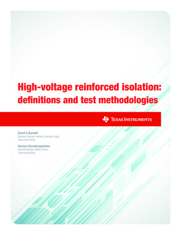
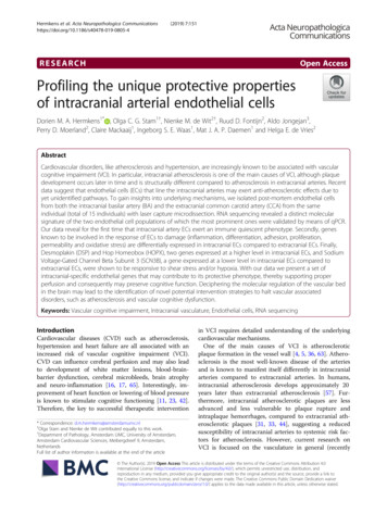
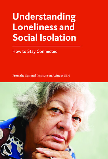
Timm et al. Microbiome(2020) 8:58Page 4 of 12Fig. 2 Phylogenetic tree of bacterial isolates. 16S tree for bacterial isolates. Sequences with 99% similarity are collapsed into a single node.Concentric columns indicate number of 99% similar 16S sequences by color-coded site. Pop-out tables indicate number of isolates by researchparticipants/site that map to common S. epidermidis strainsour collection. The Acinetobacter genus has 7 isolates from3 research participants, while the Roseomonas genus has 13isolates from 4 research participants with 8 of the strainsisolated from the forearm. Of the 83 groups collapsed bybest BLAST hit (Additional file 1 Table S4), 43 containedonly a single isolate. Of the remaining 40 groups made upof multiple isolates, only 2 groups contained all isolatesfrom the same research participant site: a group mostclosely related to Paenibacillus qingshengii from the forehead of research participants 066 and a group most closelyplaced in the Variovorax genus also from research participant 066. This result suggests that the remaining 38 groupsinclude isolates from multiple participants/sites that wouldbe interesting for strain variability studies.Collection diversityThe number of unique genera identified by culturing rangedfrom 1 to 12 for any given participant/site combination, witha total of 40 genera collected (Additional file 1 Table S3).While the research participants with the highest number ofunique genera included multiple isolation conditions, the research participants with the second and third most generahad isolates collected only from blood agar, not the additional growth conditions described above. First, our indepth sampling of four research participants (using diversemedia/growth conditions) resulted in increased diversityfrom those research participants ( 16 vs. 9 genera, p 0.01,Student’s t test). Interestingly, we observed a trend betweena number of isolates from sites within a single research participant, suggesting that diversity across the cultivable fraction of the microbiome is conserved (Fig. 3). Notable groupsinclude isolates from the Micrococcus genus which washighly represented in the isolate collection but has beenobserved at low abundance in published amplicon data forthe skin microbiome. Similarly, our collection includes 12Paenibacillus, 8 Roseomonas, and 7 Dietzia isolates distributed across multiple individuals, suggesting a role of theseorganisms in some skin communities (Additional file 1
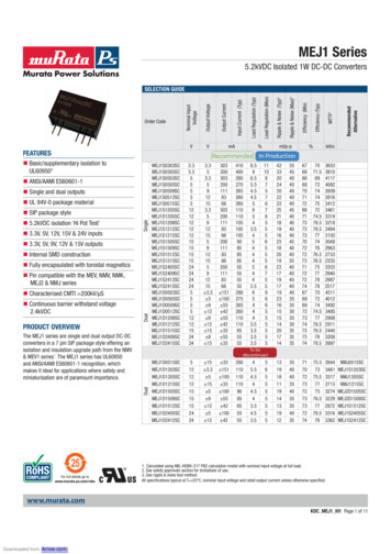
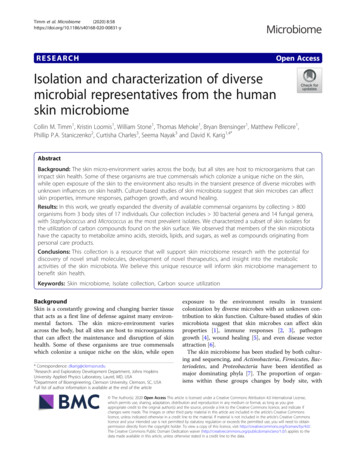
Timm et al. Microbiome(2020) 8:58Page 5 of 12Fig. 3 Bacterial diversity between subjects/sites. Correlation between number of genera by site (raw data shown in a for b antecubital fossa (AF)vs. forearm (AM), c antecubital fossa (AF) vs forehead (FM), and d forearm (AM) vs. forehead (FM). p values were calculated from Pearsoncorrelation with 17 pairs. Outlined bars and points indicate subjects with multiple isolation conditionsTable S7). Additionally, we investigated the resulting diversity from our chosen isolation conditions and observed thatafter room temperature blood agar conditions, anaerobicblood agar, R2A, and tape strips yielded the greatest additionally diversity of isolates (Table S8).Fungal isolatesIn addition to bacteria cultures which were the primary target of this study, 24 fungal isolates were collected fromthese samples. Fungal isolates were identified as moldgrowth on plates, or by microscopic examination. Fungalisolates were identified by ITS sequencing and representtaxa that are commonly found on the skin or indoor environments. For example, isolate FM063 009 closely alignedwith the genus Naganishia which has representatives foundon human skin [31] or in indoor environments [32]. A treewas built based on the ITS regions and shows the distribution of these organisms (Fig. 4, Table S5). Selected imagesof fungal plates show growth and yeast morphology.Carbon source utilization for selected strainsA subset of microbiota isolates as well as several publiclyavailable reference strains were assayed for the ability toreduce a tetrazolium-based dye, indicating metabolic
Timm et al. Microbiome(2020) 8:58Page 6 of 12Fig. 4 Fungal isolates. a ITS tree. Blue branches are Ascomycota and orange branches are Basidiomycota. b Isolate FM062 001 identified asCladosporium caldospoiroides. c Isolate AF056 018 identified as Penicillium dipodomyicola. d Isolate AF064 005 identified as Epicoccum nigrum. eIsolate AF069 007 identified as Naganishia liquefaciens. f Isolate AM063 501 identified as Naganishia liquefaciens. g Isolate AF054 013 identified asRhodotorula mucilaginosaactivity, in the presence of skin compounds as the sole carbon sources. The assay detected utilization for a variety ofcompound types, including amino acids, steroids, lipids,and molecules sourced from personal care products, suchas octocrylene, a common ingredient in sunscreens (Fig.5a). The assay also indicates clear differential metaboliccapabilities across bacteria that are phylogenetically similar.For example, the two Micrococcus isolates did not sharethe capacity to utilize any of the same carbon sources.Overall, bacteria were found to utilize a range of numbersof carbon sources present on the skin (Fig. 5b). We usedthis approach to examine the relationship between phylogeny and carbon source utilization in the skin environment. Phylogenetic distance analysis was performed on 24microbial taxa and 58 carbon sources. Mantel tests and linear regression models were run on all taxa together, as wellas a subset of taxa classed as generalists that utilized fouror more carbon sources. The Mantel test for all taxa wasnot significant (R2 0.000; p 0.357). The linear regression model including all taxon pairs was also not significant (R2 0.000; p 0.763). However, results forgeneralists only, defined as taxa using four or more carbonsources, were all significant (Fig. 5c): Mantel test (R2 0.349; p 0.009); linear regression model including alltaxon pairs (R2 0.122; p 0.002). Results were qualitatively similar when generalists were defined as utilizing fiveor more carbon sources.DiscussionIn this study, we isolated and characterized over 800 organisms from 3 body sites on 17 healthy research participants. We believe that the controlled isolation sites,
Timm et al. Microbiome(2020) 8:58Page 7 of 12Fig. 5 Skin resource utilization by skin microbiota isolates. a Resource utilization screen. Blue cells indicate a significant color change by thereduction-reporting dye; white cells indicate no significant color change not observed. b Scatterplot of the number of resources found to beutilized by each organism. c Carbon source utilization for generalists across skin isolates is positively related to phylogenetic similarity (four ormore carbon sources R2 0.169; p 0.003)paired with in-depth participant metadata, make this collection uniquely useful for the characterization of hostmicrobe, microbe-microbe, and microbe-environment interactions in the skin microbiome.Our study primarily focused on bacterial isolates.These organisms represent 25 families and 38 genera.Additionally, we collected 24 fungal isolates representing14 genera. Many of these organisms have been describedin previous studies, but some unique taxa were identified. An advantage of this study is the inclusion of distinct isolation conditions, shown in Table 1 with resultssummarized in Fig. 1 and Additional file 1 Table S8.With the caveat that these conditions were not appliedto all 17 subjects (see Table 1), and in some cases only 1subject, we investigated the ability of these methods toincrease cultural diversity. We observed that roomtemperature blood agar, anaerobic culturing on bloodagar, R2A media, and plating results of repeated tapestripping resulted in a diverse collection of organisms.This diversity is likely due to culturing of the anaerobicfraction, accommodation of slow growers using lownutrient environment (R2A), and access to potentiallydistinct fractions of the skin microbiome by successivetape stripping. Future studies should carefully considerculturing conditions for the isolation of representativepopulations. In addition, an interesting consideration forskin microbiome studies is the role of transient strains.While some of our isolates may be more commonly considered soil or environmental organisms, they nonetheless can be found on the skin of healthy individuals. Ifand how these organisms contribute to the function ofthe skin microbiome is unknown and is a meaningfularea of investigation for future research.We screened a subset of representative, aerobic, isolates for the ability to utilize carbon sources present onthe skin surface using tetrazolium dye assays with
Timm et al. Microbiome(2020) 8:58Page 8 of 12Table 1 Culture conditions for isolating organismsTargetMediaTemperatureSpecial conditionsLeading numberin isolate ID 1Researchparticipant(s)AerobesBlood agar (Hardy Diagnostics)Room temp.Aerobic0AllAerobesBlood agar (Hardy Diagnostics)37 CAerobic160, 63, 66, 70AnaerobesBlood agar (Hardy Diagnostics)37 CAnaerobic3260, 63, 66, 70Slow growersR2A (BD Biosciences)Room temp.Aerobic463, 66, 70LactobacilliMRS (Difco)Room temp.Aerobic663, 66, 70FungiPDA (BD Biosciences)Room temp.Aerobic563, 66, 70SporesBlood agar (Hardy Diagnostics)Room temp.4 h incubation in 70% EtOH, aerobic763, 66, 703Anaerobic sporesBlood agar (Hardy Diagnostics)37 C4 h incubation in 70% EtOH, anaerobic866, 70CapnophilesBlood agar (Hardy Diagnostics)37 CAerobic 5% CO2360Gram-negativeMacConkeyRoom temp.Aerobic, dark670HalophilesMedium for halophilic bacteria2Room temp.10% NaCl, aerobic370Anaerobic halophilesMedium for halophilic bacteria237 C10% NaCl, anaerobic3970Deep skin aerobesBlood agar (Hardy Diagnostics)37 CTape strips #5 and #10, aerobicTS5, TS1070TS5a, TS10a70Deep skin anaerobesBlood agar (Hardy Diagnostics)37 C3Tape strips #5 and #10, anaerobicFor example, FM060 X01 represents the first isolate from the forehead site (FM) from research participants # 60, where X is the leading number2DSMZ 73 media, 10 g/L casamino acids (Sigma), 10 g/L yeast extract (Sigma), 100 g/L NaCl, pH7.0 [37]3Anaerobic conditions were obtained using the BD GasPak EZ system (Fisher Scientific)1custom carbon sources. The 25 isolates characterized bythis assay included Firmicutes, Actinobacteria, and Proteobacteria representatives, and metabolism generallyclustered with phylogenetic relationships of strains.Lacking from the group are Cutibacterium representatives (formerly Propionibacterim), which play an important role in metabolite processing on the skin [11]. Thestrains tested utilized a total of 45 out of the 68 carboncompounds examined, which included steroids, fattyacids, sugars, amino acids, and other classes of chemicals. These results indicate that isolated organisms havethe ability to metabolize a variety of compounds foundin the skin environment. A range of specialists and generalists was observed, and the diversity of utilization patterns may play a role in the observed coexistence ofdiverse taxa in the skin environment. We found differentpatterns in resource utilization even across bacteria ofthe same genera, underscoring the need to consider species and strain level differences in microorganism function. Despite the differences observed at the species andstrain levels, phylogenetic distance analysis showed thatthere was a significant and positive relationship betweenthe phylogenetic similarity of microbial taxa and the carbon sources they utilized. This finding was strongestamong generalist taxa (those that utilized four or morecarbon sources), with more phylogenetically similar taxautilizing increasingly overlapping sets of carbon sources.Collectively, these results suggest that there is somedegree of validity to inferring community propertiesfrom phylogenetic characterization [33], yet strain levelinformation can be critical to obtaining an accuratelydetailed profile. In addition, recent work by Bouslimaniet al. [10] has revealed that skin hygiene products canremain on the skin long after use is discontinued, andthese products can influence microbiome composition[11]. Our approach may be used to develop a more detailed understanding of how and why the microbiomeshifts in response to the use of certain hygiene and cosmetic products. Towards this direction and otherin vitro characterization efforts, the size and diversity ofthe presented isolates collection make it a powerful resource for skin microbiome research.In gut microbiome research, a synergistic interplay between culturing, sequencing, and informatics approachesis exemplified by the identification of a “most wanted”list of microbes [34], followed by targeted culturing efforts which concluded that most gut microbes are likelyculturable [35]. Along similar lines, we envision a growing convergence of approaches that assimilates sequencing and informatics, microbial trait characterization[36], and in vitro studies with cultured isolates, whichwill deepen our understanding of the skin microbiomecomposition, its role in health, and ultimately methodsfor optimizing it.ConclusionUntil now, the majority of available commensal skin microorganisms were from the Human Microbiome Project(HMP) which provided valuable insight into humanmicrobiome interactions. With this publicly available collection (BEI Resources, with additional strains available bycorrespondence), we increased the number of skin
Timm et al. Microbiome(2020) 8:58commensal genera available (Additional file 2 FigureS1). Additionally, we added significant diversity to theStaphylococcus and Micrococcus genera. Our collection also includes 12 Paenibacillus, 8 Roseomonas,and 7 Dietzia isolates that were previously not cataloged by the HMP. The increase in diversity in ourcollection relative to other reports is likely due to increased throughput. Specifically, the most abundantstrains in our collection (Staphylococcus, Micrococcus,Bacillus, C. acnes) are representative of ampliconstudies and other culture collections from the skinmicrobiome, while the rarer genera in our collectionhave been described in distinct cases. We believe thiscollection is a resource that will greatly increase ourunderstanding of the skin microbiome through furthergenomic characterization and analysis, in vitro experiments, and synthetic biology endeavors.MethodsHuman research participantsHuman research participants were sampled at JohnsHopkins Bayview Medical Center according to a protocol approved by the Johns Hopkins and the U.S. ArmyHuman Research Protection Office Institutional ReviewBoards. The purpose of this study was to generate microbial ecology data for the skin microbiome. Participants were healthy volunteers, aged 18–50, with nohistory of chronic skin conditions or autoimmune diseases. Research participants were asked to not shower/bathe 2 days prior to sampling and answered a 400point questionnaire (Additional file 1 Table S1) and thentwo swabs from each site were collected. To quantifyskin properties, commercially available probes were usedadjacent to swab sites in conjunction with the Multi ProbeAdapter 10 system (Courage and Khazaka, GmbH) according to manufacturer instructions. Specifically, a Sebumeterwas used to quantify sebum content and a TewameterTM300 was used to quantify transepidermal water loss.Metadata for each volunteer is included in Additional file 1Table S1 and summarized briefly in Fig. 1. Isolates datawere not compared to subject metadata. There was no clinical intervention in this study for which to group volunteersand perform additional analyses.Sample plating, growth conditions, and isolationsHealthy human research participants were sampled fromthe forehead, forearm, or antecubital fossa (inner elbow)by swabbing with cotton swabs saturated in 50 mM Tris(Amresco), 1 mM EDTA (Amresco), 0.5% Tween 20(Sigma) in nuclease-free water for 30 s. For all participants, swabs were plated immediately on blood agar(Hardy Diagnostics) and incubated until colonies wereobserved. For four selected individuals, a second adjacentswab from the same site was added to 4 ml sterile TSBPage 9 of 12and mixed to resuspend bacterial samples for further plating, as described in Table 1.From each plate, all phenotypically distinct colonieswere picked onto fresh media for isolation. Single colonies were picked and re-streaked at least three times toisolate individual strains. Strains were grown in TSB,DSMZ 73 or on plates under corresponding aerobic/anaerobic and temperature conditions and frozen at – 80 C in 25% glycerol. A subset of representative isolates(Additional file 1 Table S2) has been deposited at BEIresources (https://www.beiresources.org) for curationand distribution to the scientific community.Isolate identification and 16S gene analysisAliquots of glycerol stocks were processed at GenewizLLC (South Plainfield, NJ, USA) by colony PCR of thefull 16S rRNA gene and subsequent Sanger sequencing.Resulting forward and reverse reads were merged usingthe EMBOSS merger [38], and then merged reads werealigned using the as
gardless of the new wealth of information on the spatial ecology of the skin microbiome [8], its temporal stability . will be essential for ultimately tapping into the therapeutic potential of microbiome manipulation. Indeed, cultured isolates have offered tremendous value to the study of multiple microbiome systems [12].