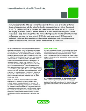
Transcription
ANTICANCER RESEARCH 38: 3273-3282 (2018)doi:10.21873/anticanres.12592An Agonistic Antibody to EPHA2 Εxhibits AntitumorEffects on Human Melanoma CellsATSUSHI SAKAMOTO1, KAZUNORI KATO2,3, TOSHIO HASEGAWA1 and SHIGAKU IKEDA1,21Departmentof Dermatology and Allergology, Juntendo University Graduate School of Medicine, Tokyo, Japan;Research Center, Juntendo University Graduate School of Medicine, Tokyo, Japan;3Department of Biomedical Engineering, Toyo University, Saitama, Japan2AtopyAbstract. Background/Aim: EPH receptor A2 (EPHA2) ishighly expressed in aggressive types of human cancer, and isexpected to be an excellent target molecule for antibodytreatments. In this study, we investigated the therapeuticpotential of antibody to EPHA2 against melanoma in vitro.Materials and Methods: We generated three monoclonalantibodies (mAbs) to EPHA2 and examined cell-surfaceexpression by flow cytometry. To investigate the ability to inhibittumor cell migration therapy with mAbs to EPHA2, weperformed a wound scratch assay and invasion assay. Weinvestigated the therapeutic effects of immunotoxins consistingof toxin-conjugated EPHA2 mAbs. Results: All humanmelanoma cell lines studied expressed EPHA2. Like naturalligand ephrin-A1, one of EPHA2 mAbs, SHM16, inhibitedmetastatic behavior of cells, such as migration and invasion. Inaddition, drastic growth inhibition and cytotoxicity were foundusing immunotoxin-conjugated SHM16. Conclusion: Theseobservations indicate a promising role for EPHA2 as a targetin antibody treatments for melanoma, and demonstrate thepotential therapeutic effects of an agonistic antibody to EPHA2.Melanoma is the most aggressive type of skin cancer. Whilemelanoma accounts for fewer than 5% of all skin cancercases, it is responsible for the large majority of deaths dueto skin cancer. The type of treatment will depend not onlyon stage and location of the melanoma, but also on thepatient’s overall health. When melanoma has not growndeeper than the epidermis, it is usually treated by surgeryThis article is freely accessible online.Correspondence to: Kazunori Kato, Ph.D., Department ofBiomedical Engineering, Toyo University, 2100 Kujirai, Kawagoe,Saitama 350-8585, Japan. Tel: 81 492391397, Fax: 81492315026, e-mail: kzkatou@juntendo.ac.jpKey Words: EPHA2, melanoma, monoclonal antibody, immunotoxin,invasion.(wide excision). When this is not possible, therapeuticoptions including injections of Bacille Calmette-Guerin(BCG) vaccine, interferon, or interleukin-2 (IL2) directlyinto the melanoma, radiation therapy, and chemotherapy (1,2). The treatment of widespread melanoma has changed inrecent years as newer forms of immunotherapy andmolecularly-targeted drugs have been shown to be moreeffective than chemotherapy (3-6).Receptor tyrosine kinases (RTKs) transmit signals thatregulate cell proliferation and differentiation, promote cellmigration and survival, and modulate cell metabolism. Theyplay critical roles in a wide range of biological processes,including embryonic development, growth of an organism,angiogenesis, synaptic plasticity, and oncogenesis (7-9). EPHreceptors, the largest subfamily of RTKs, are involved in manybiological processes including angiogenesis, tissue-borderformation, cell migration, and axon guidance (10, 11). EPHreceptors and their ephrin ligands are important mediators ofcell-to-cell communication that regulate cell attachment to theextracellular matrix, cell shape and cell motility. They havebeen studied extensively for their roles in the developmentalprocess. In recent years, EPH receptors and ephrins have beenfound to be integral players in cancer formation andprogression. EPH receptor A2 (EPHA2) and ephrin-A1 areespecially known to be involved in the development andmaintenance of many types of solid tumors (12-14).EPHA2 was originally named epithelial cell kinase, due toits widespread expression in epithelial cells. Subsequentstudies revealed that EPHA2 was overexpressed in humancancer, including melanoma, and that its overexpression wascorrelated with malignancy and poor prognosis. A largenumber of studies have demonstrated that EPHA2overexpression and activation promote tumorigenesis,suggesting a potential role as an oncogene (15). EPHA2activation by its ligand, ephrin-A1, regulates cell behavior ina manner more consistent with it being a tumor suppressor.Specifically, EPHA2 activation leads to induction of apoptosis,inhibition of cell proliferation, and suppression of cellmigration (16, 17). Accordingly, EPHA2 has been found to be3273
ANTICANCER RESEARCH 38: 3273-3282 (2018)an integral player in cancer formation, and is expected to bea good target for antibody and small-agent therapy (18-20).Recently, we established several monoclonal antibodies(mAbs) specific for human EPHA2. One of these mAbs,called SHM16, exhibited a unique function as an agonisticantibody to EPHA2. The first aim of the study was todetermine if SHM16 could mimic the effects of the naturalligand ephrin-A1 in regulating melanoma cell migration andinvasion. The second aim was to assess whether SHM16mediated targeting of toxin conjugates (immunotoxins) toEPHA2 would have antitumor effects against melanoma cells.Materials and MethodsCell lines. The melanoma cell lines A375, A2058, G361 werepurchased from JCRB Cell Bank (Osaka, Japan), and SK-MEL28 waspurchased from the American Type Culture Collection (Manassas,VA, USA). All cell lines, except fibroblasts and melanocytes(purchased from Lifeline Cell Technology, Frederick, MD, USA),were maintained in RPMI-1640 medium (Sigma-Aldrich, St. Louis,MO, USA) supplemented with 10% fetal bovine serum (FBS; Gibcoby Life Technologies, Grand Island, NY, USA), 100 units/mlpenicillin G (Gibco), 100 μg/ml streptomycin (Gibco), and 25 mmol/lHEPES (Sigma-Aldrich) at 37 C in a humidified incubator with a 5%CO2 atmosphere. Fibroblasts were maintained in Dulbecco's modifiedEagle’s medium (DMEM) (Sigma-Aldrich) supplemented with 10%FBS, penicillin G and streptomycin, and HEPES. Melanocytes weremaintained DermaLife Basal Medium (Lifeline Cell Technology) at37 C in a humidified incubator with a 5% CO2 atmosphere.Establishment of mAbs to human EPHA2. SHM16, SHM17, andSHM20 mAbs against human EPHA2 were generated by immunizingBALB/c mice with the human melanoma cell line A375. Theimmunized splenocytes were then fused to P3U1 mouse myelomacells. The supernatants were harvested at 14 days post-fusion, andscreened in a bioassay for activity against A375. This protocol forproducing and screening targeted mAbs has been described in detailelsewhere (21, 22). After cloning by limiting dilution, we selectedhybridoma cell colonies named SHM16, SHM17, and SHM20 (mouseIgG1, ĸ). EPHA2 was identified as the antigen for all three antibodiesby immunoprecipitation and biochemical analysis. To confirm thereactivity of SHM16, SHM17, and SHM20 with EPHA2, the A375cell line was transfected with short interfering RNA (siRNA) targetingEPHA2 and incubated at 37 C for 72 h. After this treatment, thereactivity of EPHA2 with A375 was examined by flow cytometry.Reagents. Anti-EPHA2 mAbs were dissolved in sterilized phosphatebuffered saline (PBS) and kept as 1 mg/ml stock solutions. Themouse immunoglobulin G1 (IgG1) isotype control was purchasedfrom BioLegend (San Diego, CA, USA). An ephrin-A1 fusion withthe human IgG fragment crystallizable (Fc) domain (ephrin-A1-Fc)was purchased from R&D systems (Minneapolis, MN, USA).Saporin-conjugated goat anti-mouse IgG (Mab-ZAP) was purchasedfrom Advanced Targeting Systems (San Diego, CA, USA).Identification of antigen recognized by SHM16. The moleculesrecognized by the mAb, SHM16 were identified by a two-step methodas previously described in detail (21, 22). First, the cell-surfaceproteins of A375 cells were biotinylated and immunoprecipitated with3274the mAbs. The precipitated proteins were separated by sodium dodecylsulfate polyacrylamide gel electrophoresis (SDS-PAGE) using 5-20%gradient polyacrylamide gels (Bio-Rad, Hercules, CA, USA) andtransferred onto nitrocellulose membranes (Millipore, MA, USA), andmolecular weights of the immunoprecipitated proteins were thendetermined. In the second step, the immunocomplexes of A375 cellsgenerated by the mAb separated by polyacrylamide gel was stainedby using a Silver Stain kit (Wako Pure Chemical Industries, Ltd,Osaka, Japan) according to the company’s recommendations. Thespecifically stained protein bands were extracted from the gel, digestedby trypsin, and analyzed by oMALDI-Qq-TOF MS/MS QSTARPulsari (Applied Biosystems Japan Ltd, Tokyo, Japan).Flow cytometric analysis. Cell-surface expression of EPHA2 wasdetermined by flow cytometry. Approximately 1 106 cells wereharvested from a monolayer culture, washed, and incubated with asaturating amount (10 μg/ml) of primary antibody in PBS (pH 7.4)with 2% FBS (staining buffer) for 30 min on ice. The cells were thenwashed and stained with Alexa Fluor 488-conjugated goat anti-mouseIgG1 secondary antibody (diluted 1:100; Invitrogen) for15 min on ice. Cells were then incubated with fluorescein-conjugatedmAb for 30 min on ice. The cell suspension was washed three timeswith PBS, and then analyzed using a FACSCalibur flow cytometer (BDImmunocytometry Systems, Franklin Lakes, NJ, USA).Cell proliferation assay. A375 cells were seeded and incubated at37 C for 4 days with ephrin-A1-Fc or one of SHM16, SHM17, orSHM20 EPHA2 mAbs. After 4 days, AlamarBlue reagent (AbDSerotec, Oxford, UK) was added to a final concentration of 10%,and incubated with cells at 37 C for 3 h. The fluorescence intensitywas measured at a wavelength of 590 nm following excitation witha wavelength of 570 nm.Competitive inhibition assay of ephrin-A1-binding to EPHA2 bymAbs. Approximately 1 106 cells (500 μl per tube) harvested froma monolayer culture were incubated with one of the anti-EPHA2mAbs (1 μg/ml SHM16, or SHM17) for 1 h on ice, and thenincubated subsequently with 3 μl per tube ephrin-A1-Fc (10 μg/ml)for 1 h at room temperature. Cells were washed, and then stainedwith goat anti-human IgG (Fc-specific) conjugated with fluorescein5-isothiocyanate (FITC) (Caltag Laboratories, Carlsbad, CA, USA)for 30 min on ice. The cell suspension was then washed three timeswith PBS, and analyzed using a FACSCalibur flow cytometer.Scratch assay. Confluent A375 cells in a six-well plate werescratched with a pipette tip, and medium that also contained either20 ng/ml of ephrin-A1-Fc, 10 μg/ml of SHM16 or SHM17 wasadded to induce migration. Cells were subsequently incubated at37 C for 24 h before being photographed. The area of the scratchgap that remained open after this time was measured using OlympusDP2-BSW software (Tokyo, Japan).Invasion assay. Biocoat Matrigel invasion chambers were purchasedfrom BD Biosciences (Bedford, MA, USA). A375 cells (2.5 104cells per well) were seeded into the upper chamber of trans-wellinserts, and were maintained at 37 C throughout the experiment.Cells were allowed to migrate toward the lower chamber in mediacontaining either 20 ng/ml of ephrin-A1-Fc, or 10 μg/ml of SHM16,for 24 h. Cell migration was evaluated after crystal violet stainingby counting cells in six randomly chosen fields.
Sakamoto et al: Antitumor Effects of Antibody to EPHA2 on Human Melanoma CellsFigure 1. Characterization of the SHM16 antibody to EPH receptor A2 (EPHA2) by immunoprecipitation, mass spectrometric analysis and siRNAmediated knockdown of EPHA2 expression in A375 melanoma cells. A: The biochemical nature of the antigen recognized by SHM16 was examinedby immunoprecipitation. The precipitates were first subjected to sodium dodecyl sulfate-polyacrylamide gel electrophoresis under reducingconditions, and then to fluorography. A band of 100 kDa (indicated by an arrow) was precipitated by ephrin-A1 (lane 2) and the SHM16 antibody(lane 3). No band was precipitated with the IgG1 isotype control monoclonal antibody (lane 1). B: High-intensity spectra indicate the peptide, thesequence of which corresponded to the amino acid sequence of human EPHA2. Bold typeface indicates the sequence of the detected peptides bymass spectrometry. C: To knock-down EPHA2 protein levels, the A375 cell line was transfected with short interfering RNA (siRNA) targeting EPHA2mRNA. After 3 days, the surface expression of EPHA2 on A375 cells was examined by flow cytometry.Internalization of the SHM16, SHM17, and SHM20 EPHA2 mAbs.A375 cells (1.0 104 cells per well) were seeded into each well offour-well imaging chambers in media that included 2 μg/ml EPHA2mAbs (SHM16, SHM17, or SHM20) and 2 μg/ml Alexa Fluor 488goat anti-mouse IgG1 secondary antibody. After 24 h of incubationat 37 C, photographs were taken using a confocal laser scanningmicroscope (FV10; OLYMPUS).Targeted delivery of immunotoxin into melanoma cells. A375 cellswere plated into a flat-bottom, 96-well plate (2,000 cells per well)and incubated for 4 days at 37 C in a humidified incubator. This cellsuspension with RPMI1640 medium included differentconcentrations of Mab-ZAP, along with either an anti-EPHA2 mAbs(SHM16, SHM17, or SHM20 at 2 μg/ml final concentration), or acontrol IgG1 mAb (2 μg/ml final concentration). After 24 h,AlamarBlue reagent was added and plates were incubated at 37 Cfor a further 48 h. The fluorescence intensity was then measured atan emission wavelength of 590 nm.ResultsSpecificity of SHM16 mAbs to human EPHA2. We establishedseveral mAbs specific for EPHA2 by immunizing BALB/cmice with the previously described A375 human melanomacell line (23). In order to examine the specificity of the mAbsto EPHA2, we performed immunoprecipitation and siRNAtransfection. The lysate of the surface biotinylated A375 wassubjected to immunoprecipitation with SHM16-coupled orephrin-A1-Fc-coupled Sepharose beads, and SDS-PAGEanalysis was performed under reducing conditions. As shown3275
ANTICANCER RESEARCH 38: 3273-3282 (2018)Figure 2. Cell surface expression of EPH receptor A2 (EPHA2) on melanoma cells versus non-transformed cells. A: The reactivity of the EPHA2antibodies to human melanoma cell lines was tested. Histograms show the results of flow cytometric analysis of (from left to right) A375, A2058,G361, and SK-MEL28 cell lines treated with (from top to bottom) SHM16, SHM17, and SHM20 antibodies. On each plot, the histogram from anisotype mouse IgG1 antibody control treatment is shown (clear histogram) along with the histogram from the respective antibody treatment (shadedhistogram). B: The reactivity of the SHM16 (top) and SHM17 (bottom) antibodies with non-transformed human melanocytes (left) and fibroblasts(right) was examined by flow cytometry. Each plot shows the histogram from a control treatment with an isotype mouse IgG1 antibody (openhistogram) and the histogram from an experimental treatment with the indicated antibody (shaded histogram). C: Expression of EPHA2 on humanlymphocytes. Mononuclear cells were isolated from human peripheral blood and stained with fluorescence isothiocianate (FITC) conjugated antiCD3 or -CD19 antibody and SHM16 followed by phycoerythrin-conjugated anti-mouse IgG.in Figure 1A, the antigens immunoprecipitated by SHM16 andephrin-A1-Fc had similar apparent molecular masses of about100 kDa. We then performed affinity purification andliquidchromatograph/tandem mass spectrometrywith the SHM16antibody, and this analysis clearly identified human EPHA2 asthe antigen (Figure 1B). To confirm the specificity of SHM16for human EPHA2, we examined the reactivity of SHM16 toA375 cells that were transfected with EPHA2-specific siRNAor a control siRNA (scrambled). Transfection with EPHA23276siRNA resulted in a significant decrease in the reactivity ofA375 with not only SHM16 mAb (Figure 1C), but also withSHM17 and SHM20 mAbs (data not shown). We, therefore,conclude that the antigen recognized by SHM16, SHM17, andSHM20 is EPHA2.Cell-surface expression of EPHA2 on human melanomacells. Flow cytometry was used to examine the expression ofEPHA2 on the surface of the human melanoma cell lines
Sakamoto et al: Antitumor Effects of Antibody to EPHA2 on Human Melanoma CellsFigure 3. Assays for competitive inhibition of ephrin-A1 binding to EPH receptor A2 (EPHA2) by SHM16 and SHM17. Approximately 1 106 A375cells (500 μl per tube) harvested from a monolayer culture were incubated with 3 μl anti-EPHA2 antibody per tube (SHM16 or SHM17, 1 μg/ml)for 1 h on ice, and subsequently incubated with 3 μl ephrin-A1-Fc per tube (10 μg/ml) for 1 h at room temperature. Cells were then washed andstained with goat anti-human IgG (Fc specific), conjugated with fluorescence isothiocianate (FITC) for 30 min on ice.A375, A2058, G361, and SK-MEL28, as well as on normalskin melanocytes and fibroblasts. Of the four cell linesexamined, EPHA2 was expressed on A375, and A2058 andfaintly expressed on G361 cells (Figure 2A). Conversely,human dermal fibroblasts were found to be negative forEPHA2 using SHM16 and SHM17 (Figure 2B). Theexpression of EPHA2 was found in normal melanocytesusing SHM17, however the reactivity of SHM16 withmelanocytes was far weaker. In addition, human T- and Blymphocytes were also negative for EPHA2 (Figure 2C).These data indicated that EPHA2 would be a suitable targetfor antibody treatment of melanoma using the SHM16 mAb.SHM17, but not SHM16, interferes with binding of ephrin-A1to EPHA2. In order to determine the difference between theepitopes recognized by SHM16 and SHM17, we next testedtheir ability to competitively inhibit ephrin-A1 binding toEPHA2 on the surface of A375 cells, using flow cytometryto detect binding. We found that SHM17 significantlyinterfered with binding of ephrin-A1 to EPHA2 (Figure 3).In contrast, SHM16 did not affect ephrin-A1 binding toEPHA2 on the surface of A375 cells. These data suggestedthat SHM17 would act as an antagonistic mAb, while SHM16recognizes an alternative epitope of EPHA2 (Figure 3).Inhibition of tumor cell migration by anti-EPHA2. It hasbeen reported that EPH receptor ligands influence cellmigration and invasion (24, 25). To investigate whetherEPHA2 mAbs have the ability to inhibit tumor cellmigration, we performed a wound scratch assay and aMatrigel invasion assay using A375 melanoma cells.Similarly to the natural ligand ephrin-A1, SHM16suppressed migration and invasion by A375 humanmelanoma cells (Figures 4). However, the antagonisticantibody SHM17 was unable to inhibit tumor cell migrationand invasion (Figure 4A). Overall, SHM16 stimulatedsignaling by EPHA2 that reduced A375 cell motility,indicating that it may be an agonistic EPHA2 mAb.Therapeutic effect of a combination of immunotoxin andanti-EPHA2. We finally investigated the therapeutic effectsof immunotoxins consisting of combinations of EPHA2mAbs with a saporin-conjugated secondary antibody. Firstly,the internalization of SHM16 and SHM17 by A375melanoma cells was analyzed using confocal laser scanningmicroscopy. As shown in Figure 5A, fluorophore- labeledSHM16 was clearly internalized by A375 cells, whereasSHM17 remained on the cell surface. Next, we examined theeffect of anti-EPHA2 immunotoxins on cell growth andsurvival. Growth inhibition and cytotoxicity were foundfollowing targeted delivery of saporin by SHM16 andSHM20 (Figure 5B). As shown in Figure 5C, the therapeuticpotential of antibody-mediated cytotoxin delivery by SHM16and SHM20 was investigated using A375, A2058, G361, andSK-MEL28 human melanoma cell lines. Drastic growthinhibition and cytotoxicity were observed following targeteddelivery of saporin by SHM16, and occurred in a dosedependent manner.DiscussionEPHA2 is a member of the EPH family of RTKs and is highlyexpressed in aggressive human cancer, including melanoma(26). By immunization with the A375 human melanoma cellline, we have established several mouse mAbs specific forhuman EPHA2. In this study, we examined whether EPHA23277
ANTICANCER RESEARCH 38: 3273-3282 (2018)Figure 4. Impact of SHM16 and SHM17 treatment on migration by A375 melanoma cells in vitro. A: Scratch assay. A confluent A375 cell culturewas scratched with a pipette tip, and medium was added to induce migration of cells in the wound. The medium was either supplemented withephrin-A1-Fc (20 ng/ml), SHM16 (10 μg/ml), SHM17 (10 μg/ml), or not at all (none). After a 24-h incubation, photographs were taken of eachcondition (left). The area of the open gap that remained was measured using the Olympus DP2-BSW software and is indicated on the bar graph(right) for each condition. *Significantly different at p 0.01 compared to the non-supplemented condition. B: Invasion assay. A375 cells wereseeded into the upper chambers of trans-well inserts. Cells were allowed to migrate for 24 h toward the lower chamber in medium containingephrin-A1-Fc (20 ng/ml) or SHM16 (10 μg/ml), or without anything added (none). Cell migration was evaluated after crystal violet staining bycounting cells in six randomly chosen fields. *Significantly different at p 0.01 compared to control.3278
Sakamoto et al: Antitumor Effects of Antibody to EPHA2 on Human Melanoma CellsFigure 5. Therapeutic effects of antibodies to EPH receptor A2 (EPHA2) when combined with animmunotoxin. A: Internalization of EPHA2 antibodies SHM16, SHM17, and SHM20 by A375 cells.Cells were seeded into each well of a four-well tissue plate in medium with one of the EPHA2antibodies (2 μg/ml), and Alexa Fluor 488-conjugated goat anti-mouse IgG1 secondary antibody (2μg/ml). A control well (None) had no primary antibody added but contained Alexa Fluor 488conjugated goat anti-mouse IgG1 secondary antibody (2 μg/ml). After 24 h of incubation, photographswere taken under confocal laser scanning microscopy. Phase-contrast images (top) and fluorescentimages (bottom) are shown for each condition. B: The cytotoxic effects of treatment with anti-EPHA2in combination with an immunotoxin. A375 cells were plated into a flat-bottom, 96-well plate (2,000cells per well) and incubated for 4 days at 37 C. Wells contained 2 μg/ml of SHM16, SHM17, SHM20,or a control IgG1 monoclonal antibody, with or without saporin-conjugated goat anti-mouse IgG(Mab-ZAP, 750 ng/ml). After 24 h, AlamarBlue reagent was added to the medium, and cells wereincubated at 37 C for 48 h. The fluorescence intensity was measured at 590 nm following excitationat 570 nm. *Significantly different at p 0.01 compared to the control. C: The therapeutic effects ofSHM16 and SHM20 against the human melanoma cell lines A375, A2058, G361, and SK-MEL28were investigated using an antibody-mediated cytotoxicity assay.3279
ANTICANCER RESEARCH 38: 3273-3282 (2018)could be an integral player in cancer formation, and whetherit could be a viable target for antibody therapy.EPHA2 pathways are now viewed as intricate signalingnetworks containing modules of multi-protein complexes thatassemble in various intracellular compartments to process,integrate, and transmit information that will ultimately specifya particular biological response (27). The interaction ofnatural ligands such as ephrin-A1 with EPHA2 receptorinduces phosphorylation of a tyrosine motif in thecytoplasmic domain of EPHA2. Downstream of EPHA2activation, an essential effector cascade required for most ofthe EPH receptor functions is the mitogen-activated proteinkinase (MAPK) cascade. The MAPK cascade is comprised ofthe MAP kinase kinase (RAF), MAPK kinase (MEK), andextracellular signal-regulated kinase (ERK) kinases. TheMAPK pathway includes several key signaling components,and phosphorylation events, that play roles in tumorigenesis.These activated kinases transmit extracellular signals thatregulate cell growth, differentiation, proliferation, andmigration, as well as apoptosis. Alteration of the RAS(oncogene of rat sarcoma)-RAF-MEK-ERK-MAPK (RASMAPK) pathway has been reported in human cancer as aresult of abnormal activation of RTKs, or gain-of-functionmutations mainly in the RAS or RAF genes (28-30).Recently, targeted therapies have emerged as promisingmodality for cancer treatment. Various cancer-associatedantigens have been targeted by immunotherapies such as cancervaccine peptides, small interfering RNAs, and recombinantantibodies (18-20, 31). It is possible that the abundantexpression of EPHA2 on the surface of malignant cells wouldmake it a suitable target molecule for various cancer therapies.We found that human melanoma cell lines expressed EPHA2on their surface, whereas normal dermal fibroblasts did not. Wealso examined the expression of EPHA2 by normalhematopoietic cells including lymphocytes, neutrophil,erythrocytes, and platelets (as shown in Figure 2C). Our dataindicate that normal cells were generally not affected due tolack of EPHA2 expression. Interestingly, significant reactivityof our SHM17 anti-EPHA2 mAb with normal culturedmelanocytes was observed, however, only faint reactivity ofSHM16 with melanocytes was observed. In order to identify theepitope recognized by these anti-EPHA2 mAbs, we performeda competitive inhibition assay with these antibodies competingagainst ephrin-A1 to bind to EPHA2. As shown in Figure 3,binding of ephrin-A1 to EPHA2 on melanoma cells wascompetitively interfered with by treatment with SHM17, but notSHM16. In addition, we found that SHM16, but not SHM17,was internalized by A375 human melanoma cells. Consideredtogether, these results strongly suggest that the epitopesrecognized by SHM16 and SHM17 are distinct.The functions of the EPHA2/ephrin-A1 system intumorigenesis and angiogenesis make it a very attractivetherapeutic target. Several studies showed that artificial3280ligands to EPHA2 inhibited tumor invasion and metastasis.Petty et al. showed that a small-molecule agonist of theEPHA2 RTK inhibited tumor cell migration in prostate cancer(20). They found that the agonistic small molecule inhibitedAKT/ERK kinase activities. Synthetic peptides designed toselectively bind EPHA2 were reported to induce tyrosinephosphorylation of EPHA2 in cultured cells (32). Based onthese findings, similarly to the natural ligand ephrin-A1,SHM16 also appears to be able to inhibit melanoma metastaticpotential by affecting cell migration and invasion.A number of these therapeutic approaches, includingantibody-drug conjugates, immunotoxins, and targetednucleic acid delivery, require antibodies that not only bind areceptor but also undergo internalization into the cell (33, 34).We previously reported the ability to generate cancer cellspecific internalized antibodies directly from antibodylibraries selected for internalization into cancer cell lines (21,35). Using an Adv-FZ33 screening system, we were able toselectively obtain different mAbs that had high affinity, andrecognized antigens of high structure. As shown in Figure 5,specific and efficient immunotoxin delivery into melanomacells was achieved following treatment with the SHM16EPHA2 mAb and a saporin-conjugated secondary antibody.Drastic dose-dependent growth inhibition and cytotoxicitywere observed using this approach, indicating that SHM16immunotoxins targeting EPHA2 exhibit antitumor effects.Overall, we have succeeded in generating an agonisticantibody to EPHA2 that might be useful as a targeted therapyagainst melanoma. Similar results have been shown incolorectal and breast cancer (data not shown). Consideringthe differences in the cell lines, further studies are needed toclarify the relative contributions of ephrin-A1-EPHA2signaling activities to tumor growth.Disclosure StatementThe Authors have no conflicts of interest in regard to this study.AcknowledgementsThe Authors wish to thank Sachie Hirai and Professor HirofumiHamada (Sapporo Medical University) for helpful discussion. TheAuthors also thank Taro Kojima and Ritsuko Harigai (Toyo University)for their helpful technical assistant. This work was supported in part byGrant-in-Aid for Scientific Research (B) from the Ministry ofEducation, Culture, Sports, Science and Technology of Japan (K. Kato).References1 Bergmann L: Malignant melanoma-prognosis and actualtreatment strategies with chemotherapy and biological responsemodifiers. Eur J Cancer Clin Oncol 25: 31-36, 1989.2 Zikich D, Schachter J and Besser MJ: Immunotherapy for thetreatment of advanced melanoma: the next steps. Am J ClinDermatol 14: 261-272, 2013.
Sakamoto et al: Antitumor Effects of Antibody to EPHA2 on Human Melanoma Cells3 Kirkwood JM, Tarhini AA, Panelli MC, Moschos SJ, Zarour HM,Butterfield LH and Gogas HJ: Next-generation of immunotherapyfor melanoma. J Clin Oncol 26: 3445-3455, 2008.4 Hodi FS, O’Day SJ, McDermott DF, Weber RW, Sosman JA,Haanen JB, Gonzalez R, Robert C, Schadendorf D, Hassel JC,Akerley W, van den Eertwegh AJ, Lutzky J, Lorigan P, VaubelJM, Linette GP, Hogg D, Ottensmeier CH, Lebbé C, Peschel C,Quirt I, Clark JI, Wolchok JD, Weber JS, Tian J, Yellin MJ,Nichol GM, Hoos A and Urba WJ: Improved survival withipilimumab in patients with metastatic melanoma. N Engl J Med363: 711-723, 2010.5 Robert C, Ribas A, Wolchok JD, Hodi FS, Hamid O, Kefford R,Weber JS, Joshua AM, Hwu WJ, Gangadhar TC, Patnaik A,Dronca R, Zarour H, Joseph RW, Boasberg P, Chmielowski B,Mateus C, Postow MA, Gergich K, Elassaiss-Schaap J and LiXN, Iannone R, Ebbinghaus SW, Kang SP and Daud A: Antiprogrammed-death-receptor-1 treatment with pembrolizumab inipilimumab-refractory advanced melanoma: a randomised dosecomparison cohort of a phase 1 trial. The Lancet 384: 11091117, 2014.6 Meier F, Busch S, Lasithiotakis K, Kulms D, Garbe C, MaczeyE, Herlyn M and Schittek B: Combined targeting of MAPK andAKT signaling pathways is a promising strategy for melanomatreatment. Br J Dermatol 156: 1204-1213, 2007.7 Sinnberg T, Lasithiotakis K, Niessner H, Schittek B, FlahertyKT, Kulms D, Maczey E, Campos M, Gogel J, Garbe C andMeier F: Inhibition of PI3K-AKT-mTOR signaling sensitizesmelanoma cells to cisplatin and temozolomide. J InvestDermatol 129: 1500-1515, 2009.8 Jiang X, Zhou J, Yuen NK, Corless CL, Heinrich MC, FletcherJA, Demetri GD, Widlund HR, Fisher DE and Hodi FS: Imatinibtargeti
with PBS, and analyzed using a FACSCalibur flow cytometer. Scratch assay. Confluent A375 cells in a six-well plate were scratched with a pipette tip, and medium that also contained either 20 ng/ml of ephrin-A1-Fc, 10 μg/ml of SHM16 or SHM17 was added to induce migration. Cells were subsequently incubated at
