Transcription
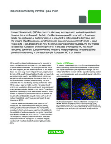
Immunohistochemistry-Paraffin Tips & TricksBulletin 0000Immunohistochemistry (IHC) is a common laboratory technique used to visualize proteins intissue or tissue sections with the help of antibodies conjugated to enzymatic or fluorescentlabels. For clarification of the terminology, it is important to differentiate this technique fromthe imaging of proteins in cells, a method referred to as immunocytochemistry (histo tissueversus cyto cell). Depending on how the immunostaining signal is visualized, the IHC methodis classed as fluorescent or chromogenic IHC. In the past, chromogenic IHC was nearlyexclusively performed, but recently due to increasing multiplexing needs (visualizing severalproteins simultaneously in one tissue sample) fluorescent IHC is on the rise.IHC is used from basic to clinical research, for example, todetermine disease states and morphological abnormalitiesof human and animal tissues. Depending on how the tissuehas been prepared, the IHC method is known as IHC-frozen(tissue has been snap frozen in liquid nitrogen, isopentane ordry ice) or IHC-paraffin (tissue has been fixed in formaldehydeand embedded in paraffin wax; FFPE). In both IHC-frozenand IHC-paraffin experiments the tissue or sections of thetissue are mounted on slides prior to staining. This is incontrast to the IHC-free-floating technique where the entireIHC procedure is performed in liquid to increase antibodybinding and penetration; slide mounting only takes place uponexperimental completion (McCollum no date). IHC-free-floatingappears to be most popular in neuroscience research. Whenanalysis of the tissue by electron microscopy is desired, thetissue is often embedded in acrylate resins such as glycolmethacrylate (GMA), a technique referred to as IHC-resin(Howat et al. 2005).Due to the significant differences in the described IHCprocedures, it is essential to confirm that your primaryantibody has been tested/validated in the intended IHCprocedure before beginning your experiment. In addition tothe availability of antibody reagents, your protein target itselfmay dictate the tissue preparation and IHC methods utilized.For example, for phosphoprotein visualization, dedicatedpreservation methods are required to conserve the posttranslationally modified state before staining (Mueller et al.2011).Staining of FFPE TissuesTo support troubleshooting and confirm the specificity of theantibody staining, we recommend inclusion of both positiveand negative controls in your experimental design. It is alsocritical to familiarize yourself with the expected staining patternand your microscope set-up to ensure that you can detect theantibody staining.Fig. 1. Localization patterns of insulin and amylin in human pancreassection. A, immunofluorescence staining of paraffin embedded humanpancreas using Guinea Pig Anti-Pig Insulin Antibody (Cat. #5330-0104G) andB, Mouse Anti-Human Amylin Antibody (#MCA1126). C, Merged image showingco-localization of the two proteins. DAPI was used as nuclear counterstain(blue).
IHC-Paraffin Tips & Tricks20-Step IHC-Paraffin Protocol Example1. Mount and section samples2. Heat sections on the specimen slide to improve adherence3. Remove paraffin and rehydrate the tissue4. (Optional) Perform heat induced or protease inducedepitope retrieval5. Wash6. Block endogenous peroxidases/phosphatases (forenzymatic labels) and biotin (for biotin/avidin detectionsystems)7. Wash 1x in phosphate buffered saline (PBS) Use Tris-buffered saline (TBS) instead of PBS for detectingphosphoproteins or when using alkaline phosphatase (AP)conjugated antibodies8. Block non-specific binding sites9. Wash 3x in PBS or TBS10. Incubate with primary antibody11. Wash 3x in PBS or TBS12. (Optional) Incubate with secondary antibody13. Wash 3x in PBS or TBS14. (Optional) Incubate with amplification reagent15. Wash 3x in PBS or TBS16. I ncubate with DAB or other substrate solution (forenzymatic labels only)17. Wash in ddH2O18. Counterstain19. D ehydrate tissue sections (for organic mounting mediaonly)20. Mount coverslipConsider these guidelines to determine if and how to perform antigenretrievalTips for Step 4 – Perform Antigen/Epitope RetrievalTissue fixation methods often result in antigen masking,which subsequently impairs antibody binding and thereforeprotein detection. The effects of tissue fixation can be partiallyreversed by performing epitope retrieval, also known asantigen unmasking. Two types of epitope retrieval have been established; heatbased (HIER; Heat-Induced Epitope Retrieval) or enzymebased (PIER; Proteolytic-Induced Epitope Retrieval).Tips for Step 6 – Block Endogenous Enzymes and BiotinTo avoid staining artifacts it is important: Table 1. Comparison of epitope retrieval methods.HIERPIERMethod Overview Can be performed usingautoclaves, heating plates,hot water baths, pressurecookers, microwaves orsteamers*Can be performed usingenzymes such as pronase,proteinase K, trypsin orpepsinMode of ActionRestores secondary andtertiary epitope structuresDegrades the peptidesmasking the epitopePopularityVery popularLess popular compared toHIERReason: may inducechanges to the specimen’smorphology* Kim et al. 2016, Ward and Rehg 2014 2017 Bio-Rad Laboratories, Inc. erform a literature search to determine what retrievalPtechniques and conditions other researchers have usedto successfully detect your protein of interest. This searchalso allows you to familiarize yourself with the expectedlocalization/distribution pattern heck if the antibody supplier or IHC resources recommendCa specific antigen retrieval protocol (for example we havea dedicated protocol for the antigen retrieval of the murineF4/80 antigen)I f no specific antigen retrieval method is recommended foryour specific protein, try HIER first (see Table 1) or HIER, initially start with a neutral pH antigen retrievalFbuffer, such as Antigen Retrieval Buffer, pH 8.0 (#BUF025A).Compare against tissue for which the antigen retrieval stephas been omitted lternatively, if no specific protocol is recommended, startAwith commonly used antigen retrieval buffers such as10 mM citrate buffer (pH 6.0) and Tris-EDTA buffer (pH 9.0).You may also want to compare different buffers to determinethe optimal conditionsI n addition to pH, consider optimizing the temperature andduration of the HIER procedure. Ideally test various pH,temperature and time parameters o eliminate staining artifacts created by the HIER process,Tcompare against a control sample for which no HIERtreatment was performed espoke treatment conditions may be required forBthe detection of certain antigens such as 5'-bromo-2'deoxyuridine (BrdU) incorporated into DNA. For BrdUstaining experiments, treatment with hydrochloric acid ornucleases is frequently performed to facilitate anti-BrdUantibody binding (Liboska et al. 2012) o block endogenous peroxidases and phosphatasesTprior to using AP/horseradish peroxidase (HRP) antibodyconjugates or blocking of endogenous peroxidase activity, useFhydrogen peroxide solutions such as Bio-Rad’s readyto-use Peroxide Blocking Reagent (#BUF017B; 3% (v/v)hydrogen peroxide). However, for the detection of certainantigens, such as CD4, this percentage of hydrogenperoxide may be too harsh (Kim et al. 2016) o block endogenous biotin when using avidin/biotin orTstreptavidin/biotin detection systems. For this specificpurpose Bio-Rad offers a ready-to-use Avidin/BiotinBlocking Reagent (#BUF016)
IHC-Paraffin Tips & Tricks o block endogenous AP activity supplement the substrateTsolution with levamisole (Ponder and Wilkinson 1981).According to Ponder and Wilkinson (1981), 20% aceticacid may also be used. However, this method may not besuitable for all antigens (Ponder and Wilkinson 1981) ever block with normal serum from the same species thatNthe primary antibody was generated in. This could lead toblocking of reactive sites or higher backgroundI f serum is unavailable, use bovine serum albumin, non-fatmilk or gelatin o not block with milk when using biotin-avidin detectionDsystems. Milk contains biotin and therefore will result instaining artifacts/high background (Kim et al. 2016)Tips for Step 7 – WashTo reduce non-specific binding and background staining,sufficient washing is essential throughout the IHC procedure. PBS is a commonly used wash buffer but not recommendedfor the detection of phosphoproteins. This is because theantibody raised against the phosphorylated protein may bindto the phospho groups present in the buffer rather than to thephospho antigen itself. TBS is a commonly used substitutein such experiments, as well as when phosphatase labeledantibodies are used.Tips for Step 10 – Incubate with Primary Antibody Check the manufacturer’s datasheet to confirm that theantibody has been tested in IHC-paraffin. If the antibodyhas not been tested in IHC-paraffin but in another methodsuch as IHC-frozen, do not assume that the antibody willautomatically work in IHC-paraffin Fig. 2. Staining of progesterone receptor phosphorylatedon serine residue 190. FFPE human breast cancer tissuestained with Mouse Anti-Progesterone Receptor (pSer190)Antibody (#MCA2406). Detection was performed using a HRPpolymer and DAB as the substrate. Antigen retrieval method:HIER, citrate buffer (pH 6.2). Tips for Step 8 – Block Non-specific Binding SitesTo prevent non-specific antibody binding to tissue surfacestructures such as Fcγ receptors, blocking is traditionallyperformed prior to incubation with the primary antibody (Kimet al. 2016).Although the impact and merit of this blocking step hasrecently been questioned by Buchwalow et al. (2011), werecommend including a blocking step in your experimentaldesign.Blocking advice: lock with serum from the same species as the tissue;Bthe immunoglobulins present in the serum will bind to thereceptors present on the tissue lternatively, use normal serum from the same species asAthe one in which the secondary antibody was generatedBlock with 10-20% normal serum 2017 Bio-Rad Laboratories, Inc. onsider using a polyclonal antibody when first establishingCan IHC protocol. Although antigen retrieval is possible, theefficiency of the process is variable and certain epitopesmight still remain inaccessible. Therefore a polyclonalantibody, which recognizes a multitude of epitopes due toits heterogeneous nature, provides a definite advantage overa monoclonal antibody which recognizes a single epitope.However, monoclonal antibodies have the advantage ofbatch-to-batch consistency and specificity, which oftenleads to lower background staining rior to performing the experiment, perform a literaturePsearch to determine the spatiotemporal localization of yourprotein of interest (in healthy and diseased tissue). For apreview of the expected staining pattern, the antibodydatasheet or other web resources may also be consulted.Knowledge of the localization patterns also assists inselecting the optimal counterstain hen using an antibody for the first time, always determineWthe optimal antibody dilution by titrating the antibody. Youmay also want to test different incubation periods andtemperature conditions (4 C versus RT or 37 C) hould you encounter high background staining or suspectSyour primary antibody to be non-specific, consider includinga concentration matched isotype control antibody (forexample Mouse IgG2b Negative Control #MCA691XZ) orpre-immune serum in your experimental designTips for Optional Steps 12 and 14 – Incubate with SecondaryAntibody and/or Amplification Reagent Especially for multiplexing experiments, it might be temptingto use directly conjugated primary antibodies to mitigate therisk of non-specific secondary antibody binding. Althoughthese antibodies are suitable for the detection of certainhigh abundant targets, we generally advise using secondaryantibodies in IHC experiments. Multiple secondaryantibodies bind to a single primary antibody, thereby leadingto signal amplification
IHC-Paraffin Tips & Tricks o reduce the risk of background staining from non-specificTsecondary antibody binding, select cross-adsorbed/preadsorbed secondary antibodies. Also, include a secondaryonly control in your experimental design to control for thistype of background or very low abundant proteins, we suggest usingFbiotinylated secondary antibodies in combination withconjugated avidin (to form an avidin-biotin complex; ABC).Since a single avidin molecule can simultaneously bind upto four biotin molecules, this method results in higher signalamplification. Labeled streptavidin is now commonly usedas a substitute for avidin in the Labeled Streptavidin Biotin(LSAB) method (Ramos-Vara 2005). Signal amplification canalso be achieved by using the Peroxidase Anti-Peroxidase(PAP) and Alkaline Phosphatase Anti-Alkaline Phosphatase(APAAP) methodsTips for Step 16 – Incubate with DAB or Other SubstratesWith the increasing multiplexing and quantification needs,more sophisticated IHC technologies such as multiplexed ionbeam imaging and mass spectrometry immunohistochemistryare being developed (Levenson et al. 2015, Angelo et al. 2014).Despite these advances, conventional chromogenic IHC is stillroutinely performed.In contrast to fluorescent labels, enzymatic labels such asHRP and AP require addition of substrates. These are alsoreferred to as chromogens, and when added to the enzyme,produce colored precipitates. hen using biotinylated primary or secondary antibodies,Wensure that you have blocked endogenous biotin prior toprimary antibody incubation (see tips for step 6) or tissues rich in Fcγ receptors, consider using fragmentFsecondary antibodies such as Rabbit F(ab’)2 Anti-MouseIgG:HRP (#STAR13B). This type of fragment antibodies lacksthe Fc region, thereby mediating Fcγ receptor interactions hen selecting fluorophore conjugates, ensure that youWare able to excite and detect the fluorophore optimally.Therefore, review the excitation and emission spectra ofyour fluorophore of interest as well as the lasers and filters ofyour microscope o reduce photobleaching (chemical destruction of aTfluorescent dye), select photostable fluorophores belongingto new generation dyes such as Alexa Fluor and DyLightFluor. Although still used in imaging applications, traditionaldyes such as FITC are highly susceptible to fading/photobleaching and should therefore not be your firstconjugate choice hile spectral separation unmixing software has significantlyWadvanced in recent years, for multiplexing experiments, westill recommend that you review the excitation and emissionspectra of fluorophores in advance to minimize spectraloverlap (Lavdas no date). Selecting fluorophores with noor very little overlap minimizes the risk of one fluorophoregetting detected in another fluorophore’s channel, a processcommonly referred to as bleed-through, cross-talk or crossoverTips for Step 13 – WashConsider adding detergents such as 0.05% Tween 20 to thewash buffer to reduce high background staining or nonspecific antibody binding. However, these detergents may notbe compatible with the markings of hydrophobic pens that aidthe compartmentalization of liquids on the slide. 2017 Bio-Rad Laboratories, Inc. ifferent enzyme/chromogen combinations produceDdifferent colored precipitates (Tables 2 and 3). A HRP/DAB (3,3'-Diaminobenzidine) reaction results in a brownprecipitate, while using TMB (3,3',5,5'-Tetramethylbenzidine)as the chromogen for HRP yields a blue-green staining (vander Loos 2010). To achieve the desired precipitate color,determine the optimal enzyme/chromogen combination atthe experimental design stage hen choosing enzyme/chromogen combinations,Wprecipitate color is not the only important selection criterion.To preserve the precipitate, ensure compatibility with yourmounting medium of choice (see tips for step 20) nzyme and substrate reaction efficiencies vary significantly.ETherefore, less efficient reactions may result in lower stainingintensities, which will impact primary antibody titrations (Vander Loos 2010). This is especially important when designingexperiments with more than one chromogenic label. Ensurethat you select the most efficient enzyme/chromogencombination for the least abundant antigen hen simultaneous detection of more than one antigenWis desired, confirm that your final precipitates andcounterstains are easily spectrally distinguishable (see tipsfor step 18)Table 2. HRP substrates and precipitate colors.HRP Substrates/ChromogensPrecipitate ColorsAEC (3-Amino-9-Ethylcarbazole)* Red*DAB (3,3’-Diaminobenzidine)* Brown*DAB with NiCl2** Purple blue**DAB with CoCl2** Dark blue**TMB (3,3’,5,5’-Tetramethylbenzidine)*Blue-green** Van der Loos 2010** Hsu and Soban 1982Table 3. AP substrates and precipitate colors.AP Substrates/ChromogensPrecipitate ColorsFast Blue BB* Blue*Fast Red TR* Red*/**New Fuchsin*** Red**** Lauter et al. 2011** IHC World, no date a*** IHC World, no date b
IHC-Paraffin Tips & TricksTips for Step 18 – CounterstainCounterstains provide contrast as well as knowledge aboutlocalization within the tissue sample (for instance by visualizingnuclei or filamentous actin).What to consider when selecting counterstains? nsure that the counterstain can be easily spectrallyEdistinguished from your precipitate color or fluorescent label.For example, when using red emitting fluorophores, select ablue nuclear counterstain such as DAPI or Hoechst 33342(Tables 4 and 5)Fluorescent counterstainsTable 4. Commonly used chromogenic rstain forEosinFast Red/KernechtrotHematoxylin (4 types Harris’s, Mayer’s,Carazzi’s and Gill’s)Methylene blueMethylene greenToluidine blueRedRedBlueProteins containing cationic uclei* Kim et al. 2016Adapted from Kim et al. 2016 and Paul no date. Fast Red/Kernechtrot has beenincluded in the table according to IHCWorld (no date c)Table 5. Commonly used fluorescent counterstains.Fig. 3. PureBlu Nuclear Fluorescent Dye Hoechst 33342.Paraffin section of human colon adenocarcinoma (heat inducedantigen retrieval with citrate buffer (pH 6), blocked with 10%FCS) stained with Mouse Anti-Human Cytokeratin 18 Antibody(#MCA1864H, green). Nuclei were counterstained with PureBluDye Hoechst 33342 (#1351304, blue).FluorescentCounterstainsColorsCounterstain forDAPIDRAQ5Hoechst 33258/33342Propidium iodideSYTOX GreenPhalloidinBlueRedBlueRedGreenConjugate dependentNucleiNucleiNucleiNucleiNucleiFilamentous actinTips for Step 20 – Mount CoverslipMounting media are essential when long term storage of slidesis required (for instance when the slides are to be stored forfuture reference purposes). They enable permanent adherenceof the coverslip to the slide, thereby protecting the tissuespecimen from damage while simultaneously adding contrastduring microscopy.Mounting media categories:1. A queous/water-based mounting media (hydrophilic;examples of aqueous mounting media include glycerineglycerol and glycerine jelly) (Ravikumar et al. 2014)2. O rganic-solvent based mounting media (hydrophobic;examples of organic solvent based media include Euparaland Canada Balsam) (Ravikumar et al. 2014).Mounting media can be further subcategorized into solidifyingmedia or those that stay liquid (Microbehunter.com no date).In general, organic-solvent based media solidify while waterbased ones remain liquid.Factors to consider:Fig. 4. PureBlu Dye DAPI. Paraffin section of humanpancreas stained with Guinea Pig Anti-Pig Insulin Antibody(#5330-0104G, red), Mouse Anti-Human Chromogranin AAntibody (#MCA4773, green) and PureBlu Dye DAPI (#1351303,blue). o save time, consider using pre-made mounting mediaTalready containing counterstains 2017 Bio-Rad Laboratories, Inc. queous-mounting media are compatible with bothAfluorescent and enzymatic labels (Ravikumar et al. 2014).It is important to confirm that the medium is clear beforeapplication as cloudiness is a possible indication ofbacterial or fungal growth (Kiernan no date). To mitigatethe risk of contamination, ensure that the medium containsbacteriostatic agents (Ravikumar et al. 2014)
IHC-Paraffin Tips & Tricks rganic mounting media should exclusively be used forOmounting chromogenic IHC slides (Ravikumar et al. 2014).However, even for these slides, exemptions exist as someprecipitates (for example by reactions with AEC) are solublein organic mounting media (Howard and Kaser 2014).For these types of precipitates and substrates aqueousmounting media should also be used or wide-field microscopy, mounting media that solidifyFshould be used (North 2006) o visualize 3D structures or to obtain 3D tissue information,Tmounting media that remain liquid are required (North 2006).When using this type of mounting media, the coverslipedges must be sealed with nail polish to avoid leaks (North2006) lides should be stored in the dark to preventSphotobleaching/fading. The risk of photobleaching may befurther reduced by selecting mounting media containingantifade reagents (North 2006). As certain fluorophores areincompatible with antifade reagents, it is important to firstconfirm suitability before using this type of mounting mediaReferencesAngelo M et al. (2014). Multiplexed ion beam imaging of human breast tumors.Nat Med 20, 436-442.Buchwalow I et al. (2011). Non-specific binding of antibodies inimmunohistochemistry: fallacies and facts. Sci. Rep.1, 28; DOI 10.1038/srep00028.Howard GC and Kaser MR, editors (2014). Making and Using Antibodies: APractical Handbook, 2nd edition (Boca Raton, Florida: Taylor & Francis Group,CRC Press) pp 311.Howat WJ et al. (2005). Resin Tissue Microarrays: a Universal Format forImmunohistochemistry. J Histochem Cytochem 53, 1189-1197.Hsu SM and Soban E (1982). Color modification of diaminobenzidine(DAB) precipitation by metallic ions and its application for doubleimmunohistochemistry. J Histochem Cytochem 30, 1079-1082.IHCWorld (no date a). Most Innovative and Complete Immunohistochemistry(IHC) Detection Systems. em/Chromogen.htm, accessed July 20, 2017.IHCWorld (no date b). New Fuchsin Alkaline Phosphatase Substrate SolutionProtocol. http://www.ihcworld.com/ protocols/chromogen substrates/APnew fuchsin red.htm, accessed July 20, 2017.IHCWorld (no date c). Nuclear Fast Red Counterstain Protocol. http://www.ihcworld.com/ protocols/counterstain solutions/nuclear fast red.htm,accessed July 03, 2017.Kiernan JA (no date). Making and using aqueous mounting media. Why buy apre-mixed medium when it’s cheap and easy to make your own? http://publish.uwo.ca/ jkiernan/aqmount.htm, accessed August 07, 2017. (This article wasoriginally published in Microscopy Today (1997) 97-10, 16-17.)Kim SW et al. (2016). Immunohistochemistry for Pathologists: Protocols, Pitfalls,and Tips. J Pathol Transl Med 50, 411-418.Lauter G et al. (2011). Two-color fluorescent in situ hybridization in theembryonic zebrafish brain using differential detection systems. BMC Dev Biol11:43.Lavdas AA (no date). Spectral Unmixing in Fluorescence nmixing-in-fluorescence-microscopy/,accessed August 07, 2017.Levenson RM (2015). Immunohistochemistry and mass spectrometry for highlymultiplexed cellular molecular imaging. Lab Invest 95, 397-405. 2017 Bio-Rad Laboratories, Inc.Liboska R et al. (2012). Most anti-BrdU antibodies react with 2'-deoxy-5'ethynyluridine – the method for the effective suppression of this cross-reactivity.PLoS One 7, e51679.Microbehunter – Amateur Microscopy Resource (no date). An overview ofmounting media for microscopy. g-media-for-microscopy/, accessed July 11, 2017.McCollum L (no date). Free-Floating Versus Slide-Mounted Sections forImmunohistochemistry. slidemounted-sections-ihc/, accessed June 29, 2017.Mueller C et al. (2011). One-Step Preservation of Phosphoproteins and TissueMorphology at Room Temperature for Diagnostic and Research Specimens.PLoS One, 6: e23780.North AJ (2006). Seeing is believing? A beginners’ guide to practical pitfalls inimage acquisition. J Cell Biol 172, 9-18.Paul C (no date). Counterstaining for Immunohistochemistry: Choices, ng-for-immunohistochemistrychoices-choices/, accessed August 07, 2017.Ponder BA and Wilkinson MM (1981). Inhibition of Endogenous TissueAlkaline Phosphatase with the Use of Alkaline Phosphatase Conjugates inImmunohistochemistry. J Histochem Cytochem 29, 981-984.Ramos-Vara JA (2005). Technical aspects of Immunohistochemistry. Vet Pathol42, 405-426.Ravikumar S et al. (2014). Mounting media: An overview. J NTR Univ Health Sci3:S1-8.Van der Loos CM (2010). Chromogens in multiple immunohistochemicalstaining used for visual assessment and spectral imaging: the colorful future.J of Histotechnol, Vol 33, 31-40.Ward JM and Rehg JE (2014). Rodent immunohistochemistry: pitfalls andtroubleshooting. Vet Pathol 51, 88-101.Visit bio-rad-antibodies.com/IHC to view other IHC relatedresources and products.
IHC-Paraffin Tips & on GuidesResourcesELISA, Flow Cytometry,IHC, Western io-rad-antibodies.com/resourcesaResearch rrImmunologyyVeterinaryCell markerselectionguidebio-rad-antibodies.comMouse andHumanbio-rad-antibodies.com/resourcesIn o experimentis completewithout all thepiecesAlexa Fluor is a registered trademark of Molecular Probes Inc. OR, USA. DRAQ5 is a trademark of Biostatus Limited. DyLight is atrademark of Thermo Fisher Scientific Inc. and its subsidiaries. SYTOX is a trademark of Life Technologies Company. Tween is aregistered trademark of Croda International plc.Bio-RadLaboratories, Inc.Life ScienceGroup38120 Ver AUS/EGWeb site bio-rad-antibodies.comAustria, Germany, Netherlands & Switzerland 49 89 80 90 95 21 Belgium & Luxembourg 44 1865 852 357 Canada & USA 1 800 424 6723France 44 1865 852 357 United Kingdom, Scandinavia & other countries 44 1865 852 700 Custom Services 49 89 80 90 95 451017 Sig 1216
staining artifacts/high background (Kim et al. 2016) Tips for Step 10 - Incubate with Primary Antibody Check the manufacturer's datasheet to confirm that the antibody has been tested in IHC-paraffin. If the antibody has not been tested in IHC-paraffin but in another method such as IHC-frozen, do not assume that the antibody will