Transcription
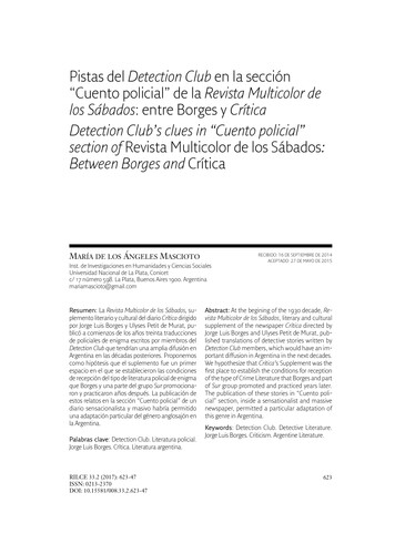
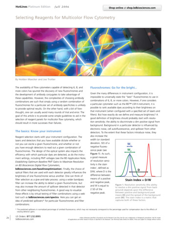
HotLines Platinum Editionfall 2006Shop online at shop.bdbiosciences.comSelecting Reagents for Multicolor Flow CytometryBy Holden Maecker and Joe TrotterThe availability of flow cytometers capable of detecting 6, 8, andmore colors has spurred the discovery of new fluorochromes andthe development of antibody conjugates to take advantage ofthese capabilities. However, the complexities of choosing antibodycombinations are such that simply using a random combination offluorochromes for a particular set of antibody specificities is unlikelyto provide optimal results. On the other hand, with a bit of forethought, one can usually avoid many rounds of trial and error. Thegoal of this article is to provide some simple guidelines to aid in theselection of reagent panels for multicolor flow cytometry, whichshould result in more successes than failures.The basics: Know your instrumentReagent selection starts with your instrument configuration. Thelasers and detectors that you have available dictate whether ornot you can excite a given fluorochrome, and whether or notyou have enough detectors to read out a given combination offluorochromes. The design of the optical system also impacts theefficiency with which particular dyes are detected, as do the instrument settings, including PMT voltages (see the BD Application Note,Establishing Optimum Baseline PMT Gains to Maximize Resolutionon BD Biosciences Digital Flow . Finally, the choice ofoptical filters that are used with each detector greatly influences thebrightness of one fluorochrome versus another. One can think offilter selection as a give-and-take process: using a wider bandpassfilter can increase the ability to detect a given fluorochrome, butmay also increase the amount of spillover detected in that detectorfrom other neighboring fluorochromes. A good way to visualizethese effects is by virtual testing of filter combinations using a webtool such as bdbiosciences.com/spectra. Here you can get anidea of predicted spillovers* for particular fluorochromes and filtercombinations.Fluorochromes: Go for the bright.Given the many differences in instrument configuration, it isimpossible to universally state the “best” fluorochromes to use incombinations of 6, 8, or more colors. However, if one considersa particular cytometer such as the BD LSR II instrument, it ispossible to rank available dyes according to their brightness onthat instrument (when configured with a specified set of lasers andfilters). But how exactly do we define and measure brightness? Agood definition of brightness should probably start with resolution sensitivity; the ability to discriminate a dim positive signal frombackground. Background in a particular detector is influenced byelectronic noise, cell autofluorescence, and spillover from otherdetectors. To the extent that these factors introduce noise, theyalso increase thewidth (or standarddeviation, SD) of aW1negative fluoresW2cence peak (seeFigure 1). As such,a good measureof resolution sensitivity is the stainindex1, defined asDD/W, where D is thedifference betweenmeans of a positiveStain Index D/Wand negative peak,Figure 1. Resolution sensitivity (the abilityand W is equal toto resolve a dim positive signal from back2 SD of theground) depends upon the differencebetween positive and background peaknegative peak.means (D) and the spread of the backgroundpeak (W). The stain index is a metric thatcaptures both of these factors.* The predicted spillovers in normalized percentage of omitted fluorescence, which may not necessarily correspond to the percentage used for compensation due to the effects ofvarying detector gains.26US Orders: 877.232.8995bdbiosciences.comUnless otherwise specified, all products are for Research Use Only. Not for use in diagnostic or therapeutic procedures. Not for resale.All applications are either tested in-house or reported in the literature. See Technical Data Sheets for details. Prices are subject to change without notice.
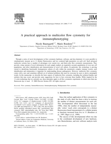
fall 2006Shop online at shop.bdbiosciences.comHotLines Platinum EditionTable 1. Stain index of various fluorochrome conjugateson a BD LSR IITable 2. Common choices for 6-, 8-, and 10-colorexperiments.ReagentCloneFilterStain Index6-color8-color10-colorPERPA-T4585/40356.3FITC orAlexa Fluor 488FITC orAlexa Fluor 488FITC orAlexa Fluor 488PEPEPEAlexa Fluor Leu-3a695/4092.7PE-Alexa Fluor 610RPA-T4610/2080.4Alexa Fluor -3a695/4064.4APC-Cy7RPA-T47801/6042.2Alexa Fluor 700RPA-T4720/4539.9Pacific Blue chromes listed with the same superscript number are read in the samedetector, and thus would not normally be used in combination.When the same antibody is conjugated to various dyes, one cancompare the stain index for those conjugates to get an idea ofthe relative brightness of the dyes on a particular instrument. Thisassumes, of course, that the conjugation chemistries for all of thereagents have been optimized. With this as a caveat, Table 1 showsthe stain index for a number of different CD4 conjugates, using thespecified filters on a standard BD LSR II flow cytometer.From this table, one can get an idea of the relative brightness ofdifferent fluorochromes on this platform. This leads to the first ruleof reagent selection, which is to pick the brightest available fluorochromes. Suppose you have a 4-color panel consisting of reagentsin FITC, PE, PerCP-Cy5.5, and APC, and you want to add a fifthcolor. PE-Cy7 is an obvious choice, since it is the brightestfluorochrome not already in your panel.But minimize spilloverBut brightness, on its own, only goes so far. The stain indexeslisted in Table 1 are calculated with the reagents run on their own,not as part of a cocktail. As soon as other reagents are added,spectral overlap (or spillover) becomes an issue. Simply stated, themore colors one attempts to resolve, the more spillover betweenthose colors that will have to be dealt with. We deal with spilloverby applying “compensation,” which results in a correction beingapplied to all signals such that a cell population fluorescing only inPE, for example, will show no FITC fluorescence, on average. Butwhile this is true for the population on average, individual cells willfall above or below the mean, and this “data spread” is higherwhen spectral overlap introduces additional noise.1 Compensation,PE-Texas Red or PEAlexa Fluor -Cy7APC orAlexa Fluor 647APC orAlexa Fluor 647APC orAlexa Fluor 647Alexa Fluor 680 or700APC-Cy7APC-Cy7APC-Cy7AmCyanAmCyanPacific Blue Pacific Blue unfortunately, does not remove this noise. The effect of data spreadis thus to reduce the resolution sensitivity, and therefore the stainindex, for a fluorescence detector that receives spillover from otherdetector(s). As a result, we can state a second rule of reagent selection, which is to minimize the potential for spectral overlap whenchoosing a reagent combination. This can be in conflict with thefirst rule, which was to choose the brightest fluorochromes. Forexample, PE-Cy5 is very bright with regard to stain index (Table 1);but it has considerable spillover into the APC detector. While thesetwo fluorochromes can be used together, the resolution sensitivityin APC will be reduced compared to, for example, a combinationof PerCP-Cy5.5 and APC. This is a case where one might wish tosacrifice a certain amount of brightness in one detector to avoidspillover (and loss of resolution sensitivity) in another.Colors and specificities: Define winning combinationsTaking the two rules above into account, one can generalize a setof fluorochromes that are reasonable choices to use for experimentsrequiring 6, 8, or more colors (Table 2). Note that these choices arebased upon BD instruments (BD LSR II, BD FACSAria cell sorter, orBD FACSCanto flow cytometer), and upon reagents that can begenerally purchased as catalog antibody conjugates.Once the fluorochromes to be used have been defined, one canbegin to match antibody specificities to particular fluorochromes.In other words, one can select the actual conjugates to be used.For this purpose, brightness and spillover remain key issues, as thefollowing example illustrates.Imagine that you want to look at CD62L staining on CD8 T cells.While CD8 is an abundant protein, and antibodies to it stain cellsvery brightly, CD62L is relatively “dim” (the protein is not abundantUnless otherwise specified, all products are for Research Use Only. Not for use in diagnostic or therapeutic procedures. Not for resale.All applications are either tested in-house or reported in the literature. See Technical Data Sheets for details. Prices are subject to change without notice.US Orders: 877.232.8995bdbiosciences.com27
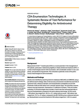
HotLines Platinum Editionfall 2006Shop online at shop.bdbiosciences.comSelecting Reagents for Multicolor Flow Cytometry(continued)Spillover affects resolution sensitivityControls to validate your panelFITC backgroundcontributes noise toPE measurementFigure 2. Spectral overlap of FITC into the PE detector. Taken from ectraon the cell surface, and/or the availableantibodies are of low affinity). Initially, onewould be tempted to use the brightestavailable fluorochrome, PE, for CD62L,while using a “dimmer” fluorochrome, likeFITC, for CD8. But FITC has considerablespillover into the PE detector (see Figure2). The result is that, for CD8 cells that arehighly stained in the FITC detector, resolution sensitivity in PE is compromised, to thepoint that CD62L resolution may be suboptimal. There are multiple possible solutionsto improve this, including:1) Move CD8 to a detector that has lessspectral overlap with PE (like PerCPCy5.5 or APC).2) Move CD62L to a detector that is stillrelatively bright, but does not overlapwith FITC (like APC or PE-Cy5). Note thatin this example, only the CD8 cells arehighly stained in the FITC detector, soonly CD8 cells contribute to data spreadin the PE detector. If one were interestedin CD62L staining only on CD4 (CD8–)cells, the original reagent combination(CD8 FITC, CD62L PE) would be fine.28US Orders: 877.232.8995bdbiosciences.comThis example illustrates two additional rulesin reagent selection: Reserve the brightestfluorochromes for “dim” antibodies, andvice versa, but avoid spillover from brightcell populations into detectors requiringhigh sensitivity for those populations.Tandem dyes: Watch out fordegradationOne final topic to consider is the potentialfor tandem dye degradation. APC-Cy7,and to a lesser extent, PE-Cy7, can degradein the presence of light, fixation, andelevated temperatures, so that they emit inthe parent dye detector (APC or PE). Thisprocess often starts with a small subpopulation of cells, leading to false positiveevents in APC or PE.1 By minimizing theexposure of samples to light, heat, andformaldehyde-based fixatives, this problemcan be largely avoided. Still, for someapplications, it may be desirable to thinkabout an additional rule: Consider theconsequences of degradation of tandemdyes and whether this will compromisesensitive readouts in the APC or PE detectors. If so, a different reagent configurationmay be in order. For situations in whichfinal fixation of samples is required (eg,biohazardous samples), there is a stabilizing fixative available that helps preventdegradation of APC-Cy7 while still fixingcells (BD Biosciences Cat. No. 338036).Once all of these considerations have beentaken into account, you are ready to testa multicolor reagent cocktail. In so doing,there are two types of controls that youmay wish to include in your initial testing:fidelity controls and fluorescence-minusone (FMO) controls.2 Fidelity controls arethose that use a given antibody by itself (orwith minimal additional gating reagents),and they compare the results to the useof that antibody in a complete cocktail.From this, one can see the effect of additional reagents on the readout of interest,to be sure that the other reagents are notcompromising that readout. FMO controlsare those that combine all the reagents ina given cocktail, except for one reagentof interest. These are useful to gauge thesensitivity of particular detectors in thecontext of the other reagents. They mayalso be used for routine gating of thosedetectors for which other means of settinggates are not possible or practical. Ingeneral, though, once a reagent panel hasbeen well validated, it is not necessary torun all of these controls on a day-to-daybasis.Unless otherwise specified, all products are for Research Use Only. Not for use in diagnostic or therapeutic procedures. Not for resale.All applications are either tested in-house or reported in the literature. See Technical Data Sheets for details. Prices are subject to change without notice.
fall 2006Shop online at shop.bdbiosciences.comTo summarize, this article has set forth“rules” for selecting reagents for multicolor flow cytometry. These rules, althoughsometimes in conflict with each other, needto be balanced to achieve the best possibleresults:Rule 1: Choose the brightest set offluorochromes for your particularinstrument configuration.Rule 2: Choose fluorochromes so as tominimize the potential for spectraloverlap.Rule 3: Reserve the brightest fluorochromesfor “dim” antibodies, and viceversa.Rule 4: Avoid spillover from bright cellpopulations into detectors requiringhigh sensitivity for thosepopulations.Rule 5: Take steps to avoid tandem dyedegradation, and consider itsimpact upon results.With time, proven panels of multicolorreagents will become available, both inthe literature and from manufacturers,such that many users will not have to start“from scratch” in designing reagent panels.Nevertheless, given the huge variety ofapplications of multicolor flow cytometry,most researchers will at least sometimesfind themselves having to select their ownmulticolor reagents, we hope that theserules will prove helpful.Multicolor Flow CytometryReagentsExpand your multicolor flow withoptions beyond your typical 5 colors B D Biosciences continues to increaseour portfolio of multicolor reagentsto support your cutting-edge researchusing the expanded fluorescentdetection capabilities of our flowcytometers. W e offer directly conjugated antibodies to fluorochromes that gobeyond your typical 5 colors byproviding colors off the Violet,Red, and Blue lasers. These includeAmCyan, Pacific Blue ,Alexa Fluor 700, and PE-Cy7conjugates to support up to9-color staining with off-the-shelfreagents. B D Biosciences Custom ConjugationProgram* offers even more choicesfor your multicolor experiment design.This program offers multiple colors, asfeatured in the table at right.HotLines Platinum EditionCustom conjugates availablefrom BD BiosciencesFluorochromeEx Max (nm)Alexa Fluor 405401Alexa Fluor 488495Alexa Fluor 594590Alexa Fluor 647650Alexa Fluor 680679Alexa Fluor 700696AmCyan457APC650APC-Cy7650FITC494Pacific Blue 405PE-Cy5496, 564PE-Cy7496, 564PerCP482PerCP-Cy5.5PE-Texas Red Texas Red 482496, 564595Please visit bdbiosciences.com/colorsfor more details about our multicolor flowcytometry reagents.Fluorochrome chart on page 30.References1. Maecker HT, Frey T, Nomura LE, and TrotterJ. Selecting fluorochrome conjugatesfor maximum sensitivity. Cytometry A.2004;62:169.2. Baumgarth N and Roederer M. A practicalapproach to multicolor flow cytometry forimmunophenotyping. J. Immunol. Methods.2000;243:77.Figure 3. Example of the use of gatingreagents (CD3, CD4, and CD19) using nontraditional fluorochrome conjugates excitedby blue, red, and violet lasers.*Please contact the Technical Service Department for more information about the Custom Conjugation Program.Unless otherwise specified, all products are for Research Use Only. Not for use in diagnostic or therapeutic procedures. Not for resale.All applications are either tested in-house or reported in the literature. See Technical Data Sheets for details. Prices are subject to change without notice.US Orders: 877.232.8995bdbiosciences.com29
HotLines Platinum Editionfall 2006Shop online at shop.bdbiosciences.comFluorochromes for combinations beyond the typical 5-color experimentBlue laser (488 nm)Red laser (633 nm)Violet laser (407 nm)PE-Cy7Alexa Fluor 700Pacific Blue AmCyanEx Max (nm): 496, 564Ex Max (nm): 696Ex Max (nm): 405Ex Max (nm): 7Hu557648CD2Hu335786CD3Hu557851, 341091557943558117CD3Bab, Cyno, Rhe557749557917558124CD3eMs552774CD4Hu557852, 90CD8Hu557746, 335787557945558207CD8Bab, Cyno, Hu557742557923558121CD16Hu557744, 335788557920558122CD19Ms552854557958CD19Hu557835, 341093557921CD20Hu335793CD25Hu557741CD25Ms557744, 45ROHu337168CD56Hu557747, Hu557844, 557643IFN-gMs557649IgG1, kHu348788IgG1, kHam552811IgG1, kMs557646, 557872IgG1, kRat557645IgG1, lHam557798IgG1, lRat552869IgG2, lHam557652IgG2a, kRat552784IgG2a, k (specific for TNP)Ms552868IgG2b, kRat552849IgMMs552867IL-4Hu557989Ly-6G and 20557963558109558118557979Applicable Patents: APC: US 4,520,110; 4,859,582; 5,055,556; Europe 76,695; Canada 1,179,942; PerCP: US 4,876,190;PE-Cy7: US 4,542,104; APC-Cy7: US 5,714,38630US Orders: 877.232.8995bdbiosciences.comUnless otherwise specified, all products are for Research Use Only. Not for use in diagnostic or therapeutic procedures. Not for resale.All applications are either tested in-house or reported in the literature. See Technical Data Sheets for details. Prices are subject to change without notice.
BD FACSCanto flow cytometer), and upon reagents that can be generally purchased as catalog antibody conjugates. Once the fluorochromes to be used have been defined, one can begin to match antibody specificities to particular fluorochromes. In other words, one can select the actual conjugates to be used.