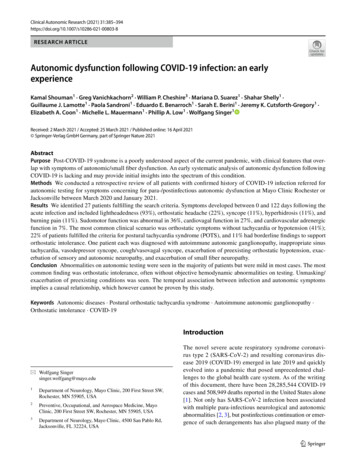
Transcription
Tao et al. Lipids in Health and Disease (2016) 15:153DOI 10.1186/s12944-016-0326-0RESEARCHOpen AccessAPOC3 induces endothelial dysfunctionthrough TNF-α and JAM-1Yun Tao1†, Yisong Xiong2†, Huimin Wang1, Shaopeng Chu1, Renqian Zhong3, Jianxin Wang1, Guihua Wang1,Xiumei Ren1 and Juan Yu1,4*AbstractBackground: The fatality rate for cardiovascular disease (CVD) has increased in recent years and higher levels oftriglyceride have been shown to be an independent risk factor for atherosclerotic CVD. Dysfunction of endothelialcells (ECs) is also a key factor of CVD. APOC3 is an important molecule in lipid metabolism that is closely associatedwith hyperlipidemia and an increased risk of developing CVD. But the direct effects of APOC3 on ECs were stillunknown. This study was aimed at determining the effects of APOC3 on inflammation, chemotaxis and exudationin ECs.Methods: ELISA, qRT-PCR, immunofluorescence, flow cytometry and transwell assays were used to investigate theeffects of APOC3 on human umbilical vein endothelial cells (HUVECs). SiRNA-induced TNF-α and JAM-1 silencingwere used to observe how APOC3 influenced the inflammatory process in the ECs.Results: Our results showed that APOC3 was closely associated with the inflammatory process in ECs, and that thisprocess was characterized by the increased expression of TNF-α. Inflammatory processes further disrupted the tightjunctions (TJs) between HUVECs by causing increased expression of JAM-1. JAM-1 was involved in maintaining theintegrity of TJs, and it promoted the assembly of platelets and the exudation of leukocytes. Changes in its expressionpromoted chemotaxis and the exudation of ECs, which contributed to atherosclerosis. While the integrity of the TJswas disrupted, the adhesion of THP-1 cells to HUVECs was also increased by APOC3.Conclusions: In this study, we describe the mechanism by which APOC3 causes inflammation, chemotaxis and theexudation of ECs, and we suggest that controlling the inflammatory reactions that are caused by APOC3 may be a newmethod to treat CVD.Keywords: APOC3, Inflammation, Cardiovascular disease, Endothelial dysfunctionBackgroundThe fatality rate for CVD has increased in recent years,and it threatens the lives of people around the world.CVD is the basis of coronary heart disease (CHD),higher levels of triglyceride have been shown to be anindependent risk factor for atherosclerotic CVD [1].While all factors related to the concentration of circulating triglycerides are important, APOC3 is of particularconcern [2, 3].* Correspondence: yujuanjs@163.com†Equal contributors1Center of Laboratory Medicine, Affiliated Hospital, Nantong University, 20 XiSi Road, Nantong 226001, People’s Republic of China4Institute of Public Health, Nantong University, 9 Se Yuan Road, Nantong226001, People’s Republic of ChinaFull list of author information is available at the end of the articleAPOC3 is a 79-amino acid glycoprotein with a molecular mass of 8.8 kDa that is synthesized mainly inthe liver and rarely in the intestines. APOC3 is a component of chylomicrons (CMs), very low density lipoproteins (VLDLs), high-density lipoproteins (HDLs)and low density lipoproteins (LDLs) [4]. It becomesmature in the endoplasmic reticulum [5]. Because it isa crucial molecule in lipid metabolism, the main effectof APOC3 is thought to be its inhibition of the functionof lipoprotein lipase (LPLs) by displacing the enzymesfrom TG-rich particles [6–8]. This process delays lipidmetabolism and causes lipid deposits. 2016 The Author(s). Open Access This article is distributed under the terms of the Creative Commons Attribution 4.0International License (http://creativecommons.org/licenses/by/4.0/), which permits unrestricted use, distribution, andreproduction in any medium, provided you give appropriate credit to the original author(s) and the source, provide a link tothe Creative Commons license, and indicate if changes were made. The Creative Commons Public Domain Dedication o/1.0/) applies to the data made available in this article, unless otherwise stated.
Tao et al. Lipids in Health and Disease (2016) 15:153Page 2 of 8It has been reported that there are clear differences inplasma concentrations of APOC3 between differentpeople. Individuals with a higher plasma level of APOC3are more likely to suffer from CVDs [9–11]. Insulin response element (IRE) is located in the promoter of theAPOC3 gene, and it has been shown that APOC3 induces diabetes and insulin resistance [10, 12].Studies have revealed a strong link between inflammation and abnormal lipid metabolism. APOC3 mediatesthe metabolism of lipids, which usually co-exists with inflammation. As previously reported, endothelial dysfunction lies at the root of AS, and inflammation occursthroughout the process of AS [13], which leads to moreserious diseases. Inflammation, chemotaxis and the exudation of ECs are indispensable in CVD. The integrityof tight junctions (TJs) is also vital. Endothelial permeability is regulated by TJs, and alterations in the expression of the proteins that form these junctions might leadto a leaky endothelial barrier [14–16]. Disruptions in TJsaffect lipid deposition, which causes CVD.However, the mechanism by which APOC3 leads toCVD and whether this process is associated with the destruction of the functions of cardiovascular ECs andinflammation remain unknown. This study was aimed atdetermining the effects of APOC3 on inflammation,chemotaxis and exudation in ECs.48 h in culture and before the cells were treated withAPOC3, some of the cells were transfected with TNF-αsiRNA or JAM-1 siRNA.To test the effect of exogenous TNF-α on HUVECs,TNF-α was added to the culture medium after 72 h in culture. After 2,4 or 16 h in culture, the secretion of JAM-1were tested by qRT-PCR.MethodsThe concentration of TNF-α was tested according to themanufacturer’s instructions. All of the samples that contained different concentrations of TNF-α were placed at37 C for 2 h. We then added working solution A andincubated the samples at 37 C for 1 h. After the plateswere washed, we added working solution B to the samples and incubated them at 37 C for 30 min. The plateswere washed again. Next, the substrate solution wasadded to the samples, and the cells were incubated at37 C for 20 min. Finally, stop buffer was added to thesamples, and the absorbance was read at 450 nm.MaterialsHUVECs and THP-1 monocytes were obtained fromScience Cell Lab (USA). APOC3 was acquired fromACADEMY Corporation (USA). Dulbecco’s modifiedEagle medium (DMEM), foetal bovine serum (FBS) andtrypsin were obtained from Science Cell Lab. The TNFα ELISA assay kit was obtained from YouershengCorporation (China). The RNA extraction reagent waspurchased from Generay Corporation. SuperReal PreMix Plus was obtained from Tiangen Corporation. TheRT-reaction Kit was purchased from Fermentas Corporation. PCR primers were purchased from ShenggongCorporation (China). All siRNAs were obtained fromJIMA Corporation (China). All of the antibodies usedin this study were purchased from Santa Cruz Biotechnology and Alexa. Transwell chambers were purchasedfrom Fisher Scientific Corporation.MethodsCell culture and treatmentHUVECs were cultured in DMEM supplemented with10 % foetal bovine serum. After the cells reached confluence, they were washed twice with PBS and isolated at37 C using a trypsin solution.To test the effect of APOC3 on HUVECs, APOC3 wasadded to the culture medium after 72 h in culture. AfterTransfection with TNF-α siRNA or JAM-1 siRNATransfection with TNF-α siRNA or JAM-1 siRNA wasperformed using Lipofectamine 2000 (Life Technologies)according to the manufacturer’s instructions. The siRNAlipid complexes were added to the cells, and the mediumwas replaced 6 h later. QRT-PCR was performed 24 hafter the cells were transfected. To confirm whether thecells had been successfully transfected, an inverse fluorescence microscope was used to confirm that the fluorescent signal of the siRNA was present in the cells. Thesequences of the siRNAs were as follows:TNF-α Forward: GCCUGUAGCCCAUGUUGUATTTNF-α Reverse: UACAACAUGGGCUACAGGCTTJAM-1 Forward: GUCGAGAGGAAACUGUUGUTTJAM-1 Reverse: ACAACAGUUUCCUCUCGACTTELISARNA purification and qRT-PCRTotal RNA was purified from the treated cells using Trizolreagent according to the manufacturer’s instructions. Thepurity and quantity of the RNA were measured using aspectrophotometer by testing its optical density at 260 nmand 280 nm, and the quality of the RNA was determinedusing agarose gel electrophoresis. Then the RNA was subjected to reverse transcription with a RT-reaction Kit.CDNA was amplified from the purified RNA and thenquantified using a CFX connect Real-Time PCR System in20-μl reaction volumes with SuperReal PreMix Plus.The reaction volumes contained 8 μl of diluted cDNAtemplate, 1 μl each of the forward and reverse primers(10 μM) and 10 μl of 2 SuperReal PreMix Plus. All ofthe reactions were performed in triplicate. The relative
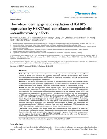
Tao et al. Lipids in Health and Disease (2016) 15:153quantities of our target mRNAs were normalized to thelevel of GAPDH. The following primers were used:Homo GAPDH Forward: AGAAGGCTGGGGCTCATTTGHomo GAPDH Reverse: AGGGGCCATCCACAGTCTTCHomo TNFα Forward: 5‘CCGAGTGACAAGCCTGTAGCC 3’Homo TNFα Reverse: 5‘TTGAAGAGGACCTGGGAGTAGATG 3’Homo JAM-1 Forward: 5‘CACGGAATGGGTATGGGACAC 3’Homo JAM-1 Reverse: 5‘CCAGGAGAATCAGGGTTACAAGGAC 3’Flow cytometryAfter the cells were washed and fixed, the HUVECs weretreated with TNF-α antibody for one night at 4 C. Theywere then incubated with a secondary antibody to analyse the expression of cell surface TNFα or JAM-1 usingan Accuri C6 Flow Cytometer (BD Biosciences).ImmunofluorescenceAfter the cells were washed and fixed, the HUVECs weretreated with JAM-1 antibodies overnight at 4 C. Theywere then incubated with separate secondary antibodies.The cells were incubated at room temperature in thedark for 1 h. Next, the cells were washed with PBS, anda DAPI solution was added to them. Then the cells wereincubated at room temperature in the dark for 20 min.After the cells were washed, a fluorescence microscopewas used to observe the expression of JAM-1.Transwell testHUVECs of the same cell density were cultured in thelower chambers of Transwell chambers for 2 h prior toPage 3 of 8the experiment. Then THP-1 cells were inoculated intothe upper chamber of the Transwell chambers. After thecells were co-cultured for 16 h, the number of THP-1cells that had migrated to the lower chamber werecounted.Statistical analysisAll of the data in these experiments are reported as themean SD. One-way ANOVA was used to analyse all ofthe data. Data with p-values that were less than 0.05 wereconsidered significant.ResultsAPOC3 stimulates the secretion of TNF-α in HUVECsTo investigate the role of APOC3 in cell inflammation,ELISA tests were used to determine whether APOC3 influences the secretion of TNF-α in HUVECs. When cellswere treated with APOC3, they secreted more TNF-α(355.3 47.114 pg/ml) than the untreated controls(103.8 16.620 pg/ml). However, when TNF-α siRNAwas transfected into cells before they were treated withAPOC3, the secretion of TNF-α was clearly lower(205.6 17.332 pg/ml) (P 0.01) (Fig. 1). In summary,APOC3 is closely associated with inflammation in ECs,and inflammatory processes are one of the causes of AS.APOC3 disrupts TJs partly via TNF-αConsistent with the results of ELISA, qRT-PCR showedthat APOC3 up-regulated the expression of TNF-α. Theexpression level of JAM-1, which is an important molecule in the TJ barrier, was also increased. The expressionof JAM-1 increased with the increasing concentration ofAPOC3 (Fig. 2).To investigate the effect of APOC3 on the expression ofJAM-1, we transfected TNF-α siRNA into HUVECs. Asthe results of qRT-PCR showed, when cells were transfected with the TNF-α siRNA, TNF-α expression wasFig. 1 Effects of APOC3 on the secretion of TNF-α in HUVECs. Blank: Untreated HUVECS; APOC3: HUVECs that were incubated with APOC3(100 μg/ml) for 24 h; APOC3 siTNF-α: HUVECs that were transfected with TNF-α siRNA before they were incubated with APOC3 (100 μg/ml) for24 h; APOC3 siNC: HUVECs that were transfected with blank siRNA before they were incubated with APOC3 (100 μg/ml) for 24 h. Each bar representsthe mean SE of 3 independent experiments. ** P 0.01 indicates a significant difference compared to the untreated HUVECs
Tao et al. Lipids in Health and Disease (2016) 15:153Page 4 of 8Fig. 2 Effect of different concentrations of APOC3 on the expression of TNF-α, JAM-1 in HUVECs according to qRT-PCR. 0: Untreated HUVECS;1: HUVECs that were incubated with APOC3 (1 μg/ml) for 24 h; 10: HUVECs that were incubated with APOC3 (10 μg/ml) for 24 h;.20: HUVECs thatwere incubated with APOC3 (20 μg/ml) for 24 h; 50: HUVECs that were incubated with APOC3 (50 μg/ml) for 24 h; 100: HUVECs that wereincubated with APOC3 (100 μg/ml) for 24 h. Each bar represents the mean SE of 3 independent experiments. ** P 0.01, significantlydifferent from untreated HUVECsattenuated (Fig. 3). Based on these data, cells were treatedwith APOC3 and incubated for 24 h. Then, qRT-PCR,flow cytometry (FCM) and immunofluorescence techniques were used to analyse the expression of JAM-1.Our results showed that APOC3 up-regulated JAM-1expression at both the mRNA and protein levels, aboutdouble the expression of untreated controls. WhenHUVECs were transfected with TNF-α siRNA, beforetreated with APOC3, the expression of JAM-1 was reduced. During this process, the expression of JAM-1 failedto return to baseline level, showing that APOC3 disruptedTJs partly via TNF-α. (Figs. 3 and 4).Considering the effect of exogenous TNF-α on the expression of JAM-1, the HUVECs were also treated withTNF-α. When cells were treated with TNF-α, the secretion of JAM-1 were up-regulated. When the cells weretreated with TNF-αfor 16 h,the secretion of JAM-1 increased to approximately 2.43 times than normal cells(Fig. 5).APOC3 promotes the adhesion of THP-1 cells to HUVECsvia TNF-α and JAM-1THP-1 cells are monocytes. In this study, THP-1 cellswere used to investigate the effect of APOC3 on theFig. 3 Effect of APOC3 on the mRNA expression levels of TNF-α, JAM-1 in HUVECs according to qRT-PCR. Blank: Untreated HUVECS; APOC3:HUVECs that were incubated with APOC3 (100 μg/ml) for 24 h; APOC3 siTNF-α: HUVECs that were transfected with TNF-α siRNA before theywere incubated with APOC3 (100 μg/ml) for 24 h; APOC3 siNC: HUVECs that were transfected with blank siRNA before they were incubated withAPOC3 (100 μg/ml) for 24 h. Each bar represents the mean SE of 3 independent experiments. ** P 0.01, significantly different from untreatedHUVECs; * P 0.05, significantly different from untreated HUVECs; ## P 0.01, significantly different from HUVECs incubated with APOC3
Tao et al. Lipids in Health and Disease (2016) 15:153Page 5 of 8Fig. 4 The expression of JAM-1 in HUVECs in protein level. A FCM results. Blank: Untreated HUVECS; APOC3: HUVECs that were incubated withAPOC3 (100 μg/ml) for 24 h; APOC3 siTNF-α: HUVECs that were transfected with TNF-α siRNA before they were incubated with APOC3 (100 μg/ml)for 24 h. Each bar represents the mean SE of 3 independent experiments. ** P 0.01, significantly different from untreated HUVECs; ## P 0.01,significantly different from HUVECs that were incubated with APOC3. B immunofluorescence results. a Untreated HUVECS; b HUVECs that wereincubated with APOC3 (100 μg/ml) for 24 h; c HUVECs that were transfected with TNF-α siRNA before they were incubated with APOC3 (100 μg/ml)for 24 h; d relative expression levels of JAM-1 in HUVECS. Blank: Untreated HUVECS; APOC3: HUVECs that were incubated with APOC3 (100 μg/ml) for24 h; APOC3 siTNF-α: HUVECs that were transfected with TNF-α siRNA before they were incubated with APOC3 (100 μg/ml) for 24 h. Each barrepresents the mean SE of 3 independent experiments. ** P 0.01, significantly different from untreated HUVECs; * P 0.05, significantlydifferent from untreated HUVECs; # P 0.05, significantly different from HUVECs that were incubated with APOC3adhesion of monocytes to ECs. Transwell chamber assayswere used to determine the extent of adhesion. After 16 hin co-culture, the number of THP-1 cells that had adheredto the HUVECs was counted. When APOC3 was added tothe HUVEC culture medium, the number of transmembrane THP-1 cells clearly increased to approximately 1.98fold the number that was observed in wells that weregrown without adding APOC3. When TNF-α siRNA orJAM-1 siRNA were transfected into the HUVECs beforeAPOC3 was added to the HUVEC culture medium, celladhesion was clearly reduced. However, the adhesion ofTHP-1 cells to HUVECS remained significantly higher inthe cells that were transfected with JAM-1 siRNA than inthe untreated cells, indicating that the effect of APOC3 onadhesion in THP-1 cells was not completely inhibitedwhen we interfered with the expression of JAM-1, indicating that other molecules are likely to affect this process.These results were consistent with our previous results.
Tao et al. Lipids in Health and Disease (2016) 15:153Page 6 of 8Fig. 5 The expression of JAM-1 in HUVECs according to qRT-PCR. Blank: Untreated HUVECS; 2 h: HUVECs that were incubated with TNF-α(1.0 mg/ml) for 2 h; 4 h: HUVECs that were incubated with TNF-α (1.0 mg/ml) for 4 h; 16 h: HUVECs that were incubated with TNF-α (1.0 mg/ml)for 16 h. Each bar represents the mean SE of 3 independent experiments. ** P 0.01, significantly different from untreated HUVECs; *P 0.05,significantly different from untreated HUVECsNevertheless, the number of migrated THP-1 cells wasstatistically lower in the cells treated with siRNA andAPOC3 than in those treated with APOC3 alone, indicating that the siRNA had an inhibitory effect on APOC3(Fig. 6).DiscussionStudies have shown that APOC3 concentrations arepositively correlated with lipid levels, and APOC3 hasbeen found to be an independent risk factor for CVD[17–19]. APOC3 modulates lipid levels in various ways.Previous data have shown that APOC3 has adverse effects in lipid metabolism [5, 20, 21]. It has also been reported that APOC3 concentrations are associated withCVDs, including hyperlipidaemia, CHD [9], and nonalcoholic fatty liver disease [22]. It has also been proposed that polymorphisms of APOC3 are associatedwith plasma lipid levels [23–28]. Loss-of function mutations, include R19X, IVS2 1G A, IVS3 1G T andA43T, also contribute to lipid metabolism. We proposethat these polymorphisms are associated with cell inflammatory processes [29–35].Atherosclerosis is an inflammatory disease [13]. Abnormal blood lipid levels can further induce inflammation. Studies have suggested that atherosclerotic lesionsare characterized by a series of cellular and molecular inflammatory responses. Endothelial dysfunction has beenproposed to be an important factor in this inflammatoryFig. 6 The adhesion of THP-1 cells to HUVECs. Blank: Untreated HUVECS; APOC3: HUVECs that were incubated with APOC3 (100 μg/ml) for 16 h;APOC3 siNC: HUVECs that were transfected with a negative control siRNA before they were incubated with APOC3 (100 μg/ml) for 16 h; APOC3 siJAM-1: HUVECs that were transfected with JAM-1 siRNA before they were incubated with APOC3 (100 μg/ml) for 16 h.; APOC3 siTNF-α:HUVECs that were transfected with TNF-α siRNA before they were incubated with APOC3 (100 μg/ml) for 16 h. HUVECS in each group were inthe same cell density. Each bar represents the mean SE of 3 independent experiments. ** P 0.01, significantly different from untreated HUVECs;## P 0.01, significantly different from HUVECs that were incubated with APOC3
Tao et al. Lipids in Health and Disease (2016) 15:153process. In this study, we examined the mechanisms bywhich APOC3 might cause endothelial dysfunctions andcellular inflammation.TNF-α is one of the most important molecules in cellular inflammation, and it has been demonstrated tohave a substantial effect on EC dysfunction. CHD patients have higher plasma TNF-α levels than healthypeople, patients who suffer from hyperlipidemia alsohave high serum concentrations of TNF-α, and TNF-αconcentrations are positively associated with VLDL-Cconcentrations and negatively associated with HDL-Cconcentrations. TNF-α regulates the expression of NOSand thereby influences the production of NO, which isassociated with prediabetic metabolic syndrome [36].In our study, we found that APOC3 caused inflammation in ECs via TNF-α. The increasing concentration ofTNF-α led to an increase in reactive oxygen species thatcaused endothelial dysfunction. We showed that the inflammation that was caused in these cells disrupted theTJs between ECs by demonstrating the presence of alterations in the expression of JAM-1. These processesresulted in EC dysfunction. Emanuela Mazzon et al.showed that TNF-α had an effect on epithelial cells,caused inflammation, and contributed to TJ permeability [37, 38]. Our results show that TNF-α also affectsTJs in ECs.JAM-1 is localized in and is an important componentof the TJs in ECs. It mainly influences cell-cell adhesion.Moreover. JAM-1 was associated with the process of exudation, which is significant in AS. JAM-1 is also locatedon the surface of platelets and leukocytes. It promotesnot only cell-cell adhesion but also the assembly ofplatelets and the exudation of leukocytes, and interactions between ECs and platelets promote the formationof AS. During the process of the adhesion of platelets toECs, the N-terminus and the 1st Ig fold domain of JAM1 play leading roles [39, 40]. It has also been reportedthat APOC3 plays an important role in neutrophilmediated responses during inflammation [41]. As our results show, TNF-α overexpression increased expressionof JAM-1, which promoted the chemotaxis and exudation of cells to cause AS. Akio Kawakami et al. reportedthat APOC3 promoted the adhesion of THP-1 cells toHUVECs through PKC-α and NF-kB [17]. In our study,we illustrated that the inflammatory reaction caused byAPOC3 also played a key role in this process.The adhesion of circulating monocytes to ECs plays acrucial role in atherogenesis and is associated with inflammatory processes in ECs. Integrins and other adhesion molecules have been reported to participate in thisprocess [5, 42]. When TJs are disrupted, interactionsoccur between cells. APOC3 has been shown to inducethe expression of PCPLC in THP-1 cells [17]. PKCα wasalso activated in these cells, and it promoted thePage 7 of 8adhesion of monocytes to ECs. We found that disruptions in the TJs of ECs were also observed in thisprocess. However, the exact mechanisms that contributeto these processes remain to be investigated.ConclusionsWith previous studies suggested that APOC3 was closelywith the development of hyperlipidemia and CVD. Inour study, we demonstrated that APOC3 is closely related to the inflammatory process in cells. It further disrupted TJs between ECs, and eventually caused thedysfunction of ECs. This process is required during thegeneration of AS. Controlling inflammatory reactionscaused by APOC3 in these patients is a promising approach to treating CVD.AbbreviationsCHD: Coronary heart disease; CM: Chylomicron; CVD: Cardiovascular disease;EC: Endothelial cell; HDL: High-density lipoprotein; HUVEC: Human umbilicalvein endothelial cell; IRE: Insulin response element; JAM: Junctional adhesionmolecule; LDL: Low density lipoprotein; NAFLD: Nonalcoholic fatty liverdisease; TAMPs: TJ associated Marvel domain proteins; TJ: Tight junction;VLDL: Very low density lipoproteinAcknowledgementsWe thank the staff of the Centre of Laboratory Medicine at the AffiliatedHospital, Nantong University for their skillful technical assistance andequipment support.FundingNational Natural Science Foundation of China (81301484, 81170263, 81302596).PhD research startup foundation of Nantong University (03080737).The State Scholarship Fund organized by the China Scholarship.Availability of data and materialsAll data generated or analyzed during this study are included in thispublished article.Authors’ contributionsJY designed the study and analyzed all the data. YT performed the qRT-PCRtests, and was a major contributor in writing the manuscript. YX and HWperformed the Transwell test. RZ and GW were in charge of flow cytometryand immunofluorescence tests. SC and XR were in charge of cell culture andtransfection of si-RNAs. JW performed ELISA tests. All authors read andapproved the final manuscript.Competing interestsThe authors declare that they have no competing interests.Ethics approval and consent to participateNot applicable.Author details1Center of Laboratory Medicine, Affiliated Hospital, Nantong University, 20 XiSi Road, Nantong 226001, People’s Republic of China. 2Department ofLaboratory Medicine, Chengdu Military General Hospital, 270 Tian Hui Road,Chengdu 610000, People’s Republic of China. 3Department of LaboratoryMedicine, Changzheng Hospital, Second Military Medical University, 415Feng Yang Road, Shanghai 200003, People’s Republic of China. 4Institute ofPublic Health, Nantong University, 9 Se Yuan Road, Nantong 226001,People’s Republic of China.Received: 14 May 2016 Accepted: 6 September 2016
Tao et al. Lipids in Health and Disease (2016) 15:153References1. Kawakami A, Osaka M, Tani M, Azuma H, Sacks FM, Shimokado K, et al.Apolipoprotein CIII links hyperlipidemia with vascular endothelial celldysfunction. Circulation. 2008;118:731–42.2. Olivieri O, Martinelli N, Girelli D, Pizzolo F, Friso S, Beltrame F, et al.Apolipoprotein C-III predicts cardiovascular mortality in severe coronaryartery disease and is associated with an enhanced plasma thrombingeneration. J Thromb Haemost. 2010;8:463–71.3. Sarwar N, Danesh J, Eiriksdottir G, Sigurdsson G, Wareham N, Bingham S,et al. Triglycerides and the risk of coronary heart disease: 10,158 incidentcases among 262,525 participants in 29 Western prospective studies.Circulation. 2007;115:450–8.4. Morita SY, Sakurai A, Nakano M, Kitagawa S, Handa T. Presence ofapolipoprotein C-III attenuates apolipoprotein E-mediated cellularuptake of cholesterol-containing lipid particles by HepG2 cells. Lipids.2011;46:323–32.5. Kawakami A, Aikawa M, Libby P, Alcaide P, Luscinskas FW, Sacks FM.Apolipoprotein CIII in apolipoprotein B lipoproteins enhances the adhesion ofhuman monocytic cells to endothelial cells. Circulation. 2006;113:691–700.6. Kersten S. Physiological regulation of lipoprotein lipase. Biochim BiophysActa. 1841;2014:919–33.7. Larsson M, Vorrsjö E, Talmud P, Lookene A, Olivecrona G. Apolipoproteins CI and C-III inhibit lipoprotein lipase activity by displacement of the enzymefrom lipid droplets. J Biol Chem. 2013;288:33997–4008.8. Heng C, Khoo C, Furtado J, Sacks FM. Apolipoprotein C-III and themetabolic basis for hypertriglyceridemia and the dense low-densitylipoprotein phenotype. Circulation. 2010;121:1722–34.9. Baldi S, Bonnet F, Laville M, Morgantini C, Monti L, Hojlund K, et al.Influence of apolipoproteins on the association between lipids and insulinsensitivity. Diabetes Care. 2013;36:4125–31.10. Ginsberg HN, Brown WV. Apolipoprotein CIII: 42 years old and even moreinteresting. Arterioscler Thromb Vasc Biol. 2011;31:471–3.11. Ooi EM, Barrett PH, Chan DC, Watts GF. Apolipoprotein C-III: understandingan emerging cardiovascular risk factor. Clin Sci. 2008;114:611–24.12. Lee HY, Birkenfeld AL, Jornayvaz FR, Jurczak MJ, Kanda S, Popov V, et al.Apolipoprotein CIII overexpressing mice are predisposed to diet-inducedhepatic steatosis and hepatic insulin resistance. Hepatolog. 2011;54:1650–60.13. Epstein FH, Ross R. Atherosclerosis-an inflammatory disease. N Engl J Med.1999;340:115–26.14. Turner JR. Intestinal mucosal barrier function in health and disease. Nat RevImmunol. 2009;9:799–809.15. Vetrano S, Ploplis VA, Sala E, Sandoval-Cooper M, Donahue DL, Correale C,et al. Unexpected role of anticoagulant protein C in controlling epithelialbarrier integrity and intestinal inflammation. Proc Natl Acad Sci U S A. 2011;108:19830–5.16. Rodgers LS, Beam MT, Anderson JM, Fanning AS. Epithelial barrier assemblyrequires coordinated activity of multiple domains of the tight junctionprotein ZO-1. J Cell Sci. 2013;126:1565–75.17. Kawakami A, Aikawa M, Nitta N, Yoshida M, Libby P, Sacks FM.Apolipoprotein CIII-induced THP-1 cell adhesion to endothelial cells involvespertussis toxin-sensitive G protein and protein kinase C alpha-mediatednuclear factor-kappaB activation. Arterioscler Thromb Vasc Biol. 2007;27:219–25.18. Riwanto M, Rohrer L, Roschitzki B, Besler C, Mocharla P, Mueller M, et al.Altered activation of endothelial anti - and proapoptotic pathways byhigh-density lipoprotein from patients with coronary artery disease: roleof high-density lipoprotein-proteome remodeling. Circulation. 2013;127:891–904.19. Silbernagel G, Genser B, Drechsler C, Scharnagl H, Grammer TB, Stojakovic T,et al. HDL cholesterol, apolipoproteins, and cardiovascular risk in hemodialysispatients. J Am Soc Nephrol. 2015;26:484–92.20. Jensen MK, Rimm EB, Furtado JD, Sacks FM. Apolipoprotein C-III as apotential modulator of the association between HDL-cholesterol andincident coronary heart disease. J Am Heart Assoc. 2012;1.21. Kawakami A, Aikawa M, Alcaide P, Luscinskas FW, Libby P, Sacks FM.Apolipoprotein CIII induces expression of vascular cell adhesion Molecule-1in vascular endothelial cells and increases adhesion of monocytic cells.Circulation. 2006;114:681–7.22. Petersen KF, Dufour S, Hariri A, Nelson-Williams C, Foo JN, Zhang XM, et al.Apolipoprotein C3 gene variants in nonalcoholic fatty liver disease. N Engl JMed. 2010;362:1082–9.Page 8 of 823. Yu J, Wang H, Yang S, Yuan J, Chen L, Chen CL, et al. The effect of APOC3promoter polymorphisms on the risk of Hypertriglyceridemia in ChineseHan population with or without Type 2 diabetes mellitus. Labmed.2010;
Flow cytometry After the cells were washed and fixed, the HUVECs were treated with TNF-α antibody for one night at 4 C. They were then incubated with a secondary antibody to ana-lyse the expression of cell surface TNFα or JAM-1 using an Accuri C6 Flow Cytometer (BD Biosciences). Immunofluorescence After the cells were washed and fixed, the .