Transcription
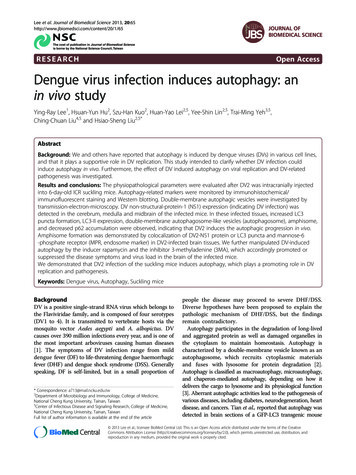
Lee et al. Journal of Biomedical Science 2013, ARCHOpen AccessDengue virus infection induces autophagy: anin vivo studyYing-Ray Lee1, Hsuan-Yun Hu2, Szu-Han Kuo2, Huan-Yao Lei2,5, Yee-Shin Lin2,5, Trai-Ming Yeh3,5,Ching-Chuan Liu4,5 and Hsiao-Sheng Liu2,5*AbstractBackground: We and others have reported that autophagy is induced by dengue viruses (DVs) in various cell lines,and that it plays a supportive role in DV replication. This study intended to clarify whether DV infection couldinduce autophagy in vivo. Furthermore, the effect of DV induced autophagy on viral replication and DV-relatedpathogenesis was investigated.Results and conclusions: The physiopathological parameters were evaluated after DV2 was intracranially injectedinto 6-day-old ICR suckling mice. Autophagy-related markers were monitored by immunohistochemical/immunofluorescent staining and Western blotting. Double-membrane autophagic vesicles were investigated bytransmission-electron-microscopy. DV non-structural-protein-1 (NS1) expression (indicating DV infection) wasdetected in the cerebrum, medulla and midbrain of the infected mice. In these infected tissues, increased LC3puncta formation, LC3-II expression, double-membrane autophagosome-like vesicles (autophagosome), amphisome,and decreased p62 accumulation were observed, indicating that DV2 induces the autophagic progression in vivo.Amphisome formation was demonstrated by colocalization of DV2-NS1 protein or LC3 puncta and mannose-6-phosphate receptor (MPR, endosome marker) in DV2-infected brain tissues. We further manipulated DV-inducedautophagy by the inducer rapamycin and the inhibitor 3-methyladenine (3MA), which accordingly promoted orsuppressed the disease symptoms and virus load in the brain of the infected mice.We demonstrated that DV2 infection of the suckling mice induces autophagy, which plays a promoting role in DVreplication and pathogenesis.Keywords: Dengue virus, Autophagy, Suckling miceBackgroundDV is a positive single-strand RNA virus which belongs tothe Flaviviridae family, and is composed of four serotypes(DV1 to 4). It is transmitted to vertebrate hosts via themosquito vector Aedes aegypti and A. albopictus. DVcauses over 390 million infections every year, and is one ofthe most important arboviruses causing human diseases[1]. The symptoms of DV infection range from milddengue fever (DF) to life-threatening dengue haemorrhagicfever (DHF) and dengue shock syndrome (DSS). Generallyspeaking, DF is self-limited, but in a small proportion of* Correspondence: a713@mail.ncku.edu.tw2Department of Microbiology and Immunology, College of Medicine,National Cheng Kung University, Tainan, Taiwan5Center of Infectious Disease and Signaling Research, College of Medicine,National Cheng Kung University, Tainan, TaiwanFull list of author information is available at the end of the articlepeople the disease may proceed to severe DHF/DSS.Diverse hypotheses have been proposed to explain thepathologic mechanism of DHF/DSS, but the findingsremain contradictory.Autophagy participates in the degradation of long-livedand aggregated protein as well as damaged organelles inthe cytoplasm to maintain homeostasis. Autophagy ischaracterized by a double-membrane vesicle known as anautophagosome, which recruits cytoplasmic materialsand fuses with lysosome for protein degradation [2].Autophagy is classified as macroautophagy, microautophagy,and chaperon-mediated autophagy, depending on how itdelivers the cargo to lysosome and its physiological function[3]. Aberrant autophagic activities lead to the pathogenesis ofvarious diseases, including diabetes, neurodegeneration, heartdisease, and cancers. Tian et al., reported that autophagy wasdetected in brain sections of a GFP-LC3 transgenic mouse 2013 Lee et al.; licensee BioMed Central Ltd. This is an Open Access article distributed under the terms of the CreativeCommons Attribution License (http://creativecommons.org/licenses/by/2.0), which permits unrestricted use, distribution, andreproduction in any medium, provided the original work is properly cited.
Lee et al. Journal of Biomedical Science 2013, l after transient cerebral ischemia and demonstrated arelationship between autophagy and apoptosis [4,5]. Denguevirus infection induces apoptosis in various cell linesand clinical patient specimens [6]. However, denguevirus-induced apoptosis and its relationship with autophagyremain to be determined.Beclin1 serves as a platform to recruit other regulatorymolecules of C3-PI3K complex, including Atg14-likeprotein (Atg14L), UV irradiation resistance-associatedgene (UVRAG), Bax-interacting factor-1 (Bif-1) andactivating molecule in Beclin-1-regulated autophagyprotein-1 (Ambra-1) [7-10]. During vesicle elongation,two ubiquitin-like conjugation systems are activated.First, Atg12 is covalently conjugated with Atg5 by E1-likeenzyme Atg7 and E2-like enzyme Atg10. Second, Atg5binds to Atg16L1, a coiled-coil domain-containing protein,to form a heterotrimeric complex, Atg5-Atg12-Atg16L1.This complex is responsible for the expansion of thephagophore and is dissociated from the membrane whenautophagosome formation is completed. Microtubuleassociated protein 1A/1B light chain 3 (LC3) (mammalianhomologue of yeast Atg8) is initially cleaved by Atg4, a cysteine protease, followed by phosphatidylethanolamine (PE)modification on the carboxyl terminus of the cleaved LC3[11]. The lipidated LC3 located on the membrane facilitatesautophagosome maturation. The autophagosome may fusewith the endosome to form the amphisome or with thelysosome to form the autophagolysosome [12-14].Autophagy is also involved in the host immunityagainst pathogen infection [15]. Autophagy acts as ananti-viral component of the innate immune system andis induced by the ligands of the toll-like receptors [16].Furthermore, autophagy enhances the presentation ofviral antigens by dendritic cells during the infection ofSendai and vesicular stomatitis viruses [17]. Autophagycan also function in the adaptive immune response byenhancing the presentation of antigen onto MHC classII molecules [18-20]. Autophagy not only plays an antiviral role, but also shows pro-viral functions [21,22].Poliovirus, coxsackievirus B3, hepatitis C virus (HCV),coronavirus, enterovirus 71 and DV activate autophagyto elevate viral replication [23-28]. HCV uses autophagyfor the early protein translation and suppresses theinnate antiviral immunity [23,29]. The double membraneof the autophagosome may support poliovirus replication[30], and the autophagic machinery is utilized for thereplication of coronaviruses [25,31]. DV infection increasesautophagic activity to enhance viral replication, indicatingthe use of autophagosome as the docking site for viral replication complex or as the organelle for lipid metabolism toprovide ATP energy for DV replication [25,32-35]. Autophagy induction by NS4A protein of DV prevents the infectedcell from death and enhances viral replication [35]. While itis known that autophagy plays an important role in DVPage 2 of 11replication in vitro, the role of autophagy in vivo has notbeen reported. This study focused on autophagic activity,virus titer and pathogenesis in DV2 infection of thesuckling mice.MethodsDengue virus and miceThe DV2 (strain PL046) was routinely maintained in A.albopictus-derived cell line C6/36 (purchased fromATCC). Breeder mice of the ICR strain were purchasedfrom the National Laboratory Animal Center, Taiwan.The mice were maintained at the Animal Facility of National Cheng Kung University, Taiwan, and were manipulated according to the animal experiment guidelines ofthe National Science Council, Taiwan. Six-day-old suckling mice were inoculated intracerebrally with 2.5 105pfu of active or heat-inactivated DV2 or controlDulbecco’s Modified Eagle Medium (DMEM) (GIBCOBRL, Gaithersburg, MD, USA) containing 2% fetal bovine serum (FBS). The mice were sacrificed and perfusedwith isotonic saline containing EDTA. For plaque assay,the brain tissues were collected, weighed and homogenized in 1 ml of DMEM containing 2% FBS. Thesupernatant was collected by centrifugation at 8000rpm for 15 min at 4 C and frozen at 70 C. ForWestern blot analysis, the brain tissues were homogenized with 1 ml of Radio-immunoprecipitation assay(RIPA) lysis buffer (50mM Tris, 150mM NaCl, 0.1%SDS, 0.5% sodium deoxycholate, 1% Triton X-100,PMSF, pH7-8) (Sigma-Aldrich, St. Louis, MO, USA).The supernatant was collected by centrifugation at 14000rpm for 20 min at 4 C and frozen at 70 C. For IFA andIHC assays, the brain tissues were embedded inTissue-Tek O. C. T. compound (Sankura Finetek,Torrance, CA, USA), frozen in liquid nitrogen andstored at 70 C. Serial coronal sections (5 μm) were cuton a cryostat (Leica CM1800, Heidelberg, Germany)and mounted on a silanized slide (Dako, Carpinteria,CA, USA).Plaque assayBHK-21 cells were plated in a 12-well plate (9 104 cells/well)and cultured in DMEM (GIBCO). After adsorptionfor 2 h with serially diluted virus solutions, the solution was replaced with fresh DMEM containing 2%FBS and 0.8% methyl cellulose (Sigma-Aldrich). Atfour days post-infection, the medium was removedand the cells were fixed and stained with the crystalviolet solution consisting of 1% crystal violet, 0.64%NaCl, and 2% formalin for 1 h at room temperature(RT). Finally, the crystal violet was removed and theplate was washed with the tap water. The viral titerwas determined by the plaque assay [25].
Lee et al. Journal of Biomedical Science 2013, 20:65http://www.jbiomedsci.com/content/20/1/65Page 3 of 11Immunohistochemostry staining (IHC) andimmunofluorescence assay (IFA)and stained with saturated aqueous uranyl acetate(Electron Microscopy Sciences) and lead citrate (ElectronMicroscopy Sciences) at RT, and then investigatedunder a Hitachi H-7650 transmission electron microscope(Hitachi, Tokyo, Japan).Mice brain sections were fixed with acetone andblocked with 3% H2O2 (Merck, Darmstadt, Germany)in methanol. The sections were blocked with SuperBlock(SuperBlock blocking buffer in PBS; Thermo Scientific,Rockford, IL, USA) or Vector M.O.M mouse immunoglobulin G blocking reagent (Vector Laboratories,Burlingame, CA, USA) for 1 h at RT. The sectionswere further incubated with the anti-DV2-NS1 antibody(ab41632, Abcam, Cambridge, MA, USA), anti-autophagyLC3 antibody (AP1802a, Abgent, San Diego, CA, USA) oranti-Beclin 1 antibody (ab62472, Abcam), which wasdiluted in blocking buffer overnight at 4 C. The sectionswere further incubated with biotinylated secondaryantibody (Dako, Glostrup, Denmark) for 1 h at RTand stained with AEC Substrate Chromogen (Dako).DAPI (4′-6-Diamidino-2-phenylindole; Sigma-Aldrich, St.Louis, MO, USA) was used to stain the nuclei of the braincells. Subsequently, the slides were immersed in hematoxylin(Merck, Darmstadt, Germany) for counterstaining andthen rinsed in tap water for 10 min. Finally, the slideswere mounted with Dako Faramount Aqueous MountingMedium (Dako).For the immunofluorescence assay, the section afterthe primary antibody treatment was incubated withthe secondary antibody conjugated with red (A11004,Invitrogen, Eugene, Oregon, USA) or green (A11008,Invitrogen) fluorescence. Subsequently, the slide wasstained with Hoechst 33258 (Sigma-Aldrich). Finally, thesections were mounted and examined under a laserconfocal scanning microscope (Olympus FluoView FV1000,CenterValley, PA, USA).Transmission electron microscopyDV2 (2.5 x 10⁵ pfu/mouse) was intracranially injectedinto the brain of six-day-old ICR suckling mice. Theclinical score and body weight were measured every day.Five mice were treated with live DV2, three were treatedwith heat inactive DV2 (iDV2), and three mice weretreated with the culture medium. At five days postinfection, mice were sacrificed after anesthesia with 7%chloral hydrate followed by perfusion with 4% paraformaldehyde in 0.1M phosphate buffer (PB). After fixation,the cervical spinal cord was removed and kept in PBsolution overnight at 4 C. The cervical cord was cut into100 μm sections and fixed with 2.5% glutaraldehyde in0.1 M cacodylate buffer (4% sucrose, 1 mM MgCl2 and 1mM CaCl2) and post-fixed in 1% osmium tetroxide(Electron Microscopy Sciences, Hatfield, PA, USA). Thesections were then dehydrated in a series dilutions ofethanol and embedded with LR White (Agar Scientific,Stansted, UK). Ultrathin sections were obtained using anultramicrotome (Reichert-Jung, Heidelberg, Germany)Western blot analysisCells were cultured and infected with DV2, and the totalcell extracts harvested at various time points weresubjected to sodium dodecyl sulfate-polyacrylamide gelelectrophoresis (SDS PAGE). The separated proteins inthe gel were electrically transferred to a PVDF membrane(Millipore, Bedford, MA, USA), followed by hybridizationwith their corresponding specific primary antibodies(anti-LC3: PM036, MBL, Woburn, MA, USA; anti-Beclin1: sc-11427, Santa Cruz, CA, USA; anti-p62: Santa Cruz;anti-β-actin: A5441, Sigma-Aldrich) and the secondaryantibodies (Goat anti-mouse IgG peroxidase conjugatedAb: AP124P and Goat anti-rabbit IgG HRP conjugated: AP132P, Chemicon, Billerica, MA, USA). Afterincubation with enhanced chemiluminescence (ECL)solution (Millipore) for 1 min, the membrane was exposedto an X-ray film (Eastman Kodak, NY, USA).Statistical analysisData are presented as the mean standard deviation. Differences between the test and control groups were analyzedby the Student’s t test using the Prism software. Significancewas set at p 0.05 (*), p 0.01 (**) and p 0.05 (***).ResultsDengue virus type 2 infection of the ICR suckling micecauses physiopathological changesWe and others have demonstrated that dengue virusinfection of various human cell lines induces autophagy,which further promotes virus replication [25,33,36]. Inorder to establish the role of DV2 infection in the inductionof autophagy as well as its roles in DV replication andDV-related pathogenesis in vivo, DV2 or UV-inactivatedDV2 (iDV2) (2.5 105 pfu/mouse) was intracraniallyinjected into six-day-old ICR suckling mice. The bodyweight, clinical score, and survival rate of the infectedmice were measured daily for seven days. The body weightof DV2-infected mice was greatly decreased from day 5to day 7 post infection (p.i) as compared to the mockinfected and iDV2-infected groups (Figure 1A). Thedisease symptoms progressed from mild sickness (losingweight and ruffled hair) at days 2 and 3 to severe paralysisand mortally ill at day 5 p.i., and mice started to die in theDV2-infected group, whereas no deaths were noted in themock- and iDV2-treated groups at day 6 p.i. (Figure 1B).The survival rate in the DV2-infected mice was 40% atday 6 p.i and all of the mice died at day 7 p.i., but no micedied in the other two groups (Figure 1C). To verify that
Lee et al. Journal of Biomedical Science 2013, 20:65http://www.jbiomedsci.com/content/20/1/65Page 4 of 11Figure 1 Dengue-2 virus infection induces physiopathological changes of the ICR mice. Six-day-old ICR suckling mice were intracraniallyinoculated with DV2 (2.5 105 pfu/mouse). The (A) body weight (B) clinical score and (C) survival rate were monitored daily after inoculation. (D)The mice were sacrificed at day 5 p.i. The brain tissues were cryosectioned and DV2-NS1 expression in various regions of the mouse brain wasdetected with anti-DV NS1 antibody by IHC staining. Arrow points NS1 labeling. Disease symptoms were scored as follows: 0 for healthy; 1 forlightly sick (losing weight and ruffled hair); 2 for slow-moving and reduced mobility; 3 for moving with difficulty and anterior limb or posteriorlimb weakness; 4 for paralysis and mortally ill; 5 for death. Mock group was treated with 2% FBS/DMEM and iDV2 group was the virus subjectedto heat treatment at 56 C for 30 min. The numbers of mice in each group were from 3 to 5. Data were analyzed using the Student’s t test.Significance was set at p 0.05 (*), p 0.01 (**), p 0.05 (***).the abovementioned disease symptoms were indeed causedby dengue virus infection, NS1 protein, an indicator of DVinfection, was detected by anti-NS1 antibody in the braintissues, including the cerebrum, medulla and midbrain ofthe infected mice, but no NS1 antigen was seen in thecerebellum and pons (Figure 1D, arrow). Altogether, DV2was shown to be capable of infecting the brain tissue of theICR mice, and of causing severe disease symptoms leadingto death.Dengue virus induces amphisome and autophagosomeformation as well as autophagic flux in the brain ofinfected miceThe aforementioned data showed that DV-NS1 antigenwas detected in the brain tissues of the infected mice(Figure 1). In order to establish that autophagy wasinduced in these DV2-infected mice, aggregation(puncta formation) of endogenous LC3 protein (a markerof autophagy) and DV2 NS1 expression in the brain tissuewere assessed. The increased green fluorescent LC3 puncta,representing autophagosome formation, and the increasedred fluorescent, representing DV-NS1 expression, weredetected in the DV2-infected mice brain as compared tolevels found in the mock-infected mice (Figure 2A, lowerpanels). Furthermore, colocalization of green LC3 punctaand red DV2-NS1 was seen in the brain sections of theinfected mice (Figure 2A, arrow), suggesting thatautophagosome formation was induced and that NS1,as a component of the replication complex workingin concert with autophagosomes, may participate in DVreplication. The expression levels of DV2 NS1 and LC3 IIprotein (a marker of autophagosome formation) in thebrain were increased only in DV2-infected mice at days 5and 6 p.i. compared with mock-and iDV2-infected mice.
Lee et al. Journal of Biomedical Science 2013, 20:65http://www.jbiomedsci.com/content/20/1/65Page 5 of 11Figure 2 Dengue-2 virus infection induces LC3 puncta formation and increases LC3 II expression in the mouse brain tissue. As inFigure 1, DV2 was injected into the six-day-old suckling mice. (A) At day 5 p.i., the brain tissues were sectioned and the expression of DV2-NS1and LC3 in the mouse brain tissue was determined with anti-DV NS1 and anti-LC3 antibodies by IFA staining under the confocal microscope(Olympus FluoView 1000). DAPI was used to stain the nucleus of the cell. Arrow points indicate the colocalization of NS1 and LC3. (B) The braintissues of the mice used in (A) were harvested and total protein lysate was collected at various times p.i. and analyzed by Western blotting tomeasure the expression of DV2-NS1 and LC3 with the anti-DV NS1 and anti-LC3 antibodies. The numbers under each band are the quantificationof LC3II band intensity after normalization with β-actin.This result further supports the finding showing thatDV induces autophagy in vivo (Figure 2B), and thesephenomena were not seen in the heat-inactivatedDV2 infection and mock control groups. This findingindicates that autophagy induction requires active DVinfection and/or replication. Beclin 1, a coiled-coilprotein, interacts with Bcl-2 and plays a critical roleduring autophagy progression [37]. However, in thisin vivo study, Beclin 1 expression was not changed eitherin infected or in mock-infected mice brains by Westernblotting at days 3, 5 and 6 p.i. and IHC assay at day 5 p.i.(Additional file 1: A and B, arrow). In summary, the roleof Beclin 1 in DV2-induced autophagy requires furtherconfirmation. The double-membrane autophagosome-likevesicles (V) were detected in the brain tissue of DV2infected mice (Figure 3C, 3D and 3E, arrow), but notin the mock-infected group (Mock) (Figure 3A and 3B)under transmission electron microscopy. This doublemembrane ultrastructure further supports the result showing that DV induces autophagy in vivo. We further detectedthe colocalization of green LC3 puncta and red MPR inthe brain tissue of DV2-infected mice, indicating fusionof endosome with autophagosome to form amphisome(Figure 4A, arrow). We also observed the colocalization ofDV2 NS1 (red) and MPR (green fluorescence, a marker ofendosome) protein in brain sections under fluorescentmicroscopy, indicating that DV2 existed in the endosome(Figure 4B, arrow), which is consistent with an in vitroinvestigation by Panyasrivanit et al. that showed denguevirus was recruited into the endosome, which was thenfused with the autophagosome to form amphisomes [36].The data described above demonstrate that DV2 infectionin vivo can induce autophagy and amphisome formation.The polyubiquitin-binding protein p62/SQSTM1interacts with the ubiquitinated cargo protein followed bybinding with LC3, and then is transported into theautophagosomes for degradation. The degradation of p62has been used as an indicator of autophagic progressionfrom autophagosome to autophagolysosome. To furtherconfirm that DV2 infection indeed induces autophagicprogression in vivo, the kinetics of LC3, p62, and NS1expression level in two representative mice were evaluatedat days 3 and day 5 p.i. by Western blotting. When theexpression level of LC3II was induced at days 3 and 5 p.i.,
Lee et al. Journal of Biomedical Science 2013, 20:65http://www.jbiomedsci.com/content/20/1/65Page 6 of 11Figure 3 DV2 infection of mice brain induces double-membrane vesicle formation. The same treatment as in Figures 1 and 2 wasconducted in this study. The brain tissues were collected and investigated by TEM. (A) Mock group was treated with DMEM containing 2% FBS.The enlargement of the square region in panel (A, 12000X) is shown in (B, 25000X). (C) DV2-infected brain tissue of the suckling mice. Theenlargements of the square regions in panels (C, 12000X) are shown in (D, 25000X) and (E, 25000X), respectively. Arrow points indicate thedouble-membrane vesicles representing the autophagosome-like vesicles. N: nucleus; V: double-membrane vesicle.the expression of p62 was decreased in the DV2-infectedgroup compared to that of the mock-infected group(Figure 5, 0.59 vs. 0.94 and 0.70 vs. 0.83, respectively).Furthermore, 3-MA was used to block DV2-inducedautophagy, and the degradation level of p62 was reversedin the DV2 infection group at days 3 and day 5 p.i.(Figure 5, 0.73 vs. 0.59 at day 3, and 1.00 vs. 0.7 atday 5). These data indicate that an autophagic fluxwas induced during DV2 infection in vivo. Taken together,these findings demonstrate formation of amphisome andautophagosome, as well as the autophagic flux wereinduced in the DV2-infected brain tissues of the mice.Regulation of autophagy affects DV-related pathogenesisof suckling mice during dengue virus infectionWe have demonstrated that 3-MA suppresses autophagyand reduces DV replication in vitro [25]. To furtherinvestigate the effect of autophagy on DV2-relatedpathogenesis, mice were pretreated with 3-MA (80μg/g),rapamycin (0.15μg/g), or PBS (Mock) by intracranialinjection two hours before DV2 infection. The expressionof LC3-II protein was suppressed by about 18% at days 3and 5 p.i. in the 3-MA treated group (DV2 3MA) ascompared to that of the DV2-infected group (Figure 5).The viral titer in the brain of the mice pre-treated with3-MA was not significantly decreased at days 3 and 5p.i. (Figure 6A). However, in the group treated withautophagy inducer rapamycin (DV2 Rapa), the viraltiter was significantly elevated as compared to that of theDV infection group at day 5 p.i. (Figure 6B). Furthermore,with the addition of 3-MA to block autophagy, the clinicalscores were decreased at days 5 and day 6 p.i. in theDV2-infected mice (DV2 3MA) as compared to that ofthe DV2 infection and mock infection groups (Figure 7A).Accordingly, in the presence of the autophagy inducerrapamycin, the clinical scores of DV2 infection group(DV2 Rapa) were significantly increased at day 5 p.i.compared to those of the DV2 infection and mock infection groups (Figure 7B). The survival rate of DV2-infectedmice that received 3-MA treatment was increased to 40%as compared to 20% in the DV2 infection group at day 6p.i. (Figure 7C). In contrast, the survival rate of therapamycin-treated group (DV2 Rapa) dropped to 40% ascompared to 71% in the DV2 infection group at day 5 p.i.
Lee et al. Journal of Biomedical Science 2013, 20:65http://www.jbiomedsci.com/content/20/1/65Page 7 of 11Figure 4 DV2 infection of mice brain induces amphisome formation. The same treatment used in the above figures was conducted. Theexpression of NS1, the endosome marker MPR, and the autophagosome marker LC3 protein in the sections of the mice brain tissue was shownby IFA staining under a confocal microscope using specific antibodies followed by secondary fluorescent conjugation. The DAPI was used toindicate the nucleus of the cells. (A) colocalization of LC3 (green) and MPR (red); (B) colocalization of NS1 (red) and MPR (green). Arrow pointsindicate colocalization of LC3 and MPR or NS1 and MPR.(Figure 7D). Both 3-MA and rapamycin showed no effecton DV2 infection-related loss of body weight in the mice(Additional file 2: A and B). In summary, regulation ofautophagy in vivo during dengue virus infection couldinfluence physiopathological parameters, including diseasesymptoms, survival rate, and viral titer.DiscussionThis study showed that in ICR suckling mice, DV2 canindeed infect the brain tissue at various regions detectedby anti-DV-NS1 antibody at five days p.i. (Figure 1D),which is consistent with the results of our previous reportthat showed DV2 antigen was detected in the brain andliver of the infected mice [38]. However, DV-NS1 antigenwas not detected in the cerebellum and pons of theinfected mice (Figure 1D). DV2 infection of the brain of2-day-old Swiss mice induces apoptosis, and denguevirus antigen was detected in the cortical and hippocampalregions [39]. Amaral et al. reported that NS3-positivecells could be visualized throughout the parenchymaincluding the cerebrum, brainstem, and cerebellum inthe 6-week-old C57BL/6 mice [40]. The discrepancybetween our results and those reported in other studiesmay be attributed to differences in the strain of mouse orFigure 5 Autophagic flux is induced in the brain tissue of DV2-infected mice. The six-day-old ICR suckling mice were pre-treated with 3-MA(80μg/g) or PBS by intracranial inoculation followed by DV2 (2.5 x 10⁵ pfu/mouse) inoculation 2 h later. The brain tissues of two mice from eachgroup were harvested and total protein lysate was collected at day 3 and day 5 p.i. and analyzed by Western blotting to measure the expressionlevels of LC3I, LC3II, p62, and DV2-NS1. The numbers under each band are the quantification and the mean of the intensity of LC3II, p62, and NS1bands after normalization with β-actin.
Lee et al. Journal of Biomedical Science 2013, 20:65http://www.jbiomedsci.com/content/20/1/65Page 8 of 11Figure 6 The DV2 titer was affected by manipulating autophagy activity in DV2-infected mice brain. Six-day-old ICR suckling mice werepre-treated with 3-MA (80μg/g), rapamycin (0.15μg/g), or PBS by intracranial inoculation, followed by DV2 (2.5 x 10⁵ pfu/mouse) inoculation 2 hlater. Plaque assay was conducted to measure the viral titer in DV2-infected mice brain tissue at day 3 and day 5 p.i. in (A) together with 3-MA(DV2 3MA) and at day 5 p.i. in (B) together with rapamycin (DV2 Rapa). PBS-treated group is shown as DV2.virus, age disparity of the mice, and inoculation titer ofthe viruses. Taken together, the findings described aboveindicate that dengue virus could infect various regions ofthe brain and cause disease symptoms. Our findings areconsistent with those of previous reports [39,40].The results showed that DV2 infection of mouse braininduces autophagy as demonstrated by increased LC3-IIprotein expression in the brain tissues of DV2-infectedmice (Figure 2B), LC3 protein aggregation, and colocalization of DV2-NS1 and LC3 in DV2-infected brainsection (Figure 2A), as well as the double-membrane(autophagosome) vesicle formation under TEM (Figure 3C,3D, and 3E), which was similar to the findings of our previous in vitro report [25]. Fusion of autophagosome withendosome to form amphisome was also detected in denguevirus-infected mouse brain (Figure 4), which was consistentwith the result of an in vitro investigation by Panyasrivanitet al. that showed that the endosome harboring dengueviruses fuses with the autophagosome to form amphisome,which serves as the docking site of the viral replicationcomplex [36]. Welsch et al. revealed that dengue virusmodifies the endoplasmic reticulum (ER) membrane structure to promote its replication and efficient encapsidationof the genome into progeny virus under electron tomography [41]. Miller and Krijnse-Locker reported that viralreplication complexes form clusters around the doublemembrane vesicles, which are formed by contiguous invagination of the ER [42]. The aforementioned reportsFigure 7 Manipulation of autophagy by 3-MA or rapamycin affects the clinical score and survival rate of DV2-infected suckling mice.The six-day-old ICR suckling mice were divided into three groups. The same treatment as in Figure 6 was conducted. (A) The clinical score in (A)for 3-MA treatment and in (B) for rapamacin treatment, as well as survival rate in (C) for 3-MA treatment and in (D) for rapamycin weremonitored daily after the treatments. The criteria of the clinical score are described in Figure 1.
Lee et al. Journal of Biomedical Science 2013, cate that DV may replicate at diverse locations,including the autophagosome membrane. Therefore,autophagy may play a role in enhancing viral replication[43]. In this study, we demonstrated the formation ofautophagosome and amphisome in vivo during DV infection (Figures 2, 3 and 4). Whether these vesicles are alsoinvolved in DV replication requires further confirmation.Autophagy is a dynamic, multi-step process thatcan be modulated at several steps, both positivelyand negatively. An accumulation of autophagosomes(measured by TEM, as
LC3 antibody (AP1802a, Abgent, San Diego, CA, USA) or anti-Beclin 1 antibody (ab62472, Abcam), which was diluted in blocking buffer overnight at 4 C. The sections were further incubated with biotinylated secondary antibody (Dako, Glostrup, Denmark) for 1 h at RT and stained with AEC Substrate Chromogen (Dako).