Transcription
Tsimbouri, P. M. et al. (2017) Stimulation of 3D osteogenesis bymesenchymal stem cells using a nanovibrational bioreactor. NatureBiomedical Engineering, 1, pp. 758-770. (doi:10.1038/s41551-017-0127-4)This is the author’s final accepted version.There may be differences between this version and the published version.You are advised to consult the publisher’s version if you wish to cite fromit.http://eprints.gla.ac.uk/145039/Deposited on: 01 August 2017Enlighten – Research publications by members of the University of Glasgowhttp://eprints.gla.ac.uk
Stimulation of 3D osteogenesis by mesenchymal stem cells via a nanovibrationalbioreactor.P. Monica Tsimbouri1†, Peter G. Childs2†, Gabriel D. Pemberton1†, Jingli Yang1,Vineetha Jayawarna1, Wich Oripiriyakul1, Karl Burgess3, Cristina Gonzalez-Garcia4,Gavin Blackburn3, Dilip Thomas5, Catalina V. Giraldo5, Manus J.P. Biggs5, Adam S.G.Curtis1, Manuel Salmeron-Sanchez4, Stuart Reid2*#, Matthew J. Dalby1*1Centre forCell Engineering, Institute for Molecular, Cell and Systems Biology, Collegeof Medical, Veterinary and Life Sciences, University of Glasgow, Glasgow G12 8QQ,UK.2SUPA, Institute of Thin Films, Sensors and Imaging, University of the West of Scotland,Paisley PA1 2BE, UK.3GlasgowPolyomics facility, College of Medical, Veterinary and Life Sciences,University of Glasgow, Wolfson Wohl Cancer Research Centre, Garscube Campus,Bearsden, G61 1QH, UK.4Microenvironmentsfor Medicine, Division of Biomedical Engineering, School ofEngineering, College of Science and Engineering, University of Glasgow, Glasgow, G128QQ, UK.5Centrefor Research in Medical Devices (CÚRAM), National University of IrelandGalway, Ireland.#Nowat SUPA, Department of Biomedical Engineering, Wolfson Centre, StrathclydeUniversity, Glasgow, G4 0NW, UK.†theseauthors contributed equally to this ndmatthew.dalby@glasgow.ac.uk1
Bone grafts are one of the most commonly transplanted tissues. However,autologous grafts are in short supply, and can be associated with pain and donorsite morbidity. The creation of tissue-engineered bone grafts could help to fulfilclinical demand and provide a crucial resource for drug screening. Here, we showthat vibrations of nanoscale amplitude provided by a newly developed bioreactorcan differentiate a potential autologous cell source, mesenchymal stem cells(MSCs), into mineralized tissue in 3D. We demonstrate that nanoscalemechanotransduction can stimulate osteogenesis independently of otherenvironmental factors, such as matrix rigidity. We show this by generatingmineralized matrix from MSCs seeded in collagen gels with stiffness an order ofmagnitude below those needed to induce bone formation in vitro. Our approach isscalable and can be compatible with 3D scaffolds.With an aging population increasing the demand for musculoskeletal surgery, there isa growing need to provide viable bone grafts for clinical use1. A wide range of surgeriesrequire bone grafts2, whose supply is limited3. Also, they are associated with donorsite morbidity4. Allogeneic decellularized grafts or synthetic ceramic alternatives,although widely accepted as substitutes, suffer from being biologically inferior in vivorelative to viable bone grafts1, 3.The creation of appropriate 3D bone mimics would also support a fail-fast, fail-cheapworkflow for the pharmaceutical sector, allowing the rapid prioritization of the bestcandidates for in vivo or on-chip testing of drug leads5 for bone-related disorders, suchas osteoporosis. A scalable method that does not require soluble factors orosteoinductive scaffolds to direct differentiation would be advantageous; it wouldprevent the confounding effects that arise from the use of differing mediaformulations or from the reliance on a limited selection of materials that are typicallyhighly engineered6-13.Tissue engineering provides a route to bioengineering bone-like environments, inwhich autologous cells with osteogenic potential, such as MSCs, are mineralizedwithin a 3D scaffold using a bioreactor to aid in vitro tissue development. To achieve2
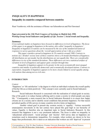
cost-effective osteogenesis without the use of defined media or osteoinductivescaffolds requires a bioreactor that itself drives osteogenesis14; such a bioreactorwould have implications for the pharmaceutical sector and the clinic15.The designs of currently available bioreactors are complex, often involving unfamiliarand hard-to-use parts, such as rotary and perfusion chambers and associated tubing.The creation and use of these parts present major sterility, upscale and cost issues 16.It would therefore be advantageous for a new bioreactor to be able to sit in a standardincubator, consist of a few parts, and to use off-the-shelf cell-culture containers thatare easy to move between the bioreactor and a laminar-flow cabinet.We have previously used nanoscale vibrational displacements to stimulateosteogenesis in MSCs cultured on tissue culture plastic in 2D17, 18. This work identifiedthat mechanical stimulation of 1000 Hz frequency with 15-nm vertical displacementpromoted MSC differentiation18. The process was shown to be RhoA kinase (ROCK)dependent, consistent with known mechanisms of MSC osteogenesis in 2D culturesvia increased adhesion-driven cytoskeletal tension18-20.Multi-sample nanovibrational bioreactor for 2D osteogenesisIn our previous 2D work18 only one sample could be stimulated at a time, and issuesexisted around vibration transmission, which meant that the nanovibrational setupwas complex and lacked scalability. In our nanovibrational bioreactor, we employed areverse piezo effect, where an applied voltage drives nanoscale displacements frompiezo ceramics. The bioreactor design incorporates a 13-piezo array, mounted ontoan aluminium base, directing expansion upwards (Figure 1a). Actuators can be wiredin series or in parallel, with parallel wiring allowing higher vibration amplitudes to beproduced. Mounted above the array is a top plate, which has a surface area capableof accommodating two 6-well culture plates. This plate is dual-layered, usingaluminium bonded to magnetic stainless steel (Figure 1a). This design provides abalance of weight, rigidity, and the ability to magnetically couple 6-well plates viaferrite magnets attached to the base of each well. The magnets allow vibration to be3
transferred to the 6-well plates, to provide extra rigidity to ensure the consistenttransfer of vibration amplitude across each well, and to facilitate the removal of theplate when cells need to be fed.When employing high-frequency vibration, it is important to consider the possibilityof deformation. Using ANSYS finite element analysis (FEA), harmonic response modelsat 1000 Hz with 30 nm amplitude predicted: (1) that the bioreactor top plate wouldprovide rigidity, minimizing average deformation at the nanometre level to 5 nm or17% (Figure 1b); and (2) that the magnetic attachment of 6-well plates maintainsamplitude accuracy across the growth surfaces (within 3.3 nm (11%) for frequencies 3600 Hz) (Figure 1c).Laser interferometry techniques developed for gravitational wave research21 wereused to validate FEA simulations. When wired in series, piezo actuators, without a topplate, gave an average vibration amplitude of 24.7 nm (8.3% accuracy betweenactuators - 1000 Hz, 80 Vpk-pk).Across 25 set points (Figure 1d), amplitudemeasurement of the top plate demonstrated the plate vibrated at 28.2 nm (11.2%amplitude consistency). Measurements made with the 6-well plates magneticallyattached to the top plate (Figure 1d) revealed a slight reduction in displacementamplitude. However, displacement amplitude across multiple wells was shown to beconsistent (21.2% accuracy in the range of 0-3000 Hz (Figure 1e)). Above 4000 Hz, theamplitudes increased towards a resonant condition (Figure 1e – denoted by largevariance), as predicted by ANSYS (Figure 1c).To confirm osteogenesis occurs in 2D, MSCs (from human bone marrow) werestimulated at 1000 Hz (80 Vpk-pk giving a 22 nm vertical displacement)18. While 1000Hz may seem fast relative to the stimulation parameters employed with traditionalmechanical bioreactors, it is noteworthy that for hydrated bone to be optimallypiezoactive, a kilohertz range (972 Hz) stimulus is required22. Furthermore, researchinto the effects of nanotopography indicates that feature sizes of 20 nm high areosteogenic23, 24; this is very close to the 22 nm displacement we use here. After 7 daysof MSC culture, quantitative (q)PCR was used to assess the expression of theosteospecific transcription factor, osterix, and significant up-regulation of its4
expression was observed following nanostimulation (Figure 1f). Furthermore, levelsof the proteins, alkaline phosphatase (ALP) at 14 days and osteopontin (OPN) at 21days, were assessed in nanostimulated MSCs and compared to mature primary humanosteoblasts using in-cell western analysis. Both the bioreactor-stimulated MSCs andthe osteoblasts produced higher levels of these bone marker proteins relative tounstimulated MSCs (Figure 1g). These time points were chosen as they map ontoclassical osteodifferentiation progression models for transcription factors (osterix),early stage markers (ALP) and later stage markers (OPN)25.The 2D paradigm for osteogenesis19, 20, 26 indicates that adhesion-based signalling,including signalling via ROCK18, is important for nanovibration-induced MSCdifferentiation. However, in 3D systems, cellular mechanoreceptors might also play arole in MSC osteogenesis. Transient receptor potential (TRP) channels function asmechanosensors, and TRP channel activation occurs via cytoskeletal linkage27. Tworecently discovered mechanosensitive cation channels, called Piezo1 and Piezo228, 29,affect cytoskeletal organization30, 31 when mechanically activated, and render cellsmechanosensitive to cell adhesion and/or cytoskeletal strain32. In non-excitable (nonneural) cells, TRP and Piezo channels have been implicated in the transduction ofvibrational force, for example in the stereocillia that signal changes in fluid pressurein the ear during hearing, indicating the sensitivity of these channels to high frequencyvibration33, 34. In addition, in a mouse model of osteoporosis, the depletion of thechannel Trpv1 (TRP vallanoid sub-family 1)35 protected mice against ovariectomyinduced bone loss36.A wide range of stimulus can activate TRP channels, including mechanical force, aslong as the energy transduction to the channel is sufficient for mechanical gating tooccur37. Thus, we hypothesised a role for these channels in the response of MSCs tonanovibrational stimulation. Currently available inhibitors of Piezo1 (GsMTX4)38 andof TRPV1 (QX314)39, designed for studies of action potential, act transiently. Thus, tomeasure their effect on nanovibration-induced MSC differentiation, a very early stagemarker of cell response was selected. Mitogen activated protein kinases, such asextracellular signal-related kinase 1/2 (ERK 1/2), provide a likely marker for the switchbetween MSC growth and osteogenic differentiation phases40, with the onset of5
differentiation typified by growth repression through high levels of ERK 1/2 activation(phosphorylation)41.Using an MSC seeding protocol, which allows 1 hour for cell attachment in 10% serum,cells were both stimulated and synchronised at G0 by applying nanovibrations andserum starvation (1% serum) for 24 hours. After synchronising, cells were releasedback into the cell cycle using 10% serum for 23 hours before the addition of GsMTX4or of QX314 for a further 1 hour (24 hours total). Active, phosphorylated, pERK1/2 wasthen assessed relative to total ERK1/2 protein levels using in-cell western analysis. Theinhibition of both the Piezo1 and TRPV1 channels repressed ERK1/2 phosphorylationin the nanovibration-stimulated cells; no repression was seen in control cultures,suggesting that these changes were mechanotransduction-driven via Piezo1 andTRPV1 (Figure 1h). From these results, we hypothesised that channel-basedmechanosensors might be important for the nano-mechanostimulation of MSCs in 3D.Nanovibration in 3D gel volumes.The ability to mechanically stimulate cells at the nanoscale in 3D is challengingbecause of the requirement to transmit nanoscale cues into 3D volumes. In order totest this, a 3D biocompatible scaffold that in itself should not initiate osteogenesis wasused; collagen type I42. The gel was set at low elastic modulus as high-modulus gelscan confer osteoinductive properties; 40 kPa (notably the stiffness of pre-calcifiedbone matrix) has been shown to be osteoinductive to MSCs7, 26, 43-45. Here, we usedgels with a bulk elastic moduli of 108 Pa without cells, and with a moduli of 73.5 Paat a loading density of 40,000 cells/ml of gel (Supplementary Table 1 and Figure 2a,2b). Local elastic modulus (as a cell might sense it) was measured using AFM, andfound to be 59 Pa without cells and 62 Pa with cells at a loading density of 40,000cells/ml (Supplementary Fig 1).To start to understand if collagen could transmit the nanovibrations, FEA harmonicresponse analysis was performed assuming that the 108 Pa collagen gel was withinthe linear elastic range and incompressible (Poisson ratio ν 0.5)46. For collagen gels6
under strain, the Poisson ratio describes the absolute ratio between transverse andlongitudinal strain. For this analysis, a Poisson ratio, ν, of less than 0.5 was chosen.Several values for ν were tested for their replication of the interferometric result withan asymptotic increase towards 0.5 improving the accuracy of the simulation (i.e. theability to transmit a full vibration amplitude of 20 nm). The value used also resulted ina bulk modulus of the same magnitude as water (Supplementary Table 1). The bulkmodulus describes a material’s resistance to uniform compression; such materialstend to have a high water content, as in the case of collagen gels. Modelling of thepolystyrene container showed it produced minimal deformation (Figure 2d).Modelling of the gel (at 2.5 ml of gel per well), at 1000 Hz with 20 nm displacement,predicted an average amplitude of 29 nm at the edge meniscus and 18 nm at the gelcentre (Figure 2e).Using these same modelling parameters, shear stress and strain within the gelproduced peak values of 3.8 mPa and 1.1 x 10⁻⁴ m/m respectively at 1000 Hz (shearstress is shown in Figure 2f). These values fall within the linear region of the shearstress-strain plot in Figure 2c (from rheological measurement). This supports thevalidity of using a constant elastic modulus value for modelling nanovibration, at thisfrequency, since the strains produced are within the linear elastic range of thematerial. Hence, the viscoelastic properties of the collagen gel are predicted to allowthe transfer of high frequency vibrations to the cells seeded in 3D.Interferometry was again used to confirm predictions. The bioreactor was set up witha 6-well plate and with cells (40,000 MG63 cells/ml –note, MG63 cells were chosen asthey can be used on the interferometer outside of the cell culture laboratory) seededwithin 2.5 ml collagen gels. Measurements were carried out both at the centre andouter edge of the gels. Amplitude calibration showed that for the frequenciesexamined, gel vibration increased linearly with input voltage to the bioreactor (Figure2h). In terms of frequency, the top surface of the gel was found to follow the surfaceof the 6-well plate consistently, showing no dampening of the vibration up to 2000 Hz.Gel samples that contained cells did not deviate in response from gels without cells(Figure 2i). Up to 2000 Hz, amplitude appeared to be increased at the edge of the gelat the meniscus curvature, thus reflecting the predicted data (Figure 1b). The vibration7
amplitude seen at the edge of the gel was 35 nm compared to 24 nm at the centre for1000 Hz (Figure 2i – inset).Resonant frequencies were recorded in the region of 2500, 3500 and 5000 Hz andwere observed in both empty and gel-filled 6-well plates. We also observed anincrease in vibration amplitude at the outer edge of each well when compared to itscentre, together with a decrease in measurement accuracy (Figure 2i). If the gelvolume is doubled to 5 ml, the data indicate that a slight amplitude-dampening effectoccurs, although the gels act similarly in terms of where resonance occurs withdifferent voltage input, and with respect to middle/edge amplitude changes(Supplementary Figure 2). Our results also indicate that cells within the 100 Pa gelsshould receive predictable and reproducible nanoscale displacements. However, thisgel environment is dramatically different to the hard environment of the plastic cellculture well (as used to generate Figure 1f & g) or to 40 kPa gels, which have previouslybeen used to stimulate osteogenesis7, 26, and this presents a significant challenge interms of achieving osteogenesis.Nanovibration driven 3D osteogenesis.To test MSCs for osteogenesis in the 108 Pa collagen gels, a range of viability andosteo-specific transcriptional, protein and mineralisation assays were performed. Interms of viability, our analysis showed that MSCs embedded in these gels (referred tohereafter as 3D) remain viable during nanostimulation (Supplementary Figure 3). Toassay for osteogenesis, qPCR at days 7, 14 and 21 of culture was performed, andshowed increased RUNX2, collagen I, ALP, OPN, osteocalcin (OCN) and bonemorphogenetic protein 2 (BMP2) expression in the nanostimulated MSCS in 3D,compared to unstimulated MSCs (Figure 3a). In MSCs that were nanostimuled, four ofthe six genes above were downregulated at day 21, as the osteogenic process wastranscriptionally completed (Figure 3a). OCN protein levels were also assayed at day21 and found to be increased following nanostimulation demonstrating progressionfrom gene to protein (Supplementary Figure 4).8
To study in vitro mineralisation, we used a combination of von Kossa staining forphosphate, Raman spectroscopy, and X-ray micro-computed tomography (µCT). VonKossa staining indicated that relatively little mineralisation had taken place withincontrol gels (Figure 3b – images are from 6 weeks of culture). However, followingnanostimulation at 15 nm displacement, significant mineralisation was detected inMSCs cultured in 3D. Mineralisation levels were higher in these cells than in MSCsafter 4 weeks of culture in osteospecific media (OSM) (Figure 3b). In longer-term, 6week, culture, MSCs cultured in 3D with OSM caught up and underwent a significantincrease in mineralisation compared to control cells (Figure 3b).Within the Raman fingerprint region of 500 to 1500 cm-1 (Figure 3c,d,f andSupplementary Figure 5), bovine cortical bone showed a dominant phosphate (PO43v1) peak at 960 cm-1 and a secondary peak at 1072 cm-1, characteristic of substitutedcarbonate (CO32-) and indicative of the mineral component of bone47, as well as ofhydroxyapatite Ca10(PO4)6(OH)2, where CO32- can substitute for OH- and/or PO43(Figure 3c). For MSCs cultured in control gels, a broad, merged peak from 1200 cm-1was observed (likely due to the large amount of collagen present), and protein-relatedcontributions from amide I (1595-1720 cm-1) and III (1243 -1269 cm-1) and from CH2(1451 cm-1). Significantly, no bone mineral peaks were noted in these cells (Figure 3d)and µCT revealed no mineral deposits (Figure 3e). In nanostimulated MSCs in 3D, twopeaks at 960 cm-1 (PO43- v1) and 1072 cm-1 (CO32-) were observed (Figure 3f), and µCTshowed the accumulation of bone mineral (Figure 3g, Supplementary Table 2).Together, these findings demonstrate that nanostimulation can drive osteogenesisand mineralisation in MSCs in an otherwise non-osteogenic 3D environment.3D nanovibrational stimulus of mechanosensitive channels.We next tested ion channel sensitivity to 3D nanostimulation in order to relate ionchannel expression to cytoskeletal tension. Temporal qPCR was performed on MSCsat days 3, 5, 7, 14 and 21 of culture to assay the expression of Piezo 1, Piezo 2, TRPV1and of potassium channel subfamily K receptor 2 (KCNK2, which is stimulated bymembrane stretch48). Nanostimulation of MSCs in 3D produced a similar trend across9
all the channels tested, specifically a bi-phasic high to low expression pattern from day3 to day 5, recovery to a higer level at days 7-14 to a lowered expression pattern atday 21, with all channel transcripts significantly downregulated by day 21, postosteogenesis (Figure 4a). From our 2D data18 and from the literature on osteospecificdifferentiation on a range of 2D surfaces19, 20, it could be postulated that intracellulartension should increase during osteogenesis; thus, if the channels were involved inosteogenesis in 3D, they might be sensitive to tension inhibitors, such as the ROCKinhibitor (Y27632) and the myosin II inhibitor (blebbistatin).To test this hypothesis, day 5 was selected as a representative time point for thedownregulation of Piezo 1, Piezo 2 and TRPV1 channel expression (Figure 4a) and, aswe were now investigating a single time point, MSCs from additional donors weresupplemented into the analysis. All three ion channels were downregulated by day 5,with Piezo 2 being significantly down-regulated. The trend was noted in OSM-treatedcells, but was more pronounced in MSCs subjected to nanostimulation (Figure 4b-e).Upon addition of Y27632 and blebbistatin, the down-regulation of Piezo 2 expressionwas reversed, indicating a relationship exists between this receptor and cellulartension. This trend was noted in both nanostimulated and OSM-treated cells, but wasmore pronounced following nanostimulation (Figure 4c). TRPV1 also showed atension-dependant link, increasing in expression with nanostimulation and OSMtreatment following treatment with Y27632. Again, this was more pronounced fornanostimulated cells, as blebbistatin treatment increased the expression of TRPV1only in nanostimulated MSCs (Figure 4e).The effect of actin and ROCK inhibition on early stage (to day 5) osteogenic transcriptswas next investigated in nanostimulated MSCs. BMP2 receptor (BMPR1a) wasdifferentially regulated in blebbistatin-inhibited MSCs (Figure 4f). The response toBMP2 is a first step in osteogenesis and it stimulates transcription factors, such asRUNX2 (the master osteogenic transcription factor, which peaks in expression at day5-10)25. At this early time point, however, RUNX2 expression was not altered inresponse to either inhibitor (Figure 4g). ALP, a later stage marker of osteogenesis(which peaks in expression day 1425, 49), was minimally expressed (Figure 4). At day10
14, blebbistatin treatment downregulated the expression of two later stage markersof osteogenesis, osteonectin (ONN) and OPN (Supplementary Figure 6).These data support the hypothesis that 3D osteogenesis via nanovibrationalstimulation is a mechanotransductive process that involves intracellular tension, withmechanoreceptors, such as Piezo, TRP and KCNK, being involved in this process.However, we note that our results are not as dramatic, in terms of response tocytoskeletal inhibition, as seen in in 2D osteogenesis studies19, 20, 26.Nanovibrational mechanotransductive signalling.The receptor that appeared most mechanoresponsive to nanostimulation at thistimepoint (day 5) was TRPV1. It has been proposed that TRPV channels influenceosteogenesis through the -catenin (Wnt pathway) activation of RUNX250, 51. In thispathway, ion influx through the TRPV channels causes the activation of protein kinaseC (PKC) and of ERK, mediating -catenin activity50, 51.To investigate the effects of nanovibrations on signalling processes in MSCs, we usedmetabolomic analysis to predict canonical signalling events as this approach providesan instant picture of phenotypic modulation. Principle component analysis showedthat 3D nanovibrated MSCs and control MSCs, had different metabolomes indicativeof different phenotypes (Supplementary Figure 7a). As expected during activedifferentiation, energy based pathways52 were upregulated in nanostimulated MSCs,including lipid (Supplementary Figure 8) and carbohydrate (Supplementary Figure 9)metabolic pathways, with lipids representing the largest group. Ingenuity pathwayanalysis (IPA) fits metabolite input data to the literature in order to be able to predictbiochemical interactions. It was used here to build networks related to lipidmetabolism, that were merged and linked to canonical signalling pathways related toadhesion, cytoskeletal tension, ERK 1/2 signalling, BMP2 signalling and calcium/Wntsignalling19, 20, 26 (Supplementary Figure 7b).To investigate if TRPV plays a role in the osteogenic process, western blotdensitometry was used to measure key members of these pathways after control and11
nanovibrational culture. Specifically, pFAK, pMyosin, pERK, pSMAD1/5/8 and active catenin, with and without inhibition of PKC with enzastaurin, were considered. Only -catenin was affected, becoming down-regulated as PKC was inhibited and thenormal functioning of TRPV channels was indirectly blocked (Figure 5a – no effect ofPKC inhibition was seen in 3D MSCs treated with OSM, Supplementary Figure 10).We next investigated the effect of channel inhibitors on nanostimulated MSCs in 3Dby measuring total ERK 1/2 and total -catenin levels in the presence and absence ofPKC and TRPV channel inhibition. MSCs in 3D underwent nanostimulation (1000 Hz),nanostimulation with PKC inhibition (1000 Hz-PKC), and nanostimulation with TRPVinhibition (1000 Hz-TRPV); protein levels were assayed after 48 hours of stimulation(Supplementary Figure 11). At the same time, a metabolomics/IPA analysis wasperformed to investigate signalling networks linked to the -catenin/Wnt pathways.Western blot analysis showed that nanostimulated MSCs treated with PKC and TRPVinhibitors downregulated the expression of ERK 1/2 and -catenin but that only catenin was significantly down-regulated in the presence of both inhibitors (Figure5b). The metabolomics analyses indicated that the Wnt/ -catenin pathway wasupregulated following nanostimulation (Figure 6a), but was moderately downregulated in the 1000 Hz-PKC MSCs (Figure 6b) and largely suppressed in the 1000 HzTRPV cells (Figure 6c) (see Supplementary Figures 12-14 for more complete networks).The metabolomics analysis also inferred that ERK signalling is downregulated in the1000 Hz-PKC and 1000 Hz-TRPV MSCs (Figure 6a-c), which is in agreement with thewestern blot data which show that total ERK protein levels are reduced (Figure 5b).Together, these findings lead us to propose that TRPV1 activity contributes to Wntmediated osteogenesis in nanovibrated MSCs in 3D. Potential mechanisms are shownin Figure 6d with the TRPV/ -catenin pathway highlighted in bold.To test if inhibition of PKC and TRPV have direct effects on osteogenesis in 3D culture,nanovibrational stimulation time was extended out to 5 days to allow for somephenotypical development and the cultures were inhibited for PKC or TRPV for thefinal 24 hours. QPCR was used to test transcript level changes; we looked at BMPR1aand BMP2 as very early markers, RUNX2 as an early marker and ALP and osteonectin12
as mid-stage osteogenic markers. For PKC inhibition, downregulation of BMP2 wasobserved indicating a phenotypical response (Figure 7). However, with TRPVinhibition, significant downregulation of BMP2, RUNX2, ALP and osteonectin wasobserved showing an immediate and dramatic effect on progression of the osteogenicphenotype. This data supports that TRPV channels are important to osteogenesis andthat PKC may also have effect (but indicating that it might not be an exclusive effect),helping build the hypothesis presented in Figure 6d.We have presented a new class of bioreactor and showed that 3D osteogenesis canbe produced without the use of chemicals and/or growth factors, or of bioactivescaffolds, sophisticated culture ware, or dedicated incubators. The use of collagen inthis study provided mechanical integration with the 6-well plate because it attachesto the dish sides. This allowed us to take advantage of its excellent mass-transferproperties, which also permit large volumes to be used, avoiding the need to usedense osteogenic scaffolds, such as calcium phosphates and other bioactive ceramics,which have certain disadvantage and limitations. In fact, on a single bioreactor with 5ml of gel fill per well, 60 ml of mineralising osteoblasts in an appropriate 3D matrixcan be generated from MSCs.Summary.In addition to the potential volumes of graft material that could be produced usingthis bioreactor, it also fits well into a good-manufacturing-practice (GMP) workflow.GMP-grade collagen, media and culture plates exist and have been used in humantrials53. In this approach, the nanovibrational bioreactor remains external to thesecomponents as it does not come into contact with cell-therapy products. This preventsthe need to, for example, meet FDA regulation for new scaffolds or inductionchemicals.We propose that, because of the soft 3D nature of the collagen matrix used in ourapproach, the magnitudes of the roles for adhesion and cytoskeletal tension werelower than those observed in 2D studies19, 20, 26. We also suggest an increased role for13
mechanoresponsive channels, which we believe stimulate enhanced osteogenesisthrough the activation of -catenin signalling.Acknowledgements. This work was supported by grants to Reid and Dalby fromBB/N012690/1, BB/P00220X/1, EP/N013905/1, EP/N012631/1, EP/P001114/1 fromBBSRC, BBSRC/SFI and EPSRC and a programme grant from Find a Better Way. Wethank Jim Hough, Habib Nikukar, Ivor Tifenbrun and Keith Robertson for theirdiscussion, Carol-Anne Smith for technical support, and Erin Manson for help withmetabolite analysis. Biggs is funded by SFI grant nos. 11/SIRG/B2135 and 13/RC/2073.Author Contributions. PMT, PGC, GDP, JY, VJ, WO, CG-G, DT and CVG performed thelaboratory experiments.
3Glasgow Polyomics facility, College of Medical, Veterinary and Life Sciences, University of Glasgow, Wolfson Wohl Cancer Research Centre, Garscube Campus, Bearsden, G61 1QH, UK. 4Microenvironments for Medicine, Division of Biomedical Engineering, School of Engineering, College of Science and Engineering, University of Glasgow, Glasgow, G12 .