Transcription
Emmert et al. International Journal of Implant 2International Journal ofImplant Dentistry(2021) 7:83RESEARCHOpen AccessAn experimental study on the effects of thecortical thickness and bone density oninitial mechanical anchorage of differentStraumann implant designsMarie Emmert1†, Aydin Gülses1*† , Eleonore Behrens1, Fatih Karayürek2, Yahya Acil1, Jörg Wiltfang1 andJohannes Heinrich Spille1AbstractBackground: The aim of the current study was to comparatively assess the primary stability of differentStraumann implant designs (BLX, Straumann Tapered Effect, Bone Level Tapered, and Standard Plus) via resonancefrequency analysis by using an implant insertion model in freshly slaughtered bovine ribs with and without corticalbone. Tapered Effect (4.1 10 mm), Bone Level Tapered (4.1 10 mm), Standard Plus (4.1 10 mm), and BLX (4.0 10 mm) implants were inserted into the distal epiphysis on the longitudinal axis of the freshly slaughtered bovineribs. As a control, implants with the same sizes were inserted into the proximal diaphysis. The stability of theimplants was examined with resonance frequency analysis.Results: BLX and Tapered Effect implants showed higher implant stability quotient values in both study and controlgroups. All implant systems showed a significant decrease of mechanical anchorage in the study group. BLX andBone Level Tapered designs had a significantly lower loss of mechanical anchorage in the lack of cortical bone.Conclusion: Both Tapered Effect and BLX designs could ensure sufficient initial stability; however, BLX implantscould be an appropriate option in the lack of cortical bone and poor bone quality at the implant recipient site.Clinical relevance: BLX is a novel implant system, which could be especially beneficial in the presence ofspongious bone type at posterior maxillae.Keywords: Anchorage, Bone, Cortical, Implant, Primary, StabilityIntroductionThe primary stability of a dental implant plays a key rolein implant integration and long-term successful clinicaloutcome. Considering the implant recipient site, primaryimplant stability could be affected by several factors suchas bone density, thickness of the cortical bone, andheight of the alveolar ridge. In order to overcome these* Correspondence: aguelses@mkg.uni-kiel.de†Marie Emmert and Aydin Gülses contributed equally to this work.1Christian Albrechts University, Department of Oral and Maxillofacial Surgery,University Hospital of Schleswig-Holstein, Campus Kiel, Arnold-Heller-Straße3, 24105 Kiel, GermanyFull list of author information is available at the end of the articlequantitative and qualitative deficits, various implantgeometries and thread designs have been developed.Several studies comparing the outer geometry of the implants found that implants with conical shape [1] and/ortapered designs [2] had superior primary stability compared to those with a parallel shape and could significantly improve the initial mechanical anchorage.It is well known that the loose structure of the trabecular bone could negatively affect mechanical anchorage [3–9]. Therefore, numerous clinical andexperimental studies have mainly focused on the effectsof bone density and geometry on implant stability. The Author(s). 2021 Open Access This article is licensed under a Creative Commons Attribution 4.0 International License,which permits use, sharing, adaptation, distribution and reproduction in any medium or format, as long as you giveappropriate credit to the original author(s) and the source, provide a link to the Creative Commons licence, and indicate ifchanges were made. The images or other third party material in this article are included in the article's Creative Commonslicence, unless indicated otherwise in a credit line to the material. If material is not included in the article's Creative Commonslicence and your intended use is not permitted by statutory regulation or exceeds the permitted use, you will need to obtainpermission directly from the copyright holder. To view a copy of this licence, visit http://creativecommons.org/licenses/by/4.0/.
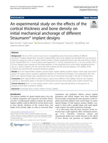
Emmert et al. International Journal of Implant Dentistry(2021) 7:83Nevertheless, the lack of cortical bone at the implant recipient site could be also challenging; thus, in additionto total bone thickness, the height of cortical bone alongthe implant has shown to have a relevant effect on primary implant stability [10, 11]. Sugiura et al. have suggested that the crestal cortical bone height couldsignificantly affect the maximum extent of micromotionand peri-implant bone strain in the low-density cancellous bone under immediate loading [6]. Several studieshave also clearly showed that improving primary stabilityvia bi-cortical anchorage plays a great role in the successof immediate loading protocols such as All-on-4 [12]and All-on-3 [13] and implant placements followingsinus floor augmentation. However, 70% of all agerelated bone loss is cortical [14] and the lack of corticalbone especially in the posterior maxillae could pose agreat challenge for the clinician [15], especially in elderlydental implant patients.The number of studies assessing the influence of cortical bone height on the mechanical anchorage regardingthe dental implant design is limited [10, 11]. The aim ofthe current study was to comparatively assess the primary stability of different Straumann implant designs(BLX, Tapered Effect [TE], Bone Level Tapered [BLT],and Standard Plus [SP]; Institute Straumann AG, Basel,Page 2 of 8Switzerland) via resonance frequency analysis by usingan implant insertion model in freshly slaughtered bovineribs with and without cortical bone.Material and methodsAll experiments were performed at room temperatureby two researchers (M.E. and A.G.). A total of 10 freshlyslaughtered bovine ribs were used. Soft tissues and periosteum were completely removed off the bone.Straumann Tapered Effect (4.1 10 mm), Bone LevelTapered (4.1 10 mm), Standard Plus (4.1 10 mm), andBLX (4.0 10 mm) implants (Fig. 1) were inserted intothe distal epiphysis on the longitudinal axis of the freshlyslaughtered bovine ribs (Fig. 2). The distance between theimplant shoulders was set to a minimum of 5 mm.As a control, implants were also individually placedinto the proximal diaphysis of the bovine ribs, where acortical bone surrounding the bone recipient site exists(Fig. 3). Implant insertions in both groups were conductedaccording to the drilling protocol with instrumentariumrequired for each system, with one exception: The use ofboth pilot and 2.0-mm drills were skipped; thus, prior topreparation of the samples, bone biopsies were taken witha trephine burr (2.3 mm) for histological evaluation to determine the cortical bone height at the correspondingFig. 1 a From left to right: BLX (4.0 10 mm), Straumann Tapered Effect (4.1 10 mm), Bone Level Tapered (4.1 10 mm), and Standard Plus(4.1 10 mm). (The figures were taken from the product catalogue with kind permission of Institute Straumann AG, Basel, Switzerland AG,2015. All rights reserved.) b The implants were inserted into the distal epiphysis on the longitudinal axis of the freshly slaughtered bovine ribs.The distance between the implant shoulders was set to a minimum of 5 mm. c Prior to implant placement, bone biopsies were taken tohistologically examine the cortical bone height at the implant recipient area. Left: Biopsy taken from the control, right: Study group. dMeasurement of the primary stability of the implants with Osstell device inserted in cancellous bone (from left to right, Straumann Tapered Effect[4,1 x 10 mm], Standard Plus [4,1 x10 mm], Bone Level Tapered [4,1 x 10 mm], and BLX [4,0 x 10 mm]). e. Implants were also individually insertedinto the proximal diaphysis of the bovine ribs to serve as control. (Tapered Effect 4.1 10 mm)
Emmert et al. International Journal of Implant Dentistry(2021) 7:83Page 3 of 8Fig. 2 The height of the bone cortex was measured on samples stained with toluidine blue (a, b) and later by micro-radiography (c, d). a Thestudy group. b The control group with the bone cortex. c The study group. d The control group with cortical boneimplant recipient site (Fig. 4). For both groups, a total of40 implant insertions were performed. Two blinded independent observers (E.B. and J. W.) have assessed the accuracy of placement. A peak insertion torque value of 30N/cm has been exceeded by all implant insertions.Measurement of the primary stabilityImplant stability quotient (ISQ) has been quantified byusing resonance frequency analysis (RFA) via Osstell device (Osstell Mentor, Integration Diagnostics Ltd., Göteborg, Sweden). For each implant design, a suitabletransducer was inserted into the implant body (SmartPeg, Osstell, Göteborg, Sweden). Measurements weretaken in four different directions perpendicular to theSmartPeg, according to the manufacturer’s guidelines.The mean value of four measurements has been calculated to determine the final ISQ value of each implant.All measurements were conducted in triplicate.Histological evaluationThe tissue samples were histologically evaluated according to the technique described by Donath et al. [16] andlater developed by Acil et al. [17, 18]. Briefly, tissue samples were placed into 10% neutral buffered formalin(NBF) for fixation for 4 days and embedded in methacrylate prior to sawing and grinding. Sawing and grinding were performed, and the samples were placed inglass vessels filled with monometric resin solution andincubated at 37–40 C for 2 to 4 days for resinimpregnation.The samples were longitudinally cut with a band saw(Exakt, Norderstedt-Germany), and sections of about100 μm were obtained via an oscillating diamond saw(Exakt, Norderstedt-Germany), grounded with the Sapphire 360 E grinder (ATM, Altenkirchen-Germany), andhighly polished with silicon carbide paper (grades 500,1200, 2400, and 4000). Staining was performed by usingtoluidine blue. The ground surface was decalcified with0.1% formic acid and 20% methanol was applied for better cell and soft tissue staining. The samples were rinsedin distilled water and stained in a toluidine blue solutionfor 2 min. The height of the cortical bone was measuredafter micro-radiography of the specimens (Fig. 5). Thepreparations were digitally photographed with the Nikonphotomicrography with a magnification of 509 andassessed manually with the Leica Q-Win Imaging program (McBain Systems, CA, USA). Relative amounts ofbone area vs total tissue of the spongious bone weremeasured as described by Takahashi et al. [19] (% bonearea/total tissue (% BA/TA)).Micro-radiographyThe samples were taken from the slide and put on highresolution micro-radiography plates in which the resolution is 2000 lines per millimeter (high-resolutionplates, Kodak , Rochester, NY 14650, USA).
Emmert et al. International Journal of Implant Dentistry(2021) 7:83Subsequently, the exposure was performed in Faxitron53855A (Hewlett-Packard, McMinnville, OR 97128,USA) at a focus distance of 16 cm. The voltage was setto 25 kV at a power of 3 mAs, exposure time of 6 minand 30 s for 70-μ samples, and up to 10 min and 30 s for110-μm film thickness. The exposure time was increasedby increments of 1 min per 10-μm thickness of the specimen. The plates were embedded for 5 min in Kodak HRP developer at 20 C under constant movementswung (HRP developers/distilled water, 1/3). In a 1%acetic acid bath, the development was stopped after 1min. For fixation, Kodak fixer 3000A was used for 10min with agitation (Kodak fixer 3000A/distilled water,1/3). This was followed for 15 min by washing withwater and a final rinse with Agepon (400 mL of distilledwater and 2 mL Agepon ) for 1 min. The plates were airdried and then covered with cover glasses (4 4.5 cm)using 1–2 drops of n-butyl acetate (xylene substitute)and a drop of Eukitt-air for 24 h. After a drying periodof 7 days under an air extractor, the preparations weredigitally photographed under a light microscope at amagnification of 1:18. The evaluation was performedusing an image editing program (Adobe Photoshop 7.0for Windows) (Fig. 2).Statistical analysisStatistical software SPSS (IBM) was used for statisticalanalysis. Power analysis was performed to determine thesmallest sample size that is suitable to detect the effectof a given test at the desired level of significance. Thedistribution of the variables was assessed by using theShapiro-Wilk test. The comparison of two variables wasPage 4 of 8performed via independent-T and paired-T tests. Oneway analysis of variance (ANOVA) was conducted forcomparative analysis of the variables. p 0.05 was considered as statistically significant.ResultsCortical bone height and trabecular structureThe average cortical bone height in the control groupranged between 2.34 and 3.21 mm (2.58 0.615 mm).The histological examination revealed that, totally, n 6(15.0%) of the bone structures correlated with type IIIbone (SP [n 1], TE [n 3], BLX [n 2]), whereas 34 (85.0%)of the specimens were identical to type IV bone (SP [n9], BLT [n 10], TE [n 7], BLX [n 8]) [20]. The evaluationof the mean primary stability values of the implantsinserted only in type IV bone in both groups (regardlessof the presence of cortical bone) revealed that the BLXdesign had significantly higher mechanical anchoragecompared to other implant systems (p 0.043) (Fig. 3).Comparison of implant designsIn the study group, the BLX implant showed higher ISQvalues compared to other groups. The significant difference was found when compared to SP and BLT groups(p 0.001 and p 0.0016, respectively). Besides that, TE implants showed statistically significant superiority compared to SP (p 0.001). The difference between BLT andSP groups was also remarkable (p 0.003) (Fig. 4a).In the control group, TE implants showed slightlyhigher values compared to BLX; however, the differencewas statistically insignificant. BLX and TE implantsshowed significantly higher ISQ values compared to BLTFig. 3 The comparative assessment of the ISQ values in type IV bone (SP [n 9], BLT [n 10], TE [n 7], and BLX [n 8]) in both groups (regardless ofthe presence of cortical bone) revealed that the BLX design had significantly higher mechanical anchorage compared to other implant systems(p 0.043)
Emmert et al. International Journal of Implant Dentistry(2021) 7:83Page 5 of 8Fig. 4 a BLX implant showed higher ISQ values compared to other groups. The significant difference was found when compared to SP and BLTgroups (p 0.001 and p 0.0016, respectively). Besides that, TE implants showed also statistically significant superiority compared to SP (p 0.001). Thedifference between BLT and SP groups was also remarkable (p 0.003). b TE implants showed slightly higher values compared to BLX; however,the difference was statistically insignificant. BLX and TE implants showed significantly higher ISQ values compared to BLT and SP groups (BLX-BLTp 0.004, BLX-SP p 0.001 and TE-BLT p 0.001, TE-SP p 0.001, respectively)and SP groups (BLX-BLT p 0.004, BLX-SP p 0.001 andTE-BLT p 0.001, TE-SP p 0.001, respectively) (Fig. 4b).Differences in ISQ values for each implant designregarding the presence of cortical boneAll implant systems showed a significant decrease ofmechanical anchorage in the study group (BLX p 0.000*,TE p 0.001*, BLT p 0.000*, and SP p 0.002*, respectively). The decrease in ISQ values was assessed in percentages (Fig. 5). According to this, the decrease waslowest in the BLX group, followed by BLT. The difference was significant compared to the loss of mechanicalanchorage obtained in the TE group (p 0.033 and p0.008, respectively).The results of the study revealed that the TE and BLXimplants showed higher ISQ values compared to BLTand SP implants in both experimental and controlgroups.DiscussionIn the literature, the influence of the height of the residual bone height on dental implant procedures hasbeen widely studied. Despite several articles have showedthat the height of the cortical bone at the implant recipient site could play a crucial role in the primary stability,the number of articles focusing on the effects of the cortical bone on the initial implant anchorage is limited [10,11]. Miyamoto et al. have suggested that the initial stability at the time of implant installation is influencedFig. 5 The decrease in percentage was lowest in the BLX group, followed by BLT. The difference was significant compared to the loss ofmechanical anchorage obtained in the TE group (p 0.033 and p 0.008, respectively)
Emmert et al. International Journal of Implant Dentistry(2021) 7:83more by cortical bone thickness than by implant lengthand is extremely important for implant stability at thetime of surgery [4]. Similarly, Ming-Gene et al. [9] havestated that the bone strain is influenced more by thecortical bone thickness than by the implant length forimmediately loaded implants.According to an anatomical study performed byKatranji et al., the average cortical height of the edentulous posterior maxilla is about 2.06 mm [21]. It is alsowell known that the thickness of the compacta of thejawbones increases up to the age of 50 years and decreases significantly thereafter [22]. Additionally, the lossof the cortical bone presents a great challenge in themanagement of female dental implant candidates. Koet al. [23] evaluated the prospective implant recipientsites by menopausal and post-menopausal women andshowed that thickness of the crestal cortical bone wasthe lowest in the posterior maxilla with an averageheight of 0.66 0.24 mm. Considering the experimentaldesign (cortical bone of 0.00 mm vs 2.58 0.615 mm)and type IV bone morphological structure, it might beproclaimed that the current model could sufficiently resemble the human posterior maxilla.Romanos et al. [2] have previously compared the primary stability of different Straumann implants (BLT, SP,and TE) and found significantly higher implant stabilityfor TE implants. The results of the current study werealso in accordance with the existing literature. Besidesthat, all implant systems showed a significant decrease inISQ values in the study group. However, despite thenearest ISQ values of BLX and TE implants in bothstudy and control groups, it was remarkable that BLXimplants showed significantly lower loss of the mechanical anchorage in the lack of cortical bone. In addition,BLT systems revealed lower ISQ values in both controland study groups; however, the decrease of the primarystability was also lower compared to TE and SP systems.This could be attributed to the conical outer design ofthe BLT implants and corresponds to the results of theprevious articles [24, 25].The BLX system is a novel “self-drilling” implant system developed by Straumann (Straumann , InstituteStraumann AG, Basel, Switzerland). It has been proclaimed that its double thread design could provide animproved initial stability in all types of bone throughuniform and controlled compaction and densification ofthe peri-implant bone [26]. The number of studies focusing on the primary stability of this novel system islimited [27].I-Chiang et al. [7] have evaluated the micromotions ofdifferent implant types and found that the maximumstress of the peri-implant bone decreased as corticalbone thickness increased. According to that, the horizontal loading component induces stress concentrationPage 6 of 8in the bone around the implant neck more easily thandoes the vertical loading component, especially by implants with a conical shape. In the literature, it has beenclearly shown that TE implants presented higher stabilityvalues compared to the conventional implants, attributedto their compression to the cortical bone, which was alsopreviously shown at implant recipient sites with poorbone quality by O’Sullivan et al. [28]. The superiority ofthe BLX system observed herein might be a result of itsdouble thread design and the presence of two sharp andhighly engaging threads at its apex, which allowed an advanced bone-to-implant contact and higher mechanicalanchorage with minimal negative effect in the lack of thecortical bone. On the other hand, the loss of primarystability in the TE group could be due to the decreasedcompression of the implant neck to the surroundingbone, where the cortical bone does not exist.It is well known that implant length and diametercould also influence the primary stability. Therefore,in the current study, implants with the same lengthand almost the same diameters were used. One of themain limitations of the current study is that the implant recipient site could exhibit different quantitativecharacteristics in its both cancellous and corticalstructures [29]. To overcome this issue, histologicalcharacteristics have been also quantitated. Accordingto that, RFA of all implant systems in both study andcontrol groups in type IV bone showed the superiority of the BLX design. Therefore, the use of this novelsystem could be preferred in the presence of a poorbone quality at the implant recipient site, regardlessof the presence and/or height of the cortical bone.It is obvious that the implant geometry can increasethe surface area of support. A threaded design implant has 30 to 200% greater surface area comparedwith a cylinder implant of the same size. Therefore,the threaded implant in poorer density bone isstrongly encouraged [30, 31]. Considering this, theuse of the BLX design in type III and type IV bonecould be a feasible option to ensure a mechanical anchorage thanks to its greater surface area. Besidesthat, the implant neck design of the TE design whichapplies additional transversal compression forces tothe marginal bone could be also favored if the cortical bone is present.From another clinical point of view, if the removal ofan implant is indicated, double thread design and thepresence of two sharp and highly engaging threads at itsapex could complicate the removal of a partially or fullyintegrated BLX implant from a type I and type II boneand a trephine or surgical bur technique should be performed, which might not be the most suitable option interms of maintaining of the bone and minimizing damage to vital structures [32].
Emmert et al. International Journal of Implant Dentistry(2021) 7:83ConclusionBLX implant systems could be an appropriate option inthe lack of cortical bone at the implant recipient site.This fact is more likely of clinical importance for implant insertions at the posterior maxilla and could bepreferred in cases where type IV bone exists. Additionally, both TE and BLX systems could ensure sufficientinitial stability. However, further studies are needed toexactly clarify the clinical advantages of the BLX design.AbbreviationsBLT: Bone Level Tapered; SP: Standard Plus; TE: Tapered Effect; ISQ: Implantstability quotient; BA: Bone area; TA: Total area; NBF: Neutral bufferedformalin; RFA: Resonance frequency analysis; USA: United States of AmericaAcknowledgementsThe authors would like to thank Gabriele Neßenius for the preparation ofhistological samples.Authors’ contributionsAll samples were prepared by A.G. Implant insertions were performed byM.E. and A.G. E.B. and J. W. have assessed the accuracy of the implantplacement. The methodology was reviewed by F.K. Histological examinationswere conducted by J.S. and Y.A. The authors read and approved the ität zu Kiel (Forschungsförderung 2020-JuniorF359929)Availability of data and materialsAll data is available in the archive of the Research Laboratories, ChristianAlbrechts University, Department of Oral and Maxillofacial Surgery.Page 7 of 84.5.6.7.8.9.10.11.12.DeclarationsEthics approval and consent to participateAll samples were prepared by using animal body parts, which werecommercially purchased at the slaughterhouse Steffen in Muxall, Germany.No animal was harmed or sacrificed in order to conduct the experimentsdescribed herein.13.Consent for publicationAll authors gave their consent for publication.15.Competing interestsMarie Emmert, Aydin Gülses, Eleonore Behrens, Fatih Karayürek, Yahya Acil,Jörg Wiltfang, and Johannes Heinrich Spille declare that they have nocompeting interests.Author details1Christian Albrechts University, Department of Oral and Maxillofacial Surgery,University Hospital of Schleswig-Holstein, Campus Kiel, Arnold-Heller-Straße3, 24105 Kiel, Germany. 2Department of Periodontology, Cankiri KaratekinUniversity, Cankiri, Turkey.14.16.17.18.Received: 31 March 2021 Accepted: 16 June 202119.References1. da Costa Valente ML, de Castro DT, Shimano AC, et al. Analysis of theinfluence of implant shape on primary stability using the correlation ofmultiple methods. Clin Oral Investig. 2015;19(8):1861–6. https://doi.org/10.1007/s00784-015-1417-4.2. Romanos GE, Ciornei G, Jucan A, Malmstrom H, Gupta B. In vitro assessmentof primary stability of Straumann implant designs. Clin Implant Dent RelatRes. 2014;16(1):89–95. . Kim D-S, Lee W-J, Choi S-C, Lee SS, Heo MS, Huh KH, et al. Comparison ofdental implant stabilities by impact response and resonance frequencies20.21.22.using artificial bone. Med Eng Phys. 2014;36(6):715–20. yamoto I, Tsuboi Y, Wada E, Suwa H, Iizuka T. Influence of cortical bonethickness and implant length on implant stability at the time of surgery-clinical, prospective, biomechanical, and imaging study. Bone. 2005;37(6):776–80. https://doi.org/10.1016/j.bone.2005.06.019.Tözüm TF, Dursun E, Uysal S. Radiographic fractal and clinical resonancefrequency analyses of posterior mandibular dental implants: their possibleassociation with mandibular cortical index with 12-month follow-up.Implant Dent. 2016;25(6):789–95. a T, Yamamoto K, Horita S, Murakami K, Tsutsumi S, Kirita T. Theeffects of bone density and crestal cortical bone thickness on micromotionand peri-implant bone strain distribution in an immediately loaded implant:a nonlinear finite element analysis. J Periodontal Implant Sci. 2016;46(3):152–65. g C, Shyh-Yuan L, Ming-Chang W, Sun CW, Jiang CP. Finite elementmodelling of implant designs and cortical bone thickness on stressdistribution in maxillary type IV bone. Comput Methods Biomech BiomedEngin. 2014;17(5):516–26. -Maior BS, Lima CDA, Senna PM, et al. Biomechanical evaluation ofsubcrestal dental implants with different bone anchorages. Braz Oral Res.2014;28(1):1–7. 23.Tu M-G, Hsu J-T, Fuh L-J, Lin DJ, Huang HL. Effects of cortical bonethickness and implant length on bone strain and interfacial micromotion inan immediately loaded implant. Int J Oral Maxillofac Implants. 2010;25(4):706–14.Rues S, Schmitter M, Kappel S, Sonntag R, Kretzer JP, Nadorf J. Effect ofbone quality and quantity on the primary stability of dental implants in asimulated bicortical placement. Clin Oral Investig. https://doi.org/10.1007/s00784-020-03432-z. 2020;25(3):1265–72.Hsu J-T, Fuh L-J, Tu M-G, Li YF, Chen KT, Huang HL. The effects of cortical bonethickness and trabecular bone strength on noninvasive measures of theimplant primary stability using synthetic bone models. Clin Implant Dent RelatRes. 2013;15(2):251–61. aló P, de Nobre M, Lopes A. Immediate loading of “All-on-4” maxillary prosthesesusing trans-sinus tilted implants without sinus bone grafting: a retrospective studyreporting the 3-year outcome. Eur J Oral Implantol. 2013;6(3):273–83.Ayna M, Sagheb K, Gutwald R, Wieker H, Flörke C, Açil Y, et al. A clinicalstudy on the 6-year outcomes of immediately loaded three implants forcompletely edentulous mandibles: “the all-on-3 concept.”. Odontology.2020;108:133–42 https://doi.org/10.1007/s10266-019-00440-8.Zebaze R, Seeman E. Cortical bone: a challenging geography. J Bone MinerRes. 2015;30(1):24–9. https://doi.org/10.1002/jbmr.2419.Linetskiy I, Demenko V, Nesvit V, Linetska L, Yefremov O, Jayakumar A. Thecritical cases of posterior maxilla: the lack of cortical bone versus shortplateau implants success. Clinical Oral Implants Research. 2020;31:115https://doi.org/10.1111/clr.57 13644.Donath K, Breuner G. A method for the study of undecalcified bones andteeth with attached soft tissues. The Säge-Schliff (sawing and grinding)technique. J Oral Pathol. 1982;11(4):318–26. Ayna M, Gulses A, Ziebart T, Neff A, Açil Y. Histopathological andmicroradiological features of peri-implantitis. A case report. Stomatologija.2017;19(3):97–100.Açil Y, Sievers J, Gülses A, Ayna M, Wiltfang J, Terheyden H. Correlationbetween resonance frequency, insertion torque and bone-implant contactin self-cutting threaded implants. Odontology. 2017;105(3):347–53. i Y, Kanou M, Ito Y, Ohmori M, Yamamoto K, Kimura Y, et al.Histological evaluation of alveolar bone ridge for dental implant placementusing a nondecalcified frozen section technique. Journal of Hard TissueBiology. 2017;26(1):61–6. https://doi.org/10.2485/jhtb.26.61.Jensen O (1989) Site classification for the osseointegrated implant. JProsthet Dent 61:228–234. https://doi.org/10.1016/0022-3913(89)90380-6, 2Katranji A, Misch K, Wang H-L. Cortical bone thickness in dentate andedentulous human cadavers. J Periodontol. 2007;78(5):874–8. https://doi.org/10.1902/jop.2007.060342.Jonasson G, Sundh V, Hakeberg M, Hassani-Nejad A, Lissner L, Ahlqwist M.Mandibular bone changes in 24 years and skeletal fracture prediction. ClinOral Investig. 2013;17(2):565–72. https://doi.org/10.1007/s00784-012-0745-x.
Emmert et al. International Journal of Implant Dentistry(2021) 7:8323. Ko Y-C, Tsai M-T, Fuh L-J, Tsai MJ, Wang XH, Huang HL, e
Straumann implant designs (BLX, Straumann Tapered Effect, Bone Level Tapered, and Standard Plus) via resonance frequency analysis by using an implant insertion model in freshly slaughtered bovine ribs with and without cortical bone. Tapered Effect (4.1 10mm), Bone Level Tapered (4.1 10mm), Standard Plus (4.1 10 mm), and BLX (4.0