Transcription
Alkaabi et al. Trials(2021) UDY PROTOCOLOpen AccessPolyphosphate (PolyP) for alveolar cleftrepair: study protocol for a pilotrandomized controlled trialS. A. Alkaabi1,2†, D. S. Natsir Kalla1,3†, G. A. Alsabri1, A. Fauzi4, A. Tajrin4, W. E. G. Müller5,6, H. C. Schröder5,6,X. G. Wang5, T. Forouzanfar1,4, M. N. Helder1,4 and M. Ruslin4*AbstractObjective: Bone grafting is an important surgical procedure to restore missing bone in patients with alveolar cleftlip/palate, aiming to stabilize either sides of the maxillary segments by inducing new bone formation, and inbilateral cleft cases also to stabilize the pre-maxilla. Polyphosphate (PolyP), a physiological polymer composed oforthophosphate units linked together with high-energy phosphate bonds, is a naturally existing compound inplatelets which, when complexed with calcium as Ca-polyP microparticles (Ca-polyP MPs), was proven to haveosteoinductive properties in preclinical studies.Aim: To evaluate the feasibility, safety, and osteoinductivity of Ca-polyP MPs as a bone-inducing graft material inhumans.Methods: This prospective non-blinded first-in-man clinical pilot study shall consist of 8 alveolar cleft patients of 13years or older to evaluate the feasibility and safety of Ca-PolyP MPs as a bone-inducing graft material. Patients willreceive Ca-polyP graft material only or Ca-polyP in combination with biphasic calcium phosphate (BCP) as a bonesubstitute carrier. During the trial, the participants will be investigated closely for safety parameters usingradiographic imaging, regular blood tests, and physical examinations. After 6 months, a hollow drill will be used toprepare the implantation site to obtain a biopsy. The radiographic imaging will be used for clinical evaluation; thebiopsy will be processed for histological/histomorphometric evaluation of bone formation.Discussion: This is the first-in-man study evaluating the safety and feasibility of the polyP as well as the potentialregenerative capacity of polyP using an alveolar cleft model.Trial registration: Indonesian Trial Registry INA-EW74C1N. Registered on 12 June 2020Keywords: Polyphosphate, Alveolar bone grafting, Bone regeneration, Regenerative medicine* Correspondence: mruslin@unhas.ac.id†Alkaabi SA and Natsir Kalla DS shared the first authorship.4Department of Oral and Maxillofacial Surgery, Faculty of Dentistry,Hasanuddin University, Makassar 90425, IndonesiaFull list of author information is available at the end of the article The Author(s). 2021 Open Access This article is licensed under a Creative Commons Attribution 4.0 International License,which permits use, sharing, adaptation, distribution and reproduction in any medium or format, as long as you giveappropriate credit to the original author(s) and the source, provide a link to the Creative Commons licence, and indicate ifchanges were made. The images or other third party material in this article are included in the article's Creative Commonslicence, unless indicated otherwise in a credit line to the material. If material is not included in the article's Creative Commonslicence and your intended use is not permitted by statutory regulation or exceeds the permitted use, you will need to obtainpermission directly from the copyright holder. To view a copy of this licence, visit http://creativecommons.org/licenses/by/4.0/.The Creative Commons Public Domain Dedication waiver ) applies to thedata made available in this article, unless otherwise stated in a credit line to the data.
Alkaabi et al. Trials(2021) 22:393Administrative informationTrials guidanceTitleTrial registrationPage 2 of 8Trials guidance (Continued)TitlePolyphosphate (PolyP) for alveolarcleft repair, study protocol for apilot randomized controlled trial. Atotal of eight patients, four patients(randomized) will receive Ca-PolyPMP as bone graft, and the other 4patients will receive a combinationof PolyP/BCP as graft materialIndonesian Trial Registry under numberINA-EW74C1N.Ethical committee of Faculty ofMedicine, Hasanuddin University,Makassar, Indonesia 1063/UN4.6.4.5.31/PP36/2019.Protocol versionVersion 1.0, dated 28 May 2019FundingNo funding was receivedAuthor details1. Alkaabi SA & Natsir Kalla DS: Dept. ofOral and Maxillofacial Surgery/OralPathology, Amsterdam UniversityMedical Centers and Academic Centrefor Dentistry Amsterdam (ACTA), VrijeUniversiteit Amsterdam, AmsterdamMovement Sciences, Amsterdam, TheNetherlands. Role: Main author andConceptualization and Writing.2. Alsabri GA: Dept. of Oral andMaxillofacial Surgery/Oral Pathology,Amsterdam University Medical Centersand Academic Centre for DentistryAmsterdam (ACTA), Vrije UniversiteitAmsterdam, Amsterdam MovementSciences, Amsterdam, The Netherlands.Role: Reviewer and editing.3. Ruslin M , Fauzi A & Tajrin A: Dept. ofOral and Maxillofacial Surgery, Facultyof Dentistry, Hasanuddin University,Makassar, Indonesia. Role: Surgicalprocedures.4. Ruslin M: Dept. of Oral andMaxillofacial Surgery, Faculty ofDentistry, Hasanuddin University,Makassar, Indonesia. Role:Correspondance.5. Müller WEG, Schröder HC & WangXG: Institut für Physiologische Chemie,Angewandte Molekularbiologie,Universitätsmedizin, JohannesGutenberg-Universität Mainz, Mainz,Germany. Role: PolyP Inventor.6. Forouzanfar T& Helder MN: Dept. ofOral and Maxillofacial Surgery/OralPathology, Amsterdam UniversityMedical Centers and Academic Centrefor Dentistry Amsterdam (ACTA), VrijeUniversiteit Amsterdam, AmsterdamMovement Sciences, Amsterdam, TheNetherlands. Role: Methodology andsupervision.Name and contactinformation for the trialsponsorMuhammad RuslinDepartment of Oral and MaxillofacialSurgeryFaculty of DentistryHasanuddin UniversityKode Pos 90425Polyphosphate (PolyP) for alveolarcleft repair, study protocol for apilot randomized controlled trial. Atotal of eight patients, four patients(randomized) will receive Ca-PolyPMP as bone graft, and the other 4patients will receive a combinationof PolyP/BCP as graft materialMakassarIndonesiaTel: 62-41-158-6012Fax: 62-41-143-3015Role of sponsorThere was no sponsor.BackgroundAlveolar cleft is a defect occurring as a result of thefailure of regular development during frontonasalprominence growth, which mostly affects the sitebetween the lateral incisor and the canine (VonEiselsberg F., 1901). In 1901, the alveolar bone cleftdefect was first reconstructed by von Eiselsberg using anautogenous bone graft, while Lexer published in 1908the first reconstruction with nonvascular graft material[1, 2]. The autogenous bone most often derived from thecancellous iliac crest is still considered as a goldenstandard for the grafting procedure. Other sources suchas the tibia, mandibular symphysis, rib, and the craniumare still being used by surgeon preference [3–7].However, the drawback of autogenous graft is that itrequires another surgical site, which may be associatedwith post-operative complications [8]. Consequently, thedevelopment of effective bone graft substitutes is currently being given high priority and attention [9, 10].Müller and colleagues identified a new bone graftbased on polyphosphate (polyP) [11, 12]. PolyP is anaturally existing compound in the platelets [13]; aphysiological polymer composed of orthophosphateunits linked together with high-energy phosphate bondssimilar to ATP [14]. Complexed with calcium as CapolyP microparticles (Ca-polyP MPs), it was proven tohave osteoinductive properties in preclinical studies [14–16]. PolyP is also used as a food additive (E 452) and incosmetics [17]. As such, polyP is considered a safe material in current human applications [18].Biphasic calcium phosphate (BCP) is a mixture ofhydroxyapatite (HA) and β-tricalcium phosphate (βTCP) with different ratios [19]. BCP in some reportsshowed intrinsic osteoinductive properties causingectopic bone formation [20, 21]. While other reportssuch as de Lange et al. showed that BCP has osteoconductive properties facilitating the bone formation and remodeling in a maxillary sinus lift model [22].
Alkaabi et al. Trials(2021) 22:393Page 3 of 8The aim of the current phase I clinical protocol studyis to test the safety and feasibility of amorphous CapolyP MPs as a graft material.ObjectiveThe protocol of this study as presented here is first-inhuman.Primary objectiveThe primary objective is to assess the safety ofamorphous Ca-polyP MPs as a graft material in the human alveolar cleft reconstruction model.Secondary objectiveThe secondary objective is to evaluate the feasibility andthe potential regenerative capacity of polyP using analveolar cleft model amorphous Ca-polyP MPs.We hypothesize that the bony reconstruction withosteoinductive Ca-polyP MPs, either or not in combination with BCP granulate, will accelerate the quantityand quality of bone formation in a timely manner. Further, it will reduce the surgical time and morbidity bythe absence of a donor site, thereby increasing the costeffectiveness and quality of care.Hasanuddin Dental Hospital, to assess the safety andfeasibility of calcium-polyphosphate microparticles (CapolyP MPs, CAS No.: 13477-39-9, EC No.: 236-769-6) asa bone graft material in an alveolar cleft model. Theaverage MP particle size diameter is 280 120 nm [12].A total of 8 patients will be included in the trial using aparallel assignment intervention. Four patients (randomized) will receive Ca-PolyP MP as a bone graft, and theother 4 patients will receive a combination of PolyP/BCPas a graft material. The primary endpoint will be set at6 months. At each follow-up visit, AE and/or SAEs willbe documented, and clinical assessments will be performed at time points specified in the “Intervention” section. All patients will be monitored closely using labtests (complete blood count (https://doi.org/10.1053/jpan.2003.50013), others if needed), radiographs, andperiodic physical examination (Table 1). After these 6months, a bone biopsy will be taken during dentalimplant preparation and processed for histological/histomorphometric analysis. Finally, a report on safety, feasibility, and potential efficacy with regard to boneformation will be made and will, irrespective of the outcomes, be published in a peer-reviewed journal.Eligibility criteriaInclusion and exclusion criteriaMethods and designEthicsThe clinical trial was approved by the Ethics andResearch Committee of Faculty of Medicine,Hasanuddin University, Makassar, Indonesia, with codenumber 1063/UN4.6.4.5.31/PP36/2019. Participants willbe recruited from general practices of HasanuddinDental Hospital and in the area around Makassar. Thetrial will be conducted in Hasanuddin Dental Hospital.All participants shall be asked to sign an informedconsent. This study complies with the principles of theDeclaration of Helsinki.Study designThis is a single-center prospective control clinical trialthat will be conducted in Hasanuddin University,After written informed consent will be obtained by aresearch team member, the participant will be screenedfurther for eligibility. Patients should be 13 years old,healthy male or female patients with an alveolar cleftbone defect, non-smoker, with no history of previousgrafting procedure(s), with a normal blood count, andwith an ASA1 regarding anesthetic risks.Patients will be excluded when they have poor oralhygiene with mouth plaque, are over 70 years old, areclassified as ASA3 and beyond, have local infection andactive systematic disease, or received radiotherapy,chemotherapy, immunosuppressive, or anticoagulanttherapy recently. Other exclusion criteria comprisehaving received bone morphogenetic protein (BMP)Table 1 Assessment table {13}Pre-operativelyConsent formPanoramaCBCT or CT Physical examinationCBCThermometerBiopsy Operative dayPost-op day1 Post-op day 8 Post-op day14 Post-op day 30 Post-op day 90 Post-op day 180 CT computed tomography, CBCT cone beam CT, CBC complete blood count
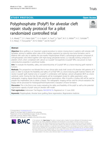
Alkaabi et al. Trials(2021) 22:393growth factors or other bone growth-promoting factortherapy, obvious malnutrition, and active influenza.Page 4 of 8feasibility of the treatment with Ca-polyP. It is assumedthat no SAEs or AEs will occur, and then, an n 4 foreach group should therefore be sufficient.Withdrawal of participantsParticipants can leave the study at any time for anyreason without any consequences. The investigator candecide to withdraw a subject from the study for urgentmedical reasons. When participants withdraw prior tografting intervention, they will be replaced. Furthermore,if a membrane has been used for any reason, the patientwill be considered as a dropout and will be replaced.RecruitmentPrior to recruitment, an audit will be carried out by thesurgical and ethical team to evaluate the safetymeasurements at the research site in the HasanuddinDental Hospital. Patients will be recruited from anexisting database of patients eligible for the proposedtreatment available from the Hasanuddin University,Hasanuddin Dental Hospital.InterventionUnder general anesthesia, and after local infiltration withadrenaline 1:100,000, an incision will be made at thecleft margin to create a pocket-like tissue towards thenose and the mouth in order to reconstruct the nasalfloor as well as the palatal tissue. The goal of this approach is to get rid of the oro-nasal fistula and to exposethe bony edges on both sides of the cleft. Under sterileconditions, either Ca-polyP MP alone (NanotecMARINGmbH, Mainz, Germany) or a combination of BCP(Straumann Bone Ceramic, Villeret, Switzerland) andPolyP will be mixed with normal saline in a ratio of 1 g:1.5 ml and 1 g:2 g:3–5 ml, respectively. A homogenousmixture should be reached before placing the graft material into the cleft defect. A good adaptation of bonegraft material should be considered while placing it inthe cleft defect. No membrane will be used. A differentgraft quantity will be considered for larger defects, however, with the same mixing ratios. Absorbable sutureswith 3/0 Vicryl for the mucosa and 4/0 Vicryl for thenasal reconstruction will be used for closure.Post-operative, suitable antibiotics and painkillers willbe prescribed to all patients.Adverse event (AE) and serious adverse event (SAE)Any adverse event will be graded with respect tointensity and classified as either serious or non-seriousaccording to the World Health Organization classification. Any change in health which occurs betweenscreening examination and first administration ofamorphous Ca-polyP microparticles or related procedures will be recorded as part of the subject’s medicalhistory, and full medical care will be given to all participants. In the case of a SAE, the sponsor will be notifiedwithin 24 h from the onset. If the SAE concerns severetoxicity or infection associated with the graft site, thetrial will be terminated immediately.Sample sizeSince this is a first-in-man trial, the current trial samplesize has been limited to only 2 4 patients, with the primary goal to gain a first insight on the safety andRandomization and treatment allocationBecause this is a first-in-human study, it is not possibleto keep all personnel blinded to the assignment group.After written informed consent will be obtained by themain surgeon, randomization will be performed with regard to the treatment group. Central randomizationusing a randomization program on a secure computerwill be used after the completion of patient enrollment.Patients will receive a unique study code, and their datawill be provided to the clinical and research evaluatorsin a patient-coded manner.BlindingThe radiologist and the histopathologist will be keptblinded to the treatment when evaluating the data(Fig. 1).Data collection and accessThe rules and responsibilities will be provided to theresearch team. The doctors and nurses of the researchteam will collect the data according to the evaluation(Table 1). All research team members will receivetraining on how to collect data at all study visits. Thepatient-coded data will be then handed over to the clinical evaluators and investigators. Each patient will befollowed up for up to 6 months. The confidentiality ofthe participant’s data will be well protected by the datamanager.OutcomesSafety assessment based on physical examination andlaboratory measurementsWhen a SAE occurs, it will be concluded that polyP isnot (yet) safe in the current setting. For AEs, if they donot occur at a higher frequency than in patients treatedwith standard care (autologous bone) and/or can beresolved by non-invasive conventional methods (e.g., analgesics, antibiotics), the polyP product will be considered safe. In all other cases, polyP will not be consideredsafe (yet).
Alkaabi et al. Trials(2021) 22:393Page 5 of 8Fig. 1 Protocol flowchartThe Chelsea scale will be used to evaluate the bone graftand the level of the bone in comparison with theadjacent teeth. This scale starts with drawing animaginary midline between the two teeth on either sideof the cleft site. Each of those teeth (mesial and distalroots) will be divided starting from the cemento-enameljunction to the root apex in four parts. A 0 score is givenwhen no bone is present up till the midline; a 0.5 scoreis given when there is bone, but it fails to reach the midline; and a 1 score is given when the bone extends fromthe root surface to the midline [23].normal procedure. The biopsies will be fixed in ylate for the evaluation of hard tissueformation. After sectioning, different stainings(Goldner’s trichrome, Toluidin blue, tartrate-resistantacid phosphatase (TRAP)) will be used, and histomorphometric parameters for bone formation will be analyzed. Two trained examiners, blinded for the treatmentmodality, will evaluate the images, and intra- and interobserver reliabilities will be determined. In case of disagreement between the observers, the specimen will bere-evaluated to reach a consensus.Histological and histomorphometric analysisMonitoringThe histological and histomorphometric analysis will beperformed in at least 3 patients from each group. Inthose patients, the dental implant site will be preparedusing a trephine burr ( 2.0 mm 10.0 mm in length)instead of a normal drill, thereby being able to collect abiopsy from the treated site without interfering with theMonitoring will be done constantly by internal monitorsof the Ethics and Research Committee of Faculty ofMedicine, Hasanuddin University. Since there is anegligible risk, a data safety monitoring board will notbe formed. A safety report will be provided to theMedical Research Ethics Committee of the Ethics andRadiographic evaluation
Alkaabi et al. Trials(2021) 22:393Research Committee of Faculty of Medicine,Hasanuddin University, every year. An interim analysiswill not be conducted.Statistical analysisA SPSS power analysis for parameter comparisonsbetween the groups will be performed. A p value lessthan 0.05 will be considered statistically significant.AmendmentsAll substantial amendments will be notified to theethical committee and competent authority to ensurethe safety and integrity of participants as well as thescientific value of the trial.Post-trial careAll participants will be kept in secondary follow-up for aperiod of 3 years to ensure their safety and to record anydelayed side effects of the Ca-polyP graft material.DiscussionThis is the first-in-man study evaluating the potential regenerative capacity of polyP using an alveolar cleftmodel. PolyP represents a completely novel type of regenerative compound, since it can be considered as arich energy source for tissue repair, which may be as pivotal for the bone regeneration process as the osteogenicfactors, which are generally believed to be the primaryactive compounds [14]. The high-energy phosphatebonds of polyP are identical to those present in the“common” cellular energy molecule ATP, and both serveas substrates for the enzyme alkaline phosphatase (ALP),a well-known marker for active bone formation [12].PolyP has also been reported to promote mineralization[24] and to increase progenitor cell differentiation intoosteoblasts [15, 25]. PolyP is present in platelets, whichplay an essential role in early wound repair. Interestingly,platelet-rich plasma (PRP), a concentrate of platelet-richplasma protein derived from the whole blood and oftenused in bone repair strategies, therefore will also containpolyP. However, the efficacy of PRP to promote bone repair is nowadays questioned, since both positive andneutral/negative effects have been published recently[26, 27]. We speculate that the much higher dose ofpolyP present in our preparations will be well above thebone regeneration threshold, and thus may have a positive effect on the bone repair process.Calcium phosphate ceramics including biphasic calciumphosphates (BCPs) have been widely used as bonesubstitutes and tissue engineering scaffolds. Calciumphosphates are highly biocompatible, proven to be safe, andsuccessfully used in many different clinical treatmentmodalities such as bone augmentation in spinal arthrodesis,maxillo- and craniofacial surgeries, orthopedics, periodontalPage 6 of 8treatment, and metallic implant coatings [28–33]. Somereports describe that BCP may also have osteoinductiveproperties [34], which implies that BCP may add to theosteoinductivity as well. Moreover, a recent clinical studyapplying microstructured β-TCP for alveolar cleft repairdemonstrated that calcium phosphate could be used safelyand effectively for this purpose as well [35]. We are therefore convinced that the Straumann Bone Ceramic used inthe current study will be a safe-to-use scaffold and mayhave a supportive or even synergistic effect on the boneformation when combined with the bioactive polyP.For the clinical evaluation of bone formation,radiographic imaging will be applied. We are well awarethat this will likely be relatively reliable in the case of thegroup that is treated only with the (radiolucent) polyPmicroparticles but will not be easy with the BCP/polyPtreatment group. The BCP scaffold will be radiopaqueand cause signal scattering, which will preclude accuratevisualization of new bone formation within the scaffoldmaterial. We will circumvent this limitation by ourhistological and histomorphometrical analysis of thebiopsies taken at the 6-month follow-up time point,during dental implant placement. This will enable usto still evaluate the bone formation at the microscopic level and to quantify multiple bone formationrelated parameters and cellular activities as demonstrated before in other bone regeneration studies performed by our group [29, 30, 36, 37].ConclusionWith this protocol, we summarized how we intend toevaluate the safety and feasibility of Ca-polyP MP as anew grafting material in an alveolar cleft model.Trial statusRecruitment started in November 2019 and is plannedto end in September 2020, with 8 patients randomized.The current protocol version is 1.0, dated 28 May 2019.Authors’ contributionsThe authors read and approved the final manuscript.DeclarationsCompeting interestsThe authors declare that they have no competing interests.Author details1Department of Oral and Maxillofacial Surgery/Oral Pathology, AmsterdamUniversity Medical Centers and Academic Centre for Dentistry Amsterdam(ACTA), Amsterdam Movement Sciences, Vrije Universiteit Amsterdam,Amsterdam, The Netherlands. 2Department of Oral and Maxillofacial Surgery,Al Kuwait Hospital, Ministry of Health, Dubai, United Arab Emirates.3Department of Biochemistry, Faculty of Medicine, Hasanuddin University,Makassar, Indonesia. 4Department of Oral and Maxillofacial Surgery, Faculty ofDentistry, Hasanuddin University, Makassar 90425, Indonesia. 5Institut fürPhysiologische Chemie, Angewandte Molekularbiologie, Universitätsmedizin,Johannes Gutenberg-Universität Mainz, Mainz, Germany. 6NanotecMARINGmbH, Mainz, Germany.
Alkaabi et al. Trials(2021) 22:393Received: 12 January 2021 Accepted: 12 May 2021References1. Lexer E. Die Verwendung der freien knochenplastik nebst versucheruber gelenkversteinfung und gelenktransplantation. Arch Klin Chir. 1908;86:939–43.2. Von Eiselsberg F. Zür technik der uranoplastik. Arch Klin Chir. 1901;64:509–29.3. Al-Sebaei MO, Papageorge MB, Woo T. Technique for in-office cranial boneharvesting. J Oral Maxillofac Surg. 2004;62(2):120–2. https://doi.org/10.1016/j.joms.2003.12.036.4. Enemark H, Jensen J, Bosch C. Mandibular bone graft material forreconstruction of alveolar cleft defects: long-term results. The Cleft Palate Craniofac J. 2001;38(2):155–63. https://doi.org/10.1597/1545-1569 2001 038 0155 mbgmfr 2.0.co 2.5. Hughes CW, Revington PJ. The proximal tibia donor site in cleft alveolarbone grafting: experience of 75 consecutive cases. J Craniomaxillofac Surg.2002;30(1):12–7. https://doi.org/10.1054/jcms.2001.0268.6. Tomar K, Sahoo NK. Evaluation of graft uptake from the iliac crest insecondary alveolar bone grafting: Bergland’s criteria revisited. J Oral BiolCraniofac Res. 2018;8(3):171–6. https://doi.org/10.1016/j.jobcr.2017.03.005.7. Witsenburg B, Peter H, Freihofer M. Autogenous rib graft for reconstructionof alveolar bone defects in cleft patients: long-term follow-up results. JCraniomaxillofac Surg. 1990;18(2):55–62. https://doi.org/10.1016/S1010-5182(05)80199-4.8. Ilankoan V, Stronczek M, Telfer M, Peterson LJ, Stassen LF, et al. Aprospective study of trephined bone grafts of the tibial shaft and iliac crest.Br J Oral Maxillofac Surg. 1998;36(6):434–9. https://doi.org/10.1016/S0266-4356(98)90459-4.9. De Ruiter A, Dik E, van Es R, van der Bilt A, Janssen N, et al. Micro-structuredcalcium phosphate ceramic for donor site repair after harvesting chin bonefor grafting alveolar clefts in children. J Craniomaxillofac Surg. 2014;42(5):460–8. https://doi.org/10.1016/j.jcms.2013.05.042.10. Lazarou SA, Contodimos GB, Gkegkes ID. Correction of alveolar cleft withcalcium-based bone substitutes. J Craniofac Surg. 2011;22(3):854–7. https://doi.org/10.1097/SCS.0b013e31820f7f19.11. Müller WEG, Achermann M, Wang S, Neufurth M, Muñoz-Espi R, et al.Inorganic polyphosphate induces accelerated tube formation of HUVECendothelial cells. Cell. Mol. Life Sci. 2013;75:21–32.12. Müller WEG, Neufurth M, Wang S, Ackermann M, Muñoz-Espí R, et al.Amorphous, smart, and bioinspired polyphosphate nano/microparticles: abiomaterial for regeneration and repair of osteo-articular impairments insitu. Int J Mol Sci. 2018;19(2):427.13. Ruiz FA, Lea CR, Oldfield E, Docampo R. Human platelet dense granulescontain polyphosphate and are similar to acidocalcisomes of bacteria andunicellular eukaryotes. J Biol Chem. 2004;279(43):44250–7. https://doi.org/10.1074/jbc.M406261200.14. Wang XH, Schröder HC, Müller WEG. Amorphous polyphosphate, a smartbioinspired nano-/bio-material for bone and cartilage regeneration: towardsa new paradigm in tissue engineering. J Mat Chem B. 00241J.15. Müller WEG, Wang XH, Diehl-Seifert B, Kropf K, Schloßmacher U, LieberwirthI, et al. Inorganic polymeric phosphate/polyphosphate as an inducer ofalkaline phosphatase and a modulator of intracellular Ca2 level inosteoblasts (SaOS- 2 cells) in vitro. Acta Biomater. 2011;7(6):2661–71. https://doi.org/10.1016/j.actbio.2011.03.007.16. Wang XH, Schröder HC, Wiens M, Ushijima H, Muller WEG. Bio-silica andbio-polyphosphate: applications in biomedicine (bone formation). Curr OpinBiotechnol. 2012;23(4):570–8. https://doi.org/10.1016/j.copbio.2012.01.018.17. Smith J, Hong-Shum L. Sodium polyphosphate, in: Food additives databook. Oxford: Blackwell Science Ltd.; 2003.18. Tsutsumi K, Saito N, Kawazoe Y, Ooi HK, Shiba T. Morphogenetic studyon the maturation of osteoblastic cell as induced by inorganicpolyphosphate. PLoS One. 2014;9(2):e86834. https://doi.org/10.1371/journal.pone.0086834.19. Greenwald AS, Boden SD, Goldberg VM, Khan Y, Laurencin CT, Rosier RN,et al. American Academy of Orthopaedic Surgeons; Bone graft substitutes:facts, fictions, and applications. J Bone Joint Surg Am. 200100022-00007.Page 7 of 820. Yuan H, van Blitterswijk CA, de Groot K, de Bruijn JD. Cross-speciescomparison of ectopic bone formation in biphasic calcium phosphate (BCP)and hydroxyapatite (HA) scaffolds. Tissue Eng. 2006;12(6):1607–15. https://doi.org/10.1089/ten.2006.12.1607.21. Yuan H, Yang Z, De Bruijn JD, De Groot K, Zhang X. Material-dependentbone induction by calcium phosphate ceramics: a 2.5-year study in dogs.Biomaterials. 2001;22(19):2617–23. https://doi.org/10.1016/S0142-9612(00)00450-6.22. De Lange GL, Overman JR, Farré-Guasch E, Korstjens CM, Hartman B, et al. Ahistomorphometric and micro–computed tomography study of boneregeneration in the maxillary sinus comparing biphasic calcium phosphateand deproteinized cancellous bovine bone in a human split-mouth model.Oral Surg Oral Med Oral Pathol Oral Radiol. 2014;117(1):8–22. https://doi.org/10.1016/j.oooo.2013.08.008.23. Witherow H, Cox S, Jones E, Carr R, Waterhouse N. A new scale to assessradiographic success of secondary alveolar bone grafts. The Cleft PalateCraniofacial Journal. 2002;39(3):255–60. https://doi.org/10.1597/1545-1569 2002 039 0255 anstar 2.0.co 2.24. Lorenz B, Schröder HC. Mammalian intestinal alkaline phosphatase acts ashighly active exopolyphosphatase. Biochim. Biophys. Acta. 2001;1547(2):254–61. https://doi.org/10.1016/S0167-4838(01)00193-5.25. Hacchou Y, Uematsu T, Ueda O, Usui Y, Uematsu S, Takahashi M, et al.Inorganic polyphosphate: a possible stimulant of bone formation. J. Dent.Res. 2007;86(9):893–7. https://doi.org/10.1177/154405910708600917.26. Kassolis JD, Rosen PS, Reynolds MA. Alveolar ridge and sinus augmentationutilizing platelet-rich plasma in combination with freeze-dried boneallograft: case series. J Periodontol. 2000;71(10):1654–61. https://doi.org/10.1902/jop.2000.71.10.1654.27. Roldán JC,
This is a single-center prospective control clinical trial that will be conducted in Hasanuddin University, Hasanuddin Dental Hospital, to assess the safety and feasibility of calcium-polyphosphate microparticles (Ca-polyP MPs, CAS No.: 13477-39-9, EC No.: 236-769-6) as a bone graft material in an alveolar cleft model. The