Transcription
Int. J. Morphol.,40(3):678-682, 2022.Distribution of Various Volumes of Methylene Blue Injection inSuprainguinal Fascia Iliaca Block Under Ultrasound Guidance:A Cadaveric StudyDistribución de Varios Volúmenes de Inyección de Azul de Metileno en el BloqueSuprainguinal de la Fascia Ilíaca Bajo Guía por Ultrasonido: Un Estudio CadavéricoPerada Kantakam1,2; Naraporn Maikong1,2; Prangmalee Leurcharusmee3; Apichat Sinthubua2,4 & Pasuk Mahakkanukrauh2,4KANTAKAM, P.; MAIKONG, N.; LEURCHARUSMEE, P.; SINTHUBUA, A. & MAHAKKANUKRAUH, P. Distribution ofvarious volumes of methylene blue injection in suprainguinal fascia iliaca block under ultrasound guidance: A cadaveric study. Int. J.Morphol., 40(3):678-682, 2022.SUMMARY: The local anesthetic volume for a single-shot suprainguinal fascia iliaca block (SFIB) is a key factor of a blocksuccess because the courses of the three target nerves from the lumbar plexus (LP), the lateral femoral cutaneous nerve (LFCN), femoralnerve (FN), and obturator nerve (ON), at the inguinal area are isolated and within striking distance. Thus, this cadaveric study aims todemonstrate the distribution of dye staining on the LFCN, FN, ON, and LP following the ultrasound-guided SFIB using 15-50 ml ofmethylene blue. A total of 40 USG-SFIBs were performed on 20 fresh adult cadavers using 15, 20, 25, 30, 35, 40, 45, and 50 ml ofmethylene blue. After the injections, the pelvic and inguinal regions were dissected to directly visualize the dye stained on the LFCN,FN, ON, and LP. All FN and LFCN were stained heavily when the 15-50 ml of dye was injected. Higher volumes of dye (40-50 ml)spread more medially and stained on the ON and LP in 60 % of cases. To increase the possibility of dye spreading to all three targetnerves and LP of the SFIB, a high volume ( 40 ml) of anesthetic is recommended. If only a blockade of the FN and LFCN is required,a low volume (15-25 ml) of anesthetic is sufficient.KEY WORDS: Ultrasonography; Fascia iliaca block; Anesthetic volume; Cadaveric study.INTRODUCTIONUltrasound-guided suprainguinal fascia iliaca block(USG-SFIB) was first described by Hebbard et al. (2011).The SFIB is an analgesic technique for hip and knee surgery,such as hip/femur fracture surgery and hip/knee arthroplasty(Hadzic, 2017). A single injection of the SFIB aims to blockthe three nerves of the lumbar plexus (LP) that innervate thelower limb. Moreover, the USG-SFIB is an anterior approachto the lumbar plexus block (Vermeylen et al., 2019). The LPis formed from the ventral rami of L1-L4. The ventral ramiare the origin of the three nerves within the psoas muscle(PM). The first nerve is the lateral femoral cutaneous nerve(LFCN). It arises from the L2-L3 nerve roots and locateslateral to the PM and the anterior superior iliac spine (ASIS).It is a sensory nerve that supplies the anterior and lateralaspects of the thigh. The LFCN travels underneath theinguinal ligament towards the sartorius muscle (Rigoard,2017; Desmet et al., 2019). The second nerve is the femoralnerve (FN). It originates from the L2, L3, and L4 nerve rootsand provides sensory and motor innervation. The sensorybranches supply the anterior aspect of the thigh and the anterior as well as the medial aspects of the knee. The motorbranches supply a group of muscles in the anterior part ofthe thigh. The FN locates lateral to the femoral artery anddeep to the fascia iliaca (Moore et al., 2010; Rigoard, 2017).The third nerve is the obturator nerve (ON). It arises fromthe L2, L3, and L4 nerve roots and travels along the medial1Program in Anatomy, Department of Anatomy, Faculty of Medicine, Chiang Mai University, Chiang Mai, 50200, Thailand.Department of Anatomy, Faculty of Medicine, Chiang Mai University, 52000, Thailand.3Department of Anesthesiology, Faculty of Medicine, Chiang Mai University, 52000, Thailand.4Excellence in Osteology Research and Training Center (ORTC), Chiang Mai University, Chiang Mai, Thailand.2Funding. The research leading to these results received funding from the Faculty of Medicine, Chiang Mai University under Grant Agreement No.54-2564Received: 2021-11-19678Accepted: 2022-03-06
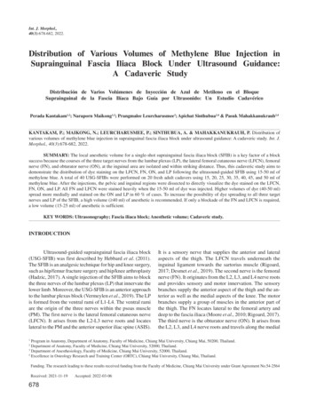
KANTAKAM, P.; MAIKONG, N.; LEURCHARUSMEE, P.; SINTHUBUA, A. & MAHAKKANUKRAUH, P. Distribution of various volumes of methylene blue injection in suprainguinal fasciailiaca block under ultrasound guidance: A cadaveric study. Int. J. Morphol., 40(3):678-682, 2022.margin of the psoas major muscle and across the obturatorforamen. It innervates the adductor muscles and suppliesthe skin at the medial part of the thigh (Rigoard, 2017).Currently, the USG-SFIB is commonly performed byanesthesiologists and emergency physicians. From 2011to 2020, the volume of a local anesthetic to achieve thesuccessful USG-SFIB varies among the literature.Vermeylen et al. (2018) suggested the effective volume ofthe USG-SFIB was 40 ml. The three target nerves wereblocked. While Hebbard et al. (2011), Bullock et al. (2017)and Eastburn et al. (2017) identified the success of the USGSFIB by using 20, 30, and 40 ml of injectate. The resultshowed that the sensory loss and the spreading of dyecovered only the LFCN and FN. However, with a 40 ml oflocal anesthetic, Desmet et al. (2017) reported the analgesicefficacy of the USG-SFIB by a reduction of pain scoresand morphine requirement after total hip arthroplasty.Recently, Gasanova et al. (2019) and Glomset et al. (2020)used 50-60 ml of local anesthetic for the USG-SFIB.Accordingly, the volume of local anesthetic for the USGSFIB ranges from 20-60 ml. As mentioned above, they usethe methylene blue that takes the place of the anesthetic inthe injection protocol for the anesthesia (Bullock et al.,2017; Vermeylen et al., 2018). Thus, this study aims todemonstrate the distribution of dye staining on the LFCN,FN, ON, and LP following the USG-SFIB using 15-50 mlof methylene blue.MATERIAL AND METHODA total of 20 fresh adult cadavers, donated to theDepartment of Anatomy, Faculty of Medicine, Chiang MaiUniversity, Thailand, were included in the study from Aprilto June 2020. The study protocol was approved by theResearch Ethics Committee, Faculty of Medicine, ChiangMai University, Thailand. (Research ID: ANA-2563-07179)The cadavers with previous surgery or pathology at the abdomen, hip, and inguinal regions were excluded.Block PerformanceAn experienced regional anesthesiologist performedthe USG-SFIB using a 6-13 MHz linear US probe (LOGIQF8, GE Healthcare, Wisconsin, USA). Each cadaver wasplaced in a supine position. The US probe was firstlypositioned in the sagittal plane on the junction between thelateral 1/3 and middle 1/3 of the imaginary line of theinguinal ligament. Then, the probe was moved medially toidentify the sartorius, iliopsoas, and internal abdominaloblique muscles. These muscles form a "bow-tie-sign"(Desmet et al., 2017). Using an in-plane approach, a blunttip needle (Stimuplex A100, B Braun Medical AG,Melsungen, Germany) was inserted in the caudal tocephalad direction. A 15-50 ml of 0.1 % methylene bluewas injected when the tip of the needle was locatedunderneath the fascia iliaca as shown in Figure 1.Fig. 1. Ultrasound image of the suprainguinal fascia iliaca block.IOM: internal abdominal oblique muscle, IM: iliacus muscle, andwhite arrows: a needle underneath the fascia iliacaCadaveric Dissection. One hour after bilateral SFIBs, allfresh cadavers were dissected using the same technique.The skin was incised along the costal margin, midaxillaryline, and inguinal ligament from the xiphoid process to thepubic tubercle on both sides. The abdominal wall, visceralorgans including stomach, intestines, and omentum, and theperitoneum of the posterior abdominal wall were removed.Carefully retract the psoas major muscle. Then, the fasciailiaca layer was dissected to identify the LFCN, FN, ON,and LP. Dye staining on the target nerves is demonstratedin Figure 2.RESULTSA mean age among the twenty cadavers was 71 13.23 years old. Eight different volume of dye between 1550 ml was injected in a total of 40 lower limbs (5 USGSFIBs for each dye volume). Twenty USG-SFIBs wereperformed on the left side and the rest were done on theright side. The result showed that the staining of low volume(15- 25 ml) of dye is sufficient for blocking two nerves(LFCN and FN). The volume of dye that used 30-35 ml ofdye spreading covered the LFCN (100 %), FN (100 %).Furthermore, the minimum volume of dye that covers theON was 30 ml (20 %) and 35 ml (40 %). A higher dyevolume (40-50 ml) was stained on the LFCN (100 %), FN(100 %), ON (60 %), and LP (60 %) of the cases (Table I).679
KANTAKAM, P.; MAIKONG, N.; LEURCHARUSMEE, P.; SINTHUBUA, A. & MAHAKKANUKRAUH, P. Distribution of various volumes of methylene blue injection in suprainguinal fasciailiaca block under ultrasound guidance: A cadaveric study. Int. J. Morphol., 40(3):678-682, 2022.DISCUSSIONThis study investigated the distributionof dye staining on the LFCN, FN, ON, and LPfollowing the USG-SFIB using 15-50 ml of dye.The result showed that only the LFCN and FNwere dyed when 15-25 ml of methylene blue wasinjected. The volume of dye that could stain allthe three nerves (i.e., successful block) was 3050 ml. The percentages of the successful blockwith 30, 35, 40-50 ml of dye were 20 %, 40 %,and 60 %, respectively. The LP was stained bythe volume (40-50 ml, 60 %) of dye (Table I).These findings are potentially applied to a localanesthetic volume selection during the USGSFIB in the clinical setting.Fig. 2. Distribution of methylene blue on the three nerve target from the LPfor USG-SFIB such as lateral femoral cutaneous nerve (LFCN), femoral nerve(FN), and obturator nerve (ON), a : 20 ml stained LFCN and FN, b : 40 mlstained LFCN, FN, and ON (fade), c : 50 ml stained LFCN,FN, and ON (deep).The nerves innervating the hip joint thatis related to the USG-SFIB are the LFCN, FN,and ON (Vermeylen et al., 2019). Our studyshowed that the volume of dye ranging from 15to 50 ml reliably stained the LFCN and FN. Onlya higher volume ( 30 ml) possibly dyed the ON.These results are consistent with previouscadaveric and clinical reports. Yamada et al.(2020) demonstrated that the 50 % and 95 %effective volume of 0.25 % ropivacaine for theUSG-SFIB was 15 and 27 ml. The definition ofa successful block in that study was a loss ofsensation of the LFCN and FN sensorydistribution. In addition, Ueshima & Otake(2018) found that the LFCN and FN weresuccessfully blocked when 20 ml oflevobupivacaine was used for USG-SFIB in acardiovascular-compromised patient undergoingfemoral neck fracture surgery. Following theUSG-SFIB with 30 ml of methylene blue,Bullock et al. (2017) and Vermeylen et al. (2018)reported that this volume was stained only onthe LFCN and FN in a cadaveric study. With 40ml of local anesthetic, a volume commonly usedin the clinical setting, the SFIB effectivelyanesthetized the LFCN, FN, ON, and LP(Vermeylen et al., 2019; Bravo et al., 2020;Table I. Number (%) of the nerves stained with dye following the the suprainguinal fascia iliaca block using 15-50 ml of methylene blue.NERVE, N(%)LFCNFNONLP68015 ml5 (100)5 (100)0 (0)0 (0)20 ml5 (100)5 (100)0 (0)0 (0)25 ml5 (100)5 (100)0 (0)0 (0)VOLUME OF DYE INJECTION (ML)30 ml35 ml40 ml5 (100)5 (100)5 (100)5 (100)5 (100)5 (100)1 (20)2 (40)3 (60)0 (0)0 (0)3 (60)45 ml5 (100)5 (100)3 (60)3 (60)50 ml5 (100)5 (100)3 (60)3 (60)
KANTAKAM, P.; MAIKONG, N.; LEURCHARUSMEE, P.; SINTHUBUA, A. & MAHAKKANUKRAUH, P. Distribution of various volumes of methylene blue injection in suprainguinal fasciailiaca block under ultrasound guidance: A cadaveric study. Int. J. Morphol., 40(3):678-682, 2022.Vergari et al., 2020). Our result showed the least volumeof dye was 40 ml that stained to the LP. Vermeylen et al.(2019) reported 80 % complete sensory blockade of themedial, lateral, and anterior regions of the thigh. They foundthat the SFIB spread toward the LP on MRI. This volumewas safe and effective for postoperative analgesia afteracetabular fracture surgery (Vergari et al., 2020). Moreover,Bravo et al. (2020) demonstrated that the SFIB and theLPB provided comparable pain control in total hiparthroplasty. The SFIB produced a longer duration of analgesia and a shorter recovery period.Determining the success of the SFIB depends mainlyon the ON block. Therefore, the anatomical course of theON deserves discussion. The ON locates deep to the medialmargin of the psoas major muscle, travels into the lesserpelvic and passes through the obturator foramen to themedial thigh (Meier & Büttner, 2015). The location of theON is adjacent to the point of needle insertion of the SFIB.For this reason, the use of a high anesthetic volume for theSFIB is necessary to reach the ON. However, a high volumeof a local anesthetic to provide effective analgesia shouldbe used with caution. The potential complications includenausea/vomit and local anesthetic systemic toxicity(LAST). Allegri et al. (2016) used the maximum volume(75 ml) of anesthetic and found ten out of 14 patientsexhibiting symptoms of the LAST. Thus, weighing the riskversus benefit of using a high anesthetic volume is aconsideration for anesthesiologists. This concern introduces analgesic options for hip surgery. Recently, thecombination of USG-SFIB with the selective obturatornerve block effectively reduced postoperative opioidconsumption and pain scores after hip arthroscopy (Lee etal., 2020). However, the efficacy of the added ON block tothe SFIB is still controversial. Nielsen et al. (2019) foundthat the ON block did not result in pain reduction after hiparthroplasty. Another alternative is the pericapsular nervegroup (PENG) block. It focuses on the articular branchesto the hip joint including the accessory of the obturatornerve (AON). The AON is located between the anteriorinferior iliac spine (AIIS) and ilio-pubic eminence (IPE).20 ml of local anesthetic for the PENG block effectivelylessened the pain of the hip joint (Girón-Arango et al.,2018). Moreover, the benefit of the PENG block over theSFIB is motor sparing. This study has some limitations.First, besides the volume of local anesthetics, theconcentration of medications and technique of blockadeare taken into account in the clinical setting (Helayel et al.,2006). Second, the spreading of dye in cadaver differs fromliving humans. Vermeylen et al. (2021) showed thatcephalad spread of dye was affected by passive movementin living subjects. Finally, our studies focused on thecommon volume used in the clinical setting. Further studiesspecifically demonstrating the effective volume of the USGSFIB have to be elucidated.In summary, to increase the possibility of dyespreading to all three target nerves and LP of the SFIB, ahigh volume ( 40 ml) of anesthetic is recommended.However, potential complications following high anestheticvolume are existing concerns. If only a blockade of the FNand LFCN is required, a low volume (15-25 ml) ofanesthetic is sufficient. Further studies specificallydemonstrating the effective volume of the USG-SFIB arenecessary to improve the efficacy of the USG-SFIB andprevent local anesthetic-related complications.ACKNOWLEDGMENTSA debt of gratitude is owed to the excellence ofOsteology Research and Training Center (ORTC), ChiangMai University. A special thanks to funding from the Facultyof Medicine, Chiang Mai University under Grant AgreementNo.54-2564 and Patara Rattanachet for linguistic review.KANTAKAM, P.; MAIKONG, N.; LEURCHARUSMEE, P.;SINTHUBUA, A. & MAHAKKANUKRAUH, P. Distribuciónde varios volúmenes de inyección de azul de metileno en el bloquesuprainguinal de la fascia ilíaca bajo guía por ultrasonido: Un estudio cadavérico. Int. J. Morphol., 40(3):678-682, 2022.RESUMEN: El volumen de anestésico local para un bloqueo de la fascia ilíaca suprainguinal (FISI) de una sola inyección es un factor clave para el éxito del bloqueo, debido a que loscursos de los tres nervios objetivo del plexo lumbar (PL), el nervio cutáneo femoral lateral (NCFL), femoral (NF) y el nervioobturador (NO), en el área inguinal están aislados y dentro de ladistancia de abordaje. Por lo tanto, este estudio cadavérico tienecomo objetivo demostrar la distribución de la tinción de tinte enNCFL, NF, NO y PL siguiendo el FISI guiado por ultrasonidousando 15-50 ml de azul de metileno. Se realizaron un total de 40USG-FISI en 20 cadáveres adultos frescos utilizando 15, 20, 25,30, 35, 40, 45 y 50 ml de azul de metileno. Después de las inyecciones, se disecaron las regiones pélvica e inguinal para visualizardirectamente el tinte teñido en NCFL, NF, NO y PL. Todos losNF y NCFL se tiñeron intensamente cuando se inyectaron los 1550 ml de colorante. Volúmenes mayores de colorante (40-50 ml)se esparcen más medialmente y tiñen el NO y la PL en el 60 % delos casos. Para aumentar la posibilidad de que el colorante sepropague a los tres nervios objetivo y al PL del FISI, se recomienda un volumen elevado ( 40 ml) de anestésico. Si solo serequiere un bloqueo de NF y NCFL, un volumen bajo (15-25 ml)de anestésico es suficiente.PALABRAS CLAVE: Ultrasonografía; Bloqueo defascia ilíaca; Volumen anestésico; Estudio cadavérico.681
KANTAKAM, P.; MAIKONG, N.; LEURCHARUSMEE, P.; SINTHUBUA, A. & MAHAKKANUKRAUH, P. Distribution of various volumes of methylene blue injection in suprainguinal fasciailiaca block under ultrasound guidance: A cadaveric study. Int. J. Morphol., 40(3):678-682, 2022.REFERENCESAllegri, M.; Bugada, D.; Grossi, P.; Manassero, A.; Pinciroli, R. L.; Zadra,N.; Fanelli, G.; Zarcone, A.; Cataldo, R.; Danelli, G.; et al. ItalianRegistry of Complications associated with Regional Anesthesia(RICALOR). An incidence analysis from a prospective clinical survey.Minerva Anestesiol., 82(4):392-402, 2016.Bravo, D.; Layera, S.; Aliste, J.; Jara, Á.; Fernández, D.; Barrientos, C.;Wulf, R.; Muñoz, G.; Finlayson, R. J. & Tran, D. Q. Lumbar plexusblock versus suprainguinal fascia iliaca block for total hip arthroplasty:A single-blinded, randomized trial. J. Clin. Anesth., 66:109907, 2020.Bullock, W. M.; Yalamuri, S. M.; Gregory, S. H.; Auyong, D. B. & Grant,S. A. Ultrasound-guided suprainguinal fascia iliaca technique providesbenefit as an analgesic adjunct for patients undergoing total hiparthroplasty. J. Ultrasound Med., 36(2):433-8, 2017.Desmet, M.; Balocco, A. L. & Belleghem, V. V. Fascia iliaca compartmentblocks: Different techniques and review of the literature. Best Pract.Res. Clin. Anaesthesiol., 33(1):57-66, 2019.Desmet, M.; Vermeylen, K.; Van Herreweghe, I.; Carlier, L.; Soetens, F.;Lambrecht, S.; Croes, K.; Pottel, H. & Van de Velde, M. A longitudinalsupra-inguinal fascia iliaca compartment block reduces morphineconsumption after total hip arthroplasty. Reg. Anesth. Pain Med.,42(3):327-33, 2017.Eastburn, E.; Hernandez, M. A. & Boretsky, K. Technical success of theultrasound-guided supra-inguinal fascia iliaca compartment block inolder children and adolescents for hip arthroscopy. Paediatr. Anaesth.,27(11):1120-4, 2017.Gasanova, I.; Alexander, J. C.; Estrera, K.; Wells, J.; Sunna, M.;Minhajuddin, A. & Joshi, G. P. Ultrasound-guided suprainguinal fasciailiaca compartment block versus periarticular infiltration for painmanagement after total hip arthroplasty: a randomized controlled trial.Reg. Anesth. Pain Med., 44(2):206-11, 2019.Girón-Arango, L.; Peng, P. W. H.; Chin, K. J.; Brull, R. & Perlas, A.Pericapsular nerve group (PENG) block for hip fracture. Reg. Anesth.Pain Med., 43(8):859-63, 2018.Glomset, J. L.; Kim, E.; Tokish, J. M.; Renfro, S. D.; Seckel, T. B.; Adams,K. J. & Folk, J. Reduction of postoperative hip arthroscopy pain withan ultrasound-guided fascia iliaca block: a prospective randomizedcontrolled trial. Am. J. Sports Med., 48(3):682-8, 2020.Hadzic, A. Hadzic's Textbook of Regional Anesthesia and Acute PainManagement. 2nd ed. New York, McGraw-Hill Education, 2017.Hebbard, P.; Ivanusic, J. & Sha, S. Ultrasound-guided supra-inguinal fasciailiaca block: a cadaveric evaluation of a novel approach. Anaesthesia,66(4):300-5, 2011.Helayel, P. E.; Lobo, G.; Vergara, R.; Conceição, D. B. D, & Oliveira Filho,G. R. D. Effective volume of local anesthetics for fascia iliaccompartment block: a double-blind, comparative study between 0.5%ropivacaine and 0.5% bupivacaine. Rev. Bras. Anestesiol., 56(5):45460, 2006.Lee, S.; Hwang, J. M.; Lee, S.; Eom, H.; Oh, C.; Chung, W.; Ko, Y. K.;Lee, W.; Hong, B. & Hwang, D. S. Implementation of the obturatornerve block into a supra-inguinal fascia iliaca compartment block basedanalgesia protocol for hip arthroscopy: Retrospective pre-post study.Medicina (Kaunas), 56(4):150, 2020.Meier, G. & Büttner, J. Atlas of Peripheral Regional Anesthesia: Anatomyand Techniques. 3rd ed. Stuttgart, Thieme, 2015.Moore, K. L.; Dalley, A. F. & Agur, A. M. R. Clinically Oriented Anatomy.6th ed. Philadelphia, Walters Kluwer Health/Lippincott Williams &Wilkins, 2010.Nielsen, N. D.; Runge, C.; Clemmesen, L.; Børglum, J.; Mikkelsen, L. R.;Larsen, J. R.; Nielsen, T. D.; Søballe, K. & Bendtsen, T. F. An obturatornerve block does not alleviate postoperative pain after total hiparthroplasty: A randomized clinical trial. Reg. Anesth. Pain Med.,44(4):466-71, 2019.682Rigoard, P. Atlas of Anatomy of the Peripheral Nerves: The Nerves of theLimbs–Student Edition. Cham, Springer, 2017.Ueshima, H. & Otake, H. Supra-inguinal fascia iliaca block under ultrasoundguidance for perioperative analgesia during bipolar hip arthroplasty ina patient with severe cardiovascular compromise: A case report. Medicine (Baltimore), 97(40):e12746, 2018.Vergari, A.; Frassanito, L.; Tamburello, E.; Nestorini, R.; Sala, F.D.; Lais,G.; Ciolli, G. & Liuzza, F. Supra-inguinal fascia iliaca compartmentblock for postoperative analgesia after Acetabular fracture surgery.Injury, 51(12):2996-8, 2020.Vermeylen, K.; Desmet, M.; Leunen, I.; Soetens, F.; Neyrinck, A.; Carens,D.; Caerts, B.; Seynaeve, P.; Hadzic, A. & Velde, M.V.D. Supra-inguinalinjection for fascia iliaca compartment block results in more consistentspread towards the lumbar plexus than an infra-inguinal injection: avolunteer study. Reg. Anesth. Pain Med., 2019. Doi: len, K.; Soetens, F.; Leunen, I.; Hadzic, A.; Boxtael, S. V.; Pomes,J.; Prats-Galino, A.; Velde, M. V. D.; Neyrinck, A. & Sala-Blanch, X.The effect of the volume of supra-inguinal injected solution on thespread of the injectate under the fascia iliaca: a preliminary study. J.Anesth., 32(6):908-13, 2018.Vermeylen, K.; Van Aken, D.; Versyck, B.; Roos, J.; Bracke, P.; Leunen, I.;Mariano, E. R. & Elsharkawy, H. The effect of passive musclemobilization on the distribution of local anesthetics after supra-inguinalfascia iliaca compartment block, a pilot case study. J. Clin. Anesth.,68:110100, 2021.Yamada, K.; Inomata, S. & Saito, S. Minimum effective volume ofropivacaine for ultrasound-guided supra-inguinal fascia iliacacompartment block. Sci. Rep., 10:21859, 2020.Corresponding authorProf. Pasuk Mahakkanukrauh, MDDepartment of AnatomyExcellence in Osteology Research and Training Center(ORTC)Faculty of MedicineChiang Mai UniversityChiang Mai, 50200THAILANDE-mail: pasuk034@gmail.com
the USG-SFIB using a 6-13 MHz linear US probe (LOGIQ F8, GE Healthcare, Wisconsin, USA). Each cadaver was placed in a supine position. The US probe was firstly positioned in the sagittal plane on the junction between the lateral 1/3 and middle 1/3 of the imaginary line of the inguinal ligament. Then, the probe was moved medially to