Transcription
REVIEWSThe distribution and targeting ofneuronal voltage-gated ion channelsHelen C. Lai* and Lily Y. Jan‡Abstract Voltage-gated ion channels have to be at the right place in the right number toendow individual neurons with their specific character. Their biophysical properties togetherwith their spatial distribution define the signalling characteristics of a neuron. Improperchannel localization could cause communication defects in a neuronal network. This reviewcovers recent studies of mechanisms for targeting voltage-gated ion channels to axons anddendrites, including trafficking, retention and endocytosis pathways for the preferentiallocalization of particular ion channels. We also discuss how the spatial localization of thesechannels might contribute to the electrical excitability of neurons, and consider the need forfuture work in this emerging field.Axon initial segment(AIS). The area of the axon nearthe soma that contains a highdensity of voltage-gated sodiumchannels, which are responsiblefor the initial depolarization thatleads to the initiation of theaction potential.Saltatory conductionThe way an action potential‘jumps’ between nodes of amyelinated axon, for fastconduction.Back-propagationThe propagation of actionpotentials ‘backward’ up thedendrites.*Center for BasicNeuroscience, University ofTexas Southwestern MedicalCenter, Dallas, Texas 75390,USA.‡Howard Hughes MedicalInstitute, University ofCalifornia,San Francisco,1550 4th Street,the Rock HallSan Francisco,California 94158, USA.Correspondence to L.J.e-mail: lily.jan@ucsf.edudoi:10.1038/nrn1938548 JULY 2006 VOLUME 7Voltage-gated ion channels were among the first ion channels to be identified when voltage-clamp recordings werefirst undertaken over half a century ago. After the landmark studies by Hodgkin and Huxley1, which establishedthe crucial roles of voltage-gated sodium and potassiumchannels in the generation and propagation of actionpotentials in the squid giant axon, the development ofpatch-clamp recording has allowed electrophysiologicalanalyses of different subcellular compartments of neurons,revealing a rich and varied consortium of voltage-gatedion channels on dendrites and axons. Molecular studiesof voltage-gated ion channels over the past quarter of acentury further unveiled the remarkably refined andmosaic-like patterns of channel distribution. Only recentlyhave we begun to appreciate just how the different channelisoforms are targeted to different parts of the neuron tocarry out specific functions. This review focuses primarilyon recent findings concerning the distribution and targeting of voltage-gated ion channels with a focus on sodium,potassium, and hyperpolarization-activated cation channels. We begin with a summary of the nomenclature andmembrane topology of various voltage-gated ion channelsto set the framework for understanding the structuralmotifs involved in targeting these channels.We will consider a model neuron that is receivingmultiple excitatory and inhibitory inputs (excitatory andinhibitory postsynaptic potentials — EPSPs and IPSPs) inthe somatodendritic region that summate and bring aboutmembrane potential changes at the axon initial segment(AIS). It is in this region that voltage-gated sodium (Nav)and certain voltage-gated potassium (Kv) channels such asthe KCNQ channel determine the threshold for firing anaction potential, thereby causing action potential generation(FIG. 1)2–5. Action potentials then propagate along the axonand, in the case of myelinated axons, ‘jump’ between thenodes of Ranvier through saltatory conduction to reach thenerve terminals, where activation of voltage-gated calcium(Cav) channels causes calcium influx and neurotransmitter release. Kv channels and hyperpolarization-activatedcyclic nucleotide-gated (HCN) cation channels on dendrites further control action potential back-propagation,and the time course and extent of the passive spread ofsynaptic potentials. Back-propagating action potentialsmight signal the occurrence of recent neuronal excitationand influence synaptic plasticity6,7, leading to long-termpotentiation (LTP) or long-term depression (LTD) depending on the timing of the back-propagating action potentialrelative to the synaptic input8,9. Action potentials mightalso be generated locally in the dendrites10–14, modulatingthe processing and integration of synaptic inputs of specific dendritic branches or segments. Synaptic integrationand the resultant pattern of action potential firing dependon the spatial distribution of various channels with different electrophysiological properties — a crucial aspectof neuronal differentiation that has recently emerged as afascinating topic for investigation.The precise distribution of voltage-gated ion channelswith specific biophysical properties that allow for thedifferent electrophysiological properties of axonal andsomatodendritic regions raises many questions. Howdo voltage-gated ion channels move to where they needto be? In how many ways can this feat be achieved inwww.nature.com/reviews/neuro
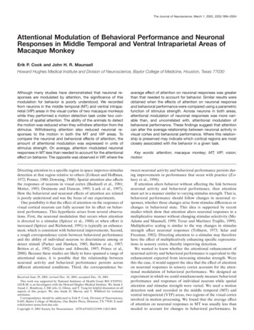
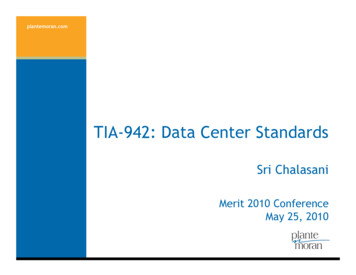
REVIEWSaSomatodendriticAxonalIntracellular recordingExcitatory inputsAction potentialsKv4.2Kv3 throughout dendriteProximaldendritesEPSPsKv2.1SomaNav, Kv1, HCNDendritic action potentialsCavAISNav, KCNQInhibitoryinputsIPSPsNodes of RanvierNav, KCNQ, Kv3.1bNeurotransmitterreleaseBack propagationbNav, KCNQ, Kv3.1bKv1.1 and Kv1.2Septate-like junctionmyelinInternodeLong-term potentiation(LTP).The prolongedstrengthening of synapticcommunication, which isinduced by patterned inputand is thought to be involved inlearning and memoryformation.Long-term depression(LTD). An enduring weakeningof synaptic strength that isthought to interact with longterm potentiation (LTP) in thecellular mechanisms of learningand memory in structures suchas the hippocampus andcerebellum. Unlike LTP, which isproduced by brief highfrequency stimulation, LTD canbe produced by long-term,low-frequency stimulation.JuxtaparanodeA region of the axon that isadjacent to the paranodes,which are adjacent to thenodes of Ranvier and arelocated underneath the gure 1 General localization of voltage-gated ion channels in a model neuron. a In general, Nav channels arefound in the axon initial segment (AIS), nodes of Ranvier and presynaptic terminals. Voltage-gated potassium Kv1channels are found at the juxtaparanodes (JXPs) in adult myelinated axons and presynaptic terminals. The Kv channelKCNQ is found at the AIS and nodes of Ranvier, and Kv3.1b channels are also found at the nodes of Ranvier. Canonically,excitatory and inhibitory inputs (EPSPs and IPSPs — excitatory and inhibitory postsynaptic potentials; yellow and bluepresynaptic nerve terminals, respectively) from the somatodendritic region spread passively to the AIS where actionpotentials are generated by depolarization, and travel by saltatory conduction to the presynaptic nerve terminals toactivate voltage-gated calcium (Cav) channels that increase intracellular calcium levels, thereby triggeringneurotransmitter release. Hyperpolarization-activated cyclic-nucleotide-gated (HCN) channels have a gradientdistribution that increases in density from the soma to the distal dendrites (dark blue shading). Kv2.1 channels are found inclusters on the soma and proximal dendrites (light yellow ovals). Kv3 channels are found throughout the dendrite. Kv4.2channels are located more prominently on distal dendrites (light blue shading). Kv channels in the dendrites contribute tocontrolling back propagation. Strong enough inputs in the dendritic region can generate dendritic action potentials.Dendritic Cav channels increase in density toward the proximal dendrites and the soma. b The left panel shows anexample of defined channel localization around the nodal region in the myelinated rat optic nerve: Nav channels in greenat the nodes; Caspr, a cell-recognition molecule, in red at the paranodes; and Kv1.2 channels in blue at the juxtaparanodes(horizontal scale bar, 10µm). The right panel depicts the channel composition surrounding a myelinated axon with Nav,KCNQ, and Kv3.1b channels at the nodes, no channels at the paranodes underlying the paranodal loops that form septatelike junctions, and Kv1.1 and Kv1.2 channels at the JXPs under the compact myelin. Panel b (left) reproduced, withpermission, from REF. 207 (2000) Blackwell Publishing.different cell types? How do the various channel typescoordinate their activities for neuronal signalling? Howdoes channel localization change during developmentand for what purposes? These are the kinds of questionsthat researchers have been trying to tackle as they workon different channel isoforms, in different model systems,and use different techniques to reach for some mechanistic insight. The determination of spatial mechanisms isintertwined with temporal considerations, as channels canoccupy different locations not only during development,but also in the mature nervous system. It will take sometime to determine what global mechanisms exist. Here wereview our current knowledge of the distribution, targeting mechanisms and motifs for several voltage-gated ionchannels.NATURE REVIEWS NEUROSCIENCEStructure of voltage-gated ion channelsVoltage-gated ion channels contain sequence motifs thatare necessary for their targeting, presumably becausethese sequences mediate interactions with proteins thatare directly or indirectly involved with channel targeting. Voltage-gated ion channels are formed by either oneα-subunit that is a contiguous polypeptide that containsfour repeats (domains I–IV), as in the case of Nav andCav channels; or four α-subunits, each with a singledomain, as in the case of Kv and HCN channels (FIG. 2).A single domain contains six α-helical transmembranesegments. The fourth transmembrane segment containsmultiple arginines that are mainly responsible for sensing changes in membrane potential. Between the fifthand sixth transmembrane segments is a re-entrant poreVOLUME 7 JULY 2006 549
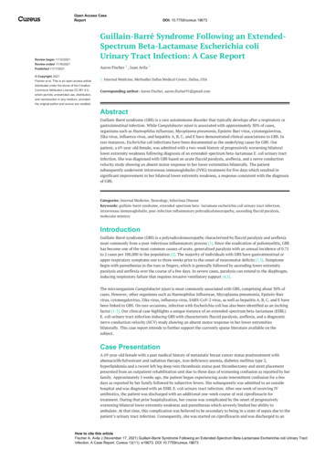
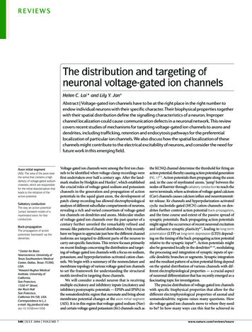
REVIEWSPore loopNav channelsβ2/4Kv and HCN channelsIIIIIIβ1/3IVPore loopDPPX for Kv4OutsideS1S2S3S4S5S6 S1S2S3S4S5S6 Insideα-subunitCα-subunitCNNCIINa NCIIIIβ-subunit for Kv1IVKChIP for Kv4IIIIIIγOutsideα1Pore loopCav channelsS1S2S3S4S5S6 α2IV δα-subunitCNInsideCNNβCFigure 2 General structural topology of voltage-gated ion channels. Voltage-gated sodium (Nav) channels are formedfrom a single polypeptide that consists of four domains (I–IV), each of which has six transmembrane segments (S1–S6).The fourth transmembrane segment of each domain contains positively charged arginines that are primarily responsible forvoltage sensing, as well as the S5-pore loop-S6 region, which forms the pore domain through which sodium ions flow. Theβ-subunits, β1/3 and β2/4, are single transmembrane proteins that have an immunoglobulin-like extracellular domain thatco-assembles with the Nav α-subunit. Voltage-gated potassium (Kv) channels and hyperpolarization-activated cyclicnucleotide-gated (HCN) cation channels have four similar or identical α-subunits, each with a single domain. Kv1 channelshave cytoplasmic β-subunits that interact with the N-terminal T1 domains. Kv4 channels have two closely associatedproteins; the intracellular protein KChIP, and the single-span membrane protein DPPX. Voltage-gated calcium (Cav) channelshave a similar topology to Nav channels in their α-subunits. Cav channels can have up to four associated auxiliary subunits: adisulphide-linked α2δ-complex, an intracellular β-subunit, and an occasional γ-subunit with four transmembrane segments.L1 CAMA cell adhesion molecule in thenervous system that isimportant for cell–cellinteractions that occur through6 immunoglobulin G-likeprotein domains and 3–5fibronectin type II domains.KChIPsβ-subunits of Kv4 channels,which have four calciumbinding EF hands withhomology to the recoverin/neuronal calcium sensor -1(NCS1) family.CD26A dipeptidyl aminopeptidaseand cell adhesion protein.550 JULY 2006 VOLUME 7loop, which forms the narrowest part of the pore. Theinteraction of these α-subunits with auxiliary subunits(α2, β, γ or δ) as well as other proteins can modulatechannel function and selectively target some channels(such as Nav, Kv1 and KCNQ) to the axon, other channels (such as HCN, Kv2 and Kv4) to somatodendriticregions, and Kv3 and various Cav isoforms to axons anddendrites.Ten genes encode the α-subunits of Nav channels inmammals; these genes encode Nav1.1 to Nav1.9, plus anatypical sodium channel that is designated Nax and hasgreater than 50% sequence identity to other Nav proteinsin its transmembrane and extracellular regions)15–17.There are four known Nav protein β-subunits (β1, β2,β3, β4)18, each with a single transmembrane segmentand an extracellular domain that is structurally homologous to the immunoglobulin G-like domains of L1 celladhesion molecules (L1 CAMs)19,20.There are approximately 40 mammalian genes forKv channel α-subunits that are grouped into 12 familiesknown as Kv1 to Kv12 (REF. 21). Different genes withina family are denoted with an additional number afterthe decimal point, such as Kv1.1 and Kv1.2, roughlyin order of their molecular characterization. Channeldiversity is greatly enhanced by the ability to formhomo- or heterotetrameric channels, with the mix andmatch of members in a subfamily Kv1, Kv3, Kv4, Kv7(KCNQ) or Kv10. Of the channels described in thisreview, Kv1 α-subunits associate with the β-subunitsKvβ1.1, Kvβ1.2, Kvβ2 and Kvβ3 through their N-terminal T1 domains, with an α4β4 stoichiometry22. Kv4subunits are associated with KChIPs, calcium-bindingproteins that bind to the N-terminus of Kv4 channels23–25, and DPPX, a single transmembrane-spanningprotein in which the extracellular domain resembles adipeptidyl aminopeptidase, as well as the cell adhesionprotein CD26 (REF. 26).HCN cation channels (HCN1–4) have the same transmembrane topology as Kv channels. However, they arenon-selective, pass both Na and K (REF. 3) and are regulated by cyclic nucleotides through a cyclic nucleotidebinding domain in their C-terminus27.www.nature.com/reviews/neuro
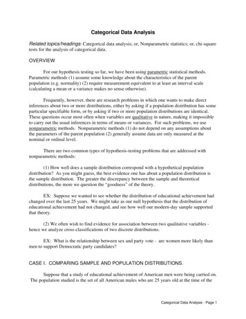
REVIEWSCav1–3 channels have an α1 subunit that formsthe ion-conduction pore. Cav1 channels give rise tothe L-type current, Cav2.1 the P/Q-type current Cav2.2the N-type current, Cav 2.3 the R-type current, and Cav3the T-type current28,29. Cav channels are associated withseveral auxiliary subunits in vivo that affect channelfunction and expression: a cytosolic β-subunit, a disulphide-linked α2δ complex and an occasional γ-subunit,which create an α1α2δβγ native Cav channel30.Targeting voltage-gated ion channels to axonsAt the nodes of Ranvier, Nav and KCNQ channelsallow currents that spread from one node to initiatean action potential at the next node. Kv1 channels atthe juxtaparanodal regions increase the fidelity of theaction potential at the nodes and reduce excitabilityduring remyelination and development31,32. In addition,Kv3 channels reside in the soma, axons and presynaptic terminals of interneurons and other neurons thatundergo high frequency firing, and probably contributeto repolarization at the end of an action potential33–35.The localization of axonal channels is shown in FIG. 1.Regarding mechanisms for axonal targeting, studiesof proteins such as neuron–glia cell adhesion molecule(NgCAM) and vesicle-associated membrane protein-2(VAMP2) have elucidated at least three feasible models36,37: directed targeting, transcytosis and selective retention. NgCAM, a chick homologue of L1 CAM, might betransported to the axonal membrane by directed targeting or by transcytosis36,37, which involves insertion ofNgCAM into the somatodendritic membrane, followedby its endocytosis and redistribution to the axonal membrane. By contrast, VAMP2, a synaptic vesicle v-SNARE(soluble N-ethylmaleimide-sensitive fusion proteinattachment protein (SNAP) receptor), was uniformlyinserted into both the somatodendritic and axonal membranes and then endocytosed from the somatodendriticmembrane, leaving VAMP2 surface channels along theaxon — a mechanism of selective retention (also knownas selective endocytosis or elimination) at the axonalmembrane37. These strategies might be used singly or incombination by axonal ion channels.Signals such as tyrosine motifs38 and di-leucine motifs39,which bind clathrin adaptor proteins and thereby linkBox 1 Myelin-dependent channel distribution during developmentDirected targetingThe specific transport ofproteins to their properlocation (axons or dendrites)after their exit from the ER.TranscytosisThe targeting of membraneproteins first to onecompartment and then, aftertheir endocytosis, to anothersubcellular compartment.Selective retentionInteraction with anchorproteins at specific sites causesmembrane proteins that aretransported to the neuronalmembrane uniformly to remainonly at those g), whilebeing internalized elsewhere.External cues that occur during myelin formation appear toLamina cribrosaKv1.1 andhave a role in nodal channel clustering. Retinal ganglionRetinaLightNav1.6Kv1.2 at JXPcells (RGCs), with their axons unmyelinated in the retina butOpticmyelinated in the optic nerve after they cross the laminanervecribrosa (see figure), have voltage-gated sodium (Nav)Unmyelinchannel 1.6 localized to a putative axon initial segmentMyelinated regionated region(AIS), which is more distal from the soma than previouslyreported, and nodes of Ranvier, whereas Nav1.2 is located inPutative AISNav1.2Nav1.6 and some Nav1.2the unmyelinated region and partially at the AIS198,199.During development, Nav1.2 channels appear first atRGCimmature nodes of Ranvier, and are eventually replaced byNav1.6 upon compact myelin formation199. Myelination affects channel localization at the nodes of Ranvier but not theAIS198, and the appearance of Nav1.6 at the AIS correlates well with the appearance of repetitive firing of rat RGCs duringdevelopment198,200.In the shiverer mice, which cannot form compact myelin due to a mutation in the myelin basic protein gene, Nav1.6channels are no longer clustered in the optic nerve199. By contrast, in myelin deficient rats that have a mutation in the genethat encodes proteolipid protein, which causes oligodendrocytic death201, Nav channel clusters also become more diffusein the optic nerve202; however, they remain clustered in the spinal cord106,109. Moreover, trembler mutant mice — which haveperipheral nerve hypomyelination, because of mutations in peripheral myelin protein-22 — retain node-like clusters of Navchannels in the sciatic nerve62,203. Consistent with these observations, for neurons in culture, myelin is important forinitiating clustering but not for maintenance of the clusters202.Myelination is required for both initiation and maintenance of voltage-gated potassium channel Kv1.1, Kv1.2 and Kvβ2clusters at the juxtaparanodal regions in the mouse optic nerve, as their distribution becomes more diffuse in both chronicdemyelinating and hypomyelinating mouse models204. Kv1 clusters colocalize with postsynaptic density protein-95 (PSD95), and the appearance of this clustering occurs concurrently with myelination during development in the mouse retina204.In a chemically induced rat model of demyelination and remyelination, Kv1.1, Kv1.2 and Kvβ2 are redistributed from theiroriginal juxtaparanodal locations in the rat sciatic nerve on demyelination31. During remyelination, these subunits clusterfirst at the nodes of Ranvier, perhaps to reduce excitability, then to paranodal, and finally to juxtaparanodal regions.As with Nav channels, Kv3.1b channels persist at the nodes in the spinal cord of myelin deficient rats202. During postnataldevelopment, Kv3.1b channels appear after Nav channels, but before the Kv1.2 channels at the juxtaparanodal regions inthe CNS109.Both direct contact with myelinating oligodendrocytes and a diffusible secreted factor have been implicated in theclustering of Nav channels199,202,205,206. This secreted factor is inactivated by heat and proteases202, but its identity remainselusive205. As with Nav channels, it is unknown what signals are provided by oligodendrocytes or Schwann cells to invokethis clustering in Kv channels31,204.The figure is a schematic of a retinal ganglion cell from which axons project into the optic nerve. Portions of the nervethat lie before the lamina cribrosa are unmyelinated and contain Nav1.2 channels, except for the putative AIS, which hasNav1.6 channels. In the myelinated regions (the post-lamina cribrosa and the optic nerve) Nav1.6 channels are at thenodal regions.NATURE REVIEWS NEUROSCIENCEVOLUME 7 JULY 2006 551
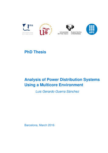
REVIEWSthese proteins to exocytic and endocytic machinery40–42,target proteins to the basolateral membrane in epithelialcells and might have a role in targeting to dendrites inneurons. However, the signals that target proteins to theapical membrane of epithelial cells do not appear to workfor axonal targeting38. In light of the identification ofnovel dendritic targeting signals for transferrin receptors,metabotropic glutamate receptors and AMPA (α-amino3-hydroxy-5-methyl-4-isoxazole propionic acid) receptors43–45, it seems likely that axonal targeting will turn outto involve novel motifs as well.ContactinA glycosylphosphatidylinositol(GPI) anchored glycoproteinwith structural homology to theL1 CAM extracellular domain.It has six IgG-like proteindomains followed by fourfibronectin type III domains.Ankyrin GOne of three types of ankyrinadaptor proteins that linkintegral membrane proteins tothe spectrin/actin membranecytoskeleton. Known as ‘‘G’’ forgiant or general, it has twomain alternative splice formsthat generate proteins of270 kDa and 480 kDa.βIV spectrinA splice form of the β-subunitof spectrin, which is a tetramerwith two α- and two β-subunitsthat form two antiparallelheterodimers.552 JULY 2006 VOLUME 7Nav channels. In axons, Nav channels are responsiblefor action potential initiation at the AIS and the nodes ofRanvier, action potential propagation along the unmyelinated axon, and action potential back-propagation in dendrites. In addition to their high concentration at the AIS,the biophysical properties of the Nav channels at the AISmight be particularly suited for action potential initiation46. The principal Nav channels in the AIS and nodesof Ranvier are Nav1.2 and Nav1.6, and their distributioncan change during development in a myelin-dependentmanner (BOX 1)17,30,47,48. Nav1.1, Nav1.2, Nav1.3 and Nav1.6are expressed mainly in the CNS, Nav1.4 and Nav1.5 arefound in the cardiac and skeletal muscle systems, andNav1.7, Nav1.8 and Nav1.9 are found in the PNS, althoughthere are exceptions (for example, Nav1.2 and Nav1.6 arefound at the nodes of Ranvier in the sciatic nerve17,30). TheNav1.1 and Nav1.3 isoforms were found to be somatodendritic for neurons in the brain30,47, and other distributionshave been reported for specific cell types. Here we focuson the channels that are targeted to the axon30,47,48.Nav channels associate, or are localized, with a numberof molecules that might have a role in anchoring or retaining these channels at the nodes of Ranvier. Nav β-subunitshave an extracellular Ig-like domain that is similar to thoseof Ig-family CAMs19,20, and these β-subunits colocalizewith several CAMs of this family — neuronal cell adhesion molecule (NrCAM), neurofascin-186 (Nf186) andcontactin — around the nodes of Ranvier49,50. The β1 andβ3 subunits interact with Nf186 through their extracellulardomains51, whereas NrCAM and Nf186 bind to ankyrin Gthrough a conserved FIGQY motif in their cytoplasmic Ctermini, which connects these CAMs to the actin cytoskeleton through βIV spectrin52. Contactin interacts with theNav β1 subunit and increases the surface expression ofNav1.2, Nav1.3 and Nav1.9 in mammalian cell lines53–55.Nav α-subunits also interact with the extracellular matrixproteins tenascin-C, tenascin-R, and phosphacan — probably through the Nav β-subunits19,56–58. A summary of protein interactions and motifs is given in FIG. 3.The precise mechanisms of these CAMs in anchoring or retaining Nav channels at nodes has been probedby assessing their appearance at nodes during development49,50. The localization of Nav channels to the AISand nodes of Ranvier is highly correlated with that ofankyrin G and βIV spectrin59,60, which appear at theAIS of Purkinje cells early in development, followed byL1 CAMs and Nav channels61. This contrasts with thenodes of Ranvier in the rodent sciatic nerve where the L1CAMs, NrCAM and neurofascin appear before ankyrinNf186Fibronectin type IIIlike domainIgG-like domainContactinNrCAMMucin-like PAT Preferentialsomatodendriticendocytosis regionSix ankyrinrepeatsII–IIIlinkerCCCAnkyrin Gbinding motif/AIS clusteringmotifβIV spectrinbinding domainAnkyrin GβIV spectrinActinFigure 3 Voltage-gated sodium channels. Contactin,Nf186 (Neurofascin-186), and NrCAM (neuronal celladhesion molecule) are cell adhesion molecules thatinteract and/or colocalize with voltage-gated sodium (Nav)channels. The glycosylphosphatidylinositol (GPI)-anchoredcontactin molecules interact only with the β1 subunit ofthe Nav channel. Only the β1 subunit (indicated by anasterisk) interacts with the S5–S6 loop in domain IV (DIV) ofNav channels. β2 and β4 subunits are linked by disulphidebonds to Nav channels. A region in the II–III linker isresponsible for preferential somatodendritic endocytosis.An ankyrin-G-binding motif that also serves as a clusteringmotif is also in the II–III linker. Nf186 and NrCAM alsointeract with ankyrin G, which, through βIV spectrin,connects to the actin cytoskeleton. A di-leucine motif inthe C-terminus controls axonal compartmentalization. TheN-terminal membrane-binding domain of ankyrin G hasfour subdomains that consist of six ankyrin repeats thatbind other proteins, mainly membrane proteins, followedby a spectrin-binding domain60. Actin mostly interacts withthe N-terminal region of one β-subunit in theheterotetramer of βIV spectrin60. Interactions are indicatedby arrows. AIS, axon initial segment, PAT, domain rich inproline, alanine and threonine residues.G and Nav channels during development62. Whetherthese differences signify differences between axon initialsegments and nodes of Ranvier, or between the CNS andPNS, remains to be determined; however, in both cases,ankyrin G and the CAMs are present before the Navchannels, raising the possibility that these proteins mightbe the first to be targeted to the axon, followed by Navchannels, which are then retained through association.www.nature.com/reviews/neuro
REVIEWSChimeric fusion proteinA polypeptide that is createdby fusing an amino acidsequence of interest to areporter protein.CD4A single-span transmembraneprotein that tends to yield auniform distribution in axonsand dendrites when it isexpressed in neurons.ER export or retentionmotifsAmino acid sequences thathave been identified in anumber of proteins to beresponsible for either exit from,or retention in, the ER (forexample, RXR).Endocytic motifA common amino acidsequence (for example, YXXφ)that signals clathrin-mediatedendocytosis.The effect of ankyrin G and βIV spectrin on the distribution of Nav channels has been studied in knockoutmice. In mice lacking the cerebellum-specific form ofankyrin G, Nav channels, neurofascin and βIV spectrinare no longer concentrated at the AIS; and their Purkinjecells showed a decreased ability to initiate action potentials and maintain repetitive firing63,64. Moreover, βIVspectrin knockout mice have aberrant ankyrin G andNav localization64. It therefore appears that βIV spectrinand ankyrin G act together to stabilize Nav channels atthe AIS and nodes of Ranvier.Even though Nav channels are associated with CAMsthat can interact with ankyrin G, Nav channels themselves interact with ankyrin G through a highly conserved nine amino acid motif — residues 1105 to 1113in Nav1.2 — in the II–III loop65–67. Chimeric fusion proteinsthat are composed of the entire Nav1.2 II–III linker andproteins not normally localized to the AIS are targetedto the AIS in hippocampal cultured neurons66, whilereplacing E1111 with alanine causes a uniform distribution of CD4–Nav1.2 II–III chimaeras to the axons, somasand dendrites68. An additional motif in the II–III linker— residues 1010 to 1030 in Nav1.2 — is responsible forselective endocytosis from somatodendritic domains inhippocampal cultured neurons68. This suggests that Navchannels are uniformly inserted into the membrane butare retained at the AIS through tethering to ankyrin G,while the non-tethered channels in the somatodendriticdomains are preferentially endocytosed68.The motifs in the II–III linker of Nav1.2 mightfurther work together with a di-leucine-based motifin the C-terminus for channel targeting to the axon. Achimaera of CD4 and the C-terminus of Nav1.2 localizes to the axon in cultured hippocampal neurons, eventhough it does not contain the ankyrin G-binding regionfor sequestration in the AIS69. This chimaera is selectively endocytosed at dendritic sites, which suggests amechanism of selective elimination for axonal localization similar to the mechanism proposed for VAMP2 thatis described above37. Indeed, the C-terminus of Nav1.2 isrecognized by components of the clathrin endocytosispathway. Axonal localization and endocytosis are compromised when the di-leucine motif within a nine aminoacid region is mutated to di-alanine. The C-terminus ofNav1.6 apparently lacks this motif, and its fusion to CD4results in a somatodendritic or non-polarized distribution of the fusion protein. In this case, perhaps the motifsin the II–III linker, which are conserved in Nav1.6, aresufficient for localization of this channel to the axon.Kv1 channels. In the mammalian nervous sy
ion channels on dendrites and axons. Molecular studies of voltage-gated ion channels over the past quarter of a century further unveiled the remarkably refined and mosaic-like patterns of channel distribution. Only recently have we begun to appreciate just how the different channel isoforms are targeted to different parts of the neuron to