Transcription
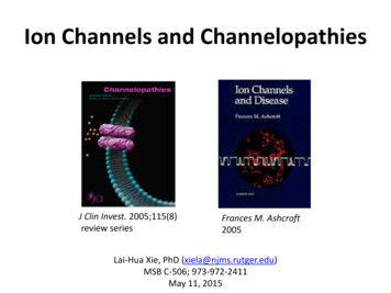
Ion Channels and ChannelopathiesJ Clin Invest. 2005;115(8)review seriesFrances M. Ashcroft2005Lai-Hua Xie, PhD (xiela@njms.rutger.edu)MSB C-506; 973-972-2411May 11, 2015
OutlinePart I: Ion ctureFunctionPart II: Channelopathies– Long QT syndromes Type 1 and 2 : LQT1 and LQT2:delayed K channel– Long QT syndrome type 3: LQT3: Na channel– Epilepsy: Voltage-gated Ca2 channel– Diabetes Mellitus: ATP-sensitive K channel– Cystic fibrosis: CFTR, Cl- channel
OutlinePart I: Ion ctureFunctionPart II: Channelopathies– Long QT syndrome Type 1 and 2 : LQT1 and LQT2:delayed K channel– Long QT syndrome type 3: LQT3: Na channel– Epilepsy: Voltage-gated Ca2 channel– Diabetes Mellitus: ATP-sensitive K channel– Cystic fibrosis: CFTR, Cl- channel
What are Ion Channels ? Ion channels - structure– are proteins that span (or traverse) the membrane– have water-filled ‘channel’ that runs through the protein– ions move through channel, and so through membrane Ion channel - properties– Selectivity: Each specific ion crosses through specific channels– Gating: transition between states (closed open Inactivation)Voltage-gated ; Ligand-gated– Channels mediate ion movement down electrochemicalgradients.– Activation of channel permeable to ion X shifts membranepotential towards to its Equilibrium Potential, EX
Equilibrium Potential or Nernst PotentialThe voltage at which there is zero net flux of a given ion(Electrical gradient a chemical concentration gradient)For K : -90 mVK current (IK1) is the major contributor for RMP
Four Milestones in Ion Channel Research1. Ionic conductanceNoble 1963 (Physiol/Medicine)Alan L. HodgkinAndrew F. Huxley3. Channel cloning sequencing(Ach receptor, Na, Ca channels)2. Patch clamp methodologyNoble 1991 (Physiol/Medicine)Erwin NeherBert Sakmann4. K channel structureNoble 2003 (Chemistry)Japan AcademyPrize 1985Shosaku Numa (沼 正作)Rod MacKinnon
Hodgkin-Huxley Model Predicted the Existenceof Ion ChannelsThe Giant Axon of SquidCdV g Na m 3 h (VNa V ) g K n 4 (VK V ) g L (VL V ) I extdtNa channelgatingK channelgating1963 noble Prize
Patch-Clamp Techniques1976βCCOαO1991 Nobel Prize
Channel cloning sequencingNumaSakmann
Nobel Prize for Chemistry 2003Protein x-ray crystallography1) Purification2) Crystallization3) X-Ray DiffractionThe Nobel Prize in Chemistry 2003Peter Agre, Roderick MacKinnonCrystal structure of ion channel
Classification of Ion Channels1) Based on ion selectivity:K , Na , Ca2 , Cl- channels2) Based on gating:Voltage-gated : ionsLigand-gated: Glutamate, GABA, ACh, ATP, cAMP3) Based on rectification:Inwardly or outwardly rectifying 40 mV0 mV-80 mVA. c/a
Structure of KCSA Channels:Selectivity Filter and GatingprofileDoyle et al. Science 1998;
Open-Close GatingopenclosedDoyle et al. Science 1998;gateBacterial K channelselective filter: P-loop; Gating: intracellular side of the pore bundle crossingBacterial Na channel pore in the closed and “open” conformation
Ligand-Gated ChannelsACh receptor channelATP-sensitive K channelAChC-ATP ATPOAlso a weak inward rectifier Open when a signal molecule (ligand) binds to an extracellular receptorregion of the channel protein. This binding changes the structural arrangements of the channel protein,which then causes the channels to open or close in response to the binding of aligand such as a neurotransmitter. This ligand-gated ion channel, allows specific ions (Na , K , Ca2 , or Cl-) toflow in and out of the membrane.
Models for Voltage GateThe conventional modelA new ‘paddle model’The transporter-like modelthe S4 segment is responsible for detecting voltage changes.The movement of positively-charged S4 segments within the membrane electric field
Transition between Close, Open, andInactivation StatesC O I
Inactivation Gating of Voltage-Gated Channels-Ball and Chain(Gulbis et al, Science 2000)N-terminal inactivation gateA positively charged inactivation particle (ball) has to pass through one of the lateral windows andbind in the hydrophobic binding pocket of the pore's central cavity. This blocks the flow ofpotassium ions through the pore. There are four balls and chains to each channel, but only one isneeded for inactivation.
Structural Basis of Gatingin a Voltage-gated ChannelA: a subunit containing six transmembrane-spanning motifs. S5 and S6 andthe pore loop are responsible for ion conduction (channel pore). S4 is the thevoltage sensor, which bears positively charged amino acids (Arg) that relocateupon changes in the membrane electric field. N-terminal ball-and-chain isresponsible for inactivationB: four such subunits assembled to form a potassium channel.
Channel Function:Single Channel and Whole-cell Current Ion channels are not open continuously but open andclose in a stochastic or random fashion. Ion channel function may be decreased by––––decreasing the open time (O),increasing the closed time (C),decreasing the single channel current amplitude (i)or decreasing the number of channels (n).CβαOCO3 pAI n*Po*iPO τoτo τc
Channel Function:Single Channel and Whole-cell CurrentActivationDepolarizing voltage pulsesresult in brief openings in theseven successive recordings ofmembrane currentInactivationClose correlation between thetime courses of microscopic andmacroscopic Na currents
Physiological Function of Ion Channels Maintain cell resting membrane potential: inward rectifier Kand Cl channels. Action potential and Conduction of electrical signal: Na, K,and Ca channels of nerve axons and muscles Excitation-contraction (E-C) coupling: Ca channels ofskeletal and heart muscles Synaptic transmission at nerve terminals: glutamate, Achreceptor channels Intracellular transfer of ion, metabolite, propagation: gapjunctions Cell volume regulation: Cl channel, aquaporins Sensory perception: cyclic necleotide gated channels of rods,cones Oscillators: pacemaker channels of the heart and centralneurons Stimulation-secretion coupling: release of insulin formpancreas (ATP sensitive K channel)
OutlinePart I: Ion tureFunctionPart II: Channelopathies– Long QT syndrome Type 1 and 2 : LQT1 and LQT2:delayed K channel– Long QT syndrome type 3: LQT3: Na channel– Epilepsy: Voltage-gated Ca2 channel– Diabetes Mellitus: ATP-sensitive K channel– Cystic fibrosis: CFTR, Cl- channel
Channelopathies?1. Definition: Disorders of ion channels or ion channel diseaseDiseases that result from defects in ion channel function. Mostly causedby mutations of ion channels.2. Channelopathies can be inherited or acquired:a. Inherited channelopathies result from mutations in genes encodingchannel proteins (major)b. Acquired channelopathies result from de novo mutations, actions ofdrugs/toxins, or autoimmune attack of ion channels Drug/Toxin - e.g. Drugs that cause long QT syndrome3. Increasingly recognized as important cause of disease ( 30diseases).4. Numerous mutation sites may cause similar channelopathye.g. cystic fibrosis where 1000 different mutations of CFTRdescribed
Molecular Mechanisms of Channel DisruptionIV. GatingI. ProductionIII. ConductionII. Processing
Consequences of Ion ChannelMutations- Mutation of ion channel can alter–Activation–Inactivation–Ion selectivity/Conduction- Abnormal gain of function- Loss of function
Cardiac Channelopathies Long QT Syndrome (types 1-12, various genes) Short QT Syndrome (Kir2.1, L-type Ca2 channel) Burgada Syndrome (Ito, Na , Ca2 channels) Catecholaminergic Polymorphic VentricularTachycardia (CPVT) (RyR2, SR Ca release)
ECG and QT intervalQT IntervalBazett's Formula:
FYI: ECG Recording 120 Years AgoFirst recorded in 1887
FYI: ECG Recording 120 Years AgoAnd Now!
AP Correlation to ECG Waveform P wave: Electrical activation(depolarization) of the atrialmyocardium. PR segment: This is a time ofelectrical quiescence duringwhich the wave of electricalexcitation (depolarization)passes through mainly the AVnode. QRS wave: Depolarization ofthe ventricular myocardium. T wave: Ending of ventricularmyocardium repolarization ST segment: Ventricularrepolarization
LQTS-facts Normal QT interval: 360-440 ms Delayed repolarization of the myocardium, QT prolongation( 450 in man; 470 in women). Increased risk for syncope, seizures, and SCD in the settingof a structurally normal heart 1/2500 persons. Usually asymptomatic, certain triggers leads to potentiallylife-threatening arrhythmias, such as Torsades de Pointes(TdP)
FYI: QT Interval Ranges
FYI: Genetic Basis for LQT syndromes
Cardiac action potential Phase 0. Influx of Na (INa). Induces membrane depolarization Phase 1. Efflux of K (Ito). Limits the Na spike Phase 2. Influx of Ca2 (ICa). Activation of IK. Balance between Ca2 influx and K efflux.Ca2 enters the cell to trigger the Ca2 -induced Ca2 release. Phase 3. Efflux of K (IK) increases. Repolarization starts Phase 4. Restoration of the resting potential: equilibrium potential of K via IK1.and Na / K pump, Na / Ca2 pump.INaIKrIKs
Pathophysiology of LQT (1, 2, 3)
LQT syndromes: proarrhythmic mechanisms Upregulation ofinward currentsOr Downregulation ofoutward currents EADs triggers Dispersion of APDs substrates reentryDrugsormutations
Example 1:LQT1 and LQT2Downregualtion of delayed K channel, IKs and IKr
LQT1: KCNQ1 (KvLQT1) mutationsIKs: Slow component of the delayed rectifier potassium current
LQT2: KCNH2(HERG) MUTATIONSKCNH2(HERG)IKr: Rapid component of the delayed rectifier potassium current
LQT 1 and 2: IKs and IKr downregulationIKKCNQ1 or KCNE2 gene mutations(IKr)(IKs)
Example 2:LQT3Inactivation of Na channel
LQT3: Increased persistent Na CurrentDel KPQWT: normal inactivation KPQ:
Functional mechanisms in LQT3LQT3
Example 3:Epilepsy - a CNS ChannelopathiesEpileptic seizureEpilepsy is a disorder marked by disturbed electrical rhythmsin the central nervous system
FYI: Ion Channels Implicated in Epilepsy
Voltage-gated Ca Channels:Subunit Assembly and Subtypes
Epilepsy: Voltage-gated Ca2 Channel
Enhancement of T-type Ca current in thalamocorticalnetworks produces spike wave absence epilepsygain-of-function
Epilepsy: Pathology and SymptomIn humanIn miceElectroencephalogram (EEG)disturbed electrical rhythms
Example 4:ATP-Sensitive K Channel andDiabetes
Discovery of KATP Channel
ATP-Sensitive Potassium ChannelIs composed of Kir6.x and sulfonylurea receptors (SURs) Inhibited by ATP Inhibited by sulfonylurea via SURs
ATP-Sensitive K channelInhibited by ATPATPATPCloseC- ATP ATPO
Role of the KATP Channel in Insulin Secretionin Pancreatic β Cell Glucose enters the cell via the GLUT2 transporter Glycolytic and mitochondrial metabolism leads to an increase in ATP This results in KATP channel closure, membrane depolarization, Opening of voltage-gated Ca2 channels, Ca2 influx, Exocytosis of insulin granules (insulin secretion).Gloyn AL et al. N Engl J Med 2004;350:1838-1849.
KATP Channel Mutations CausingLower ATP Sensitivity and DiabetesMutantWT
The KATP Channel Couples Glucose Metabolismto Insulin Secretion
Example 5:Cystic Fibrosis: Cl- Channel Disease
Cystic Fibrosis: Facts- Cystic fibrosis (CF) is autosomal recessivedisease- CF is a chronic, progressive, life threateninggenetic disorder of pediatrics.- It affect white population (1 in 3200 livebirths) but is uncommon among Asian andAfrican population- It affects exocrine glands (mainly sweatglands) and mucus gland present on theepithelial lining of lungs, pancreas, intestine,and reproductive system.- CF is a defect in epithelial chloridechannel protein, causes membrane tobecome impermeable to Chloride ion.
CFTR gene encode for the CFTR protein channelCF occurs due to the deletion of 3 nucleotides which code for the phenylalaninefrom the CFTR (cystic fibrosis transmembrane conductance regulator) genelocated on chromosome no.7 at position 508. This mutation is known as ΔF 508
Structure of the CFTR proteinCFTR protein is a cAMP inducedChannel made up of five domains:Two membrane-spanning domain(MSD1 & MSD2) that form Cl- ionchannel.Two nucleotide binding domains(NBD1 & NBD2) that bind andhydrolyze ATP.A regulatory R domain.
CFTR mutation: Loss of Cl- Channel Function
Pathology of Cystic Fibrosis - 1In sweat glands:CFTR is responsible for re-absorption of Cl- along with Na throughepithelial Na channel (ENaC).Impaired function of CFTR cause the production of hypertonic saltysweat, and ultimately dehydration.SWEAT GLANDS
Pathology of Cystic Fibrosis - 2In lung mucus glands: Loss of CFTR function to secrete chloride ion Loss or reduction of Cl- ion in luminal secretion Followed by active luminal Na absorption through ENaC Increases passive water absorption from the lumen Impaired mucociliary action, accumulation of thick, viscous, dehydrated mucus Obstruction of air passage and recurrent pulmonary infections
Channelopathies: Summary Channel mutations are an increasingly recognizedcause of disease. Many channelopathies are episodic despitepersistently abnormal channel. Abnormalities in same channel may present withdifferent disease states Mutations/ abnormalities in different channels maylead to same disease e.g. periodic paralysis orepilepsy Disease mechanism often unclear despiteidentification of mutation.
Thank you!
FYI: Human ChannelopathiesJ Clin Invest. 2005;115(8)and MORE
Disorders of ion channelsor ion channel disease. Diseases that result from defects in ion channel function. Mostly caused by mutations of ion channels. 2. Channelopathies can be inherited or acquired: a. Inherited channelopathies result from mutations in genes encoding channel proteins (major) b. Acquired channelopathies result from . de novo .