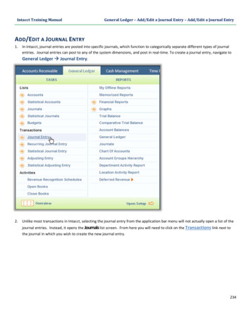
Transcription
Journal of Blood Group Serology and Molecular GeneticsV o l u m e 3 4, N u m b e r 3 , 2018
This issue of Immunohematologyis supported by a contribution fromGrifols Diagnostics Solutions, Inc.Dedicated to advancement and education inmolecular and serologic immunohematology
ImmunohematologyJournal of Blood Group Serology and Molecular GeneticsVolume 34, Number 3, 2018CO N TEN TS85ReviewProceedings from the International Society of Blood Transfusion Working Party onImmunohaematology, Workshop on the Clinical Significance of Red Blood Cell Alloantibodies,September 2, 2016, DubaiClinical significance of antibodies to antigens in the Raph, JohnMilton Hagen, I, Globoside, Gill, Rh-associated glycoprotein,FORS, JR, LAN, Vel, CD59, and Augustine blood group systemsM. Moghaddam and A.A. Naghi91S ero lo g i c M e t h o d R e v i e wRouleaux and saline replacementK.L. Waider93Original ReportMethod-specific and unexplained reactivity in automated solidphase testing and their association with specific antibodiesM.E. Harach, J.M. Gould, R.P. Brown, T. Sanders and J.H. Herman98S ero lo g i c M e t h o d R e v i e wUtility of chloroquine diphosphate in the blood bank laboratoryT. Aye and P.A. Arndt103ReviewProceedings from the International Society of Blood Transfusion Working Party onImmunohaematology, Workshop on the Clinical Significance of Red Blood Cell Alloantibodies,Friday, September 2, 2016, DubaiClinical significance of antibodies to antigens in the Scianna,Dombrock, Colton, Landsteiner-Weiner, Chido/Rodgers, H, Kx,Cromer, Gerbich, Knops, Indian, and Ok blood group systemsS. Lejon Crottet109Case ReportA delayed and acute hemolytic transfusion reaction mediated byan anti-c in a patient with variant RH allelesT.K. Walters and T. Lightfoot113S ero lo g i c M e t h o d R e v i e wDetecting polyagglutinable red blood cellsC. Melland and C. Hintz118In MemoriamMarjory Stroup WaltersJ. Hegarty and T.S. Casina120127131134AnnouncementsA dv ertisementsInstructionsf o r A u t h o rsSubscriptionI n f o rm at i o n
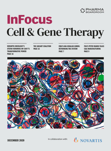
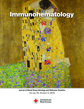
Editor-in- ChiefEdi to rial BoardSandra Nance, MS, MT(ASCP)SBBPhiladelphia, PennsylvaniaPatricia Arndt, MT(ASCP)SBBGeralyn M. Meny, MDBarbara J. Bryant, MDGalveston, TexasPaul M. Ness, MDWilmington, DelawareTe c h n i c a l E d i t o rsLilian M. Castilho, PhDThierry Peyrard, PharmD, PhDMartha R. Combs, MT(ASCP)SBBS. Gerald Sandler, MDGeoffrey Daniels, PhDIra A. Shulman, MDAnne F. Eder, MDJill R. Storry, PhDMelissa R. George, DO, FCAPNicole ThorntonManaging Edi torCynthia Flickinger, MT(ASCP)SBBJanis R. Hamilton, MS, MT(ASCP)SBBDetroit, MichiganChristine Lomas-Francis, MScNew York City, New YorkJoyce Poole, FIBMSBristol, United KingdomDawn M. Rumsey, ART(CSMLT)Norcross, GeorgiaTiffany Walters, MT(ASCP)SBBCMCharlotte, North CarolinaSenior M edi cal EditorDavid Moolten, MDPhiladelphia, PennsylvaniaA ss o c i at e M e d i c a l E d i t o rsPomona, CaliforniaSan Antonio, TexasBaltimore, MarylandCampinas, BrazilParis, FranceDurham, North CarolinaBristol, United KingdomWashington, District of ColumbiaLos Angeles, CaliforniaWashington, District of ColumbiaLund, SwedenHershey, PennsylvaniaJulie K. Karp, MDBristol, United KingdomPhiladelphia, PennsylvaniaE m e r i t u s E d i t o rsJose Lima, MDDelores Mallory, MT(ASCP)SBBDouglassville, GeorgiaSupply, North CarolinaChristine Lomas-Francis, MScMarion E. Reid, PhD, FIBMSNew York City, New YorkBristol, United KingdomP. Dayand Borge, MDPhiladelphia, PennsylvaniaCorinne L. Goldberg, MDDurham, North CarolinaM olecul ar EditorMargaret A. Keller, PhDPhiladelphia, PennsylvaniaE d i t o r i a l A ss i s ta n tLinda FrazierP r o d u c t i o n A ss i s ta n tLinda FrazierC opy EditorFrederique Courard-HouriProofreaderWendy Martin-ShumaElectronic PublisherPaul DuquetteImmunohematology is published quarterly (March, June, September, and December) by theAmerican Red Cross, National Headquarters, Washington, DC 20006.Immunohematology is indexed and included in Index Medicus and MEDLINE on theMEDLARS system. The contents are also cited in the EBASE/Excerpta Medica and ElsevierBIOBASE/Current Awareness in Biological Sciences (CABS) databases.The subscription price is 50 for individual, 100 for institution (U.S.), and 60 for individual, 100 for institution (foreign), per year.Subscriptions, Change of Address, and Extra Copies:Immunohematology, P.O. Box 40325Philadelphia, PA 19106Or call (215) 451-4902Web site: pyright 2018 by The American National Red CrossISSN 0894-203XO n O ur C ov erPerhaps Gustav Klimt’s best-known work, The Kiss (1907), dazzlingly melds the sensual withthe abstract. The painting depicts a man and woman intertwined, he standing, bowed, while shekneels on an idealized quilt-like meadow of flowers. Their proximity to the top of the paintingheightens the sense of intimacy and also suggests the possibility of transcending worldlyconstraints. We see little of the lovers—the back of his head, her face, their hands and feet, simplyrendered but wrapped in golden raiment decorated with distinct mosaics. The man’s figure iscomprised of juxtaposed rectangles and the woman’s of clustered circles, a geometry that hintsat both the contrast and complement of their union. Despite its shapelessness, the gilded mass ofclothing serves to intensify and exalt the physical act of the kiss and thus consecrates the coupleand love itself. This issue includes Waider’s review on the use of saline replacement to identifyrouleaux—the stacked coin appearance of the red cells likened to “clustered circles.”David Moolten, MDii I M M U N O H E M ATO LO GY, Vo l u m e 3 4, N u m b e r 3 , 2 018
ReviewProceedings from the International Society of Blood Transfusion Working Party on Immunohaematology,Workshop on the Clinical Significance of Red Blood Cell Alloantibodies, September 2, 2016, DubaiClinical significance of antibodies to antigens inthe Raph, John Milton Hagen, I, Globoside, Gill,Rh-associated glycoprotein, FORS, JR, LAN, Vel,CD59, and Augustine blood group systemsM. Moghaddam and A.A. NaghiThis article reviews information on the clinical significanceof antibodies to antigens in the Raph, John Milton Hagen, I,Globoside, Gill, Rh-associated glycoprotein, FORS, JR, LAN, Vel,CD59, and Augustine blood group systems. Antibodies to many ofthe antigens in these groups are rarely encountered because of thehigh prevalence of the associated antigens in most populations.For many of these antibodies, the clinical significance—that is,the potential to cause reduced survival of transfused antigenpositive red blood cells or a transfusion reaction (e.g., anti-P,anti-Jra, and anti-Lan), and/or hemolytic disease of the fetus andnewborn (e.g., anti-RHAG4 and anti-Vel)—has been documented.For other antibodies, their prevalence is so rare that informationon the clinical significance of their antibodies is not available(e.g., anti-FORS1). Immunohematology 2018;34:85–90.Key Words: clinical significance, antibodies to red bloodcell antigens, Raph, John Milton Hagen, I, Globoside, Gill, Rhassociated glycoprotein, FORS, JR, LAN, Vel, CD59, AugustineRaph Blood Group SystemThe Raph blood group system contains just oneantigen, MER2 (RAPH1), located on the tetraspanin CD151glycoprotein (TM4SF).1,2 The true MER2– phenotype,associated with the presence of anti-MER2, is very rare andresults from mutations in CD151, but there is a quantitativered blood cell (RBC) polymorphism in which RBCs of about 8percent of white individuals are serologically MER2–.2Clinical SignificanceThere have been six reports of human alloantibodies toMER2. Three of the subjects were found to have a stop codonin the CD151 gene, which encodes a member of the tetraspaninfamily of proteins. These three individuals had nephropathy anddeafness, and two of the three, who were siblings, also had skinlesions, deafness, and β-thalassemia minor. The fourth subjecthad missense mutation c.533G A (p.Argl78His). Subjects 5I M M U N O H E M ATO LO GY, Vo l u m e 3 4, N u m b e r 3 , 2 018 and 6 shared missense mutation c.511C T (p.Argl71Cys) aswell as a synonymous single-nucleotide mutation (c.579A G)and had no clinical features. Although the CD151 protein iscritical to cell adhesion and signaling and is implicated incancer progression, its significance in transfusion medicine islimited to only one report of a hemolytic transfusion reaction(HTR).3 Least-incompatible RBC units should be selectedfor transfusion to patients with anti-MER2.2 No informationon anti-MER2 causing hemolytic disease of the fetus andnewborn (HDFN) is available.4John Milton Hagen Blood Group SystemThe John Milton Hagen (JMH) blood group systemconsists of six high-prevalence antigens that are recognized bythe International Society of Blood Transfusion (ISBT) and arenumbered sequentially from JMH1 through JMH6. ConfirmedJMH variants are named with the first letter from the antibodymaker’s first name following JMH (JMH2 named JMHK,JMH3 named JMHL, JMH4 named JMHG, JMH5 namedJMHM, JMH6 named JMHQ).5 These antigens are located onthe Sema7A protein.5,6 The chromosomal location of SEMA7Ais 15q22.3-q23. JMH1 is the primary antigen in the systemand is present in greater than 99 percent of all individuals. TheJMH1– phenotype is more commonly acquired by depressionof the antigen. This finding might explain the serologicobservation of a positive direct antiglobulin test (DAT) seen inmany individuals with anti-JMH.5Clinical SignificanceJMH1, commonly known as JMH, is most notable becausetransient depression of the antigen occurs, and (auto)anti-JMHmay develop.5 JMH– patients with anti-JMH often have nohistory of transfusion or pregnancy. Of seven JMH antibodies,85
M. Moghaddam and A.A. Naghifive were IgG4 and two were IgG1, although IgG3 anti-JMHhas been described. There are numerous cases where patientswith anti-JMH have been transfused with JMH bloodwith no adverse effects. One such patient received 20 unitsof JMH blood in 10 months, with the expected hemoglobinrise. There are no reports of JMH antibodies causing HDFN,which is unsurprising considering that JMH antigens areexpressed very weakly on cord cells. One patient with antiJMH is reported to have experienced an acute intravascularHTR, but evidence that the JMH antibody was responsible islimited. Antibodies developed in the rare JMH variant typesmay cause reduced RBC survival.2 Today, rapid detection ofJMH antibodies with recombinant SEMA7A protein and theparticle gel immunoassay has been developed.7I Blood Group SystemThe I antigen, together with i, used to be part of theIi blood group collection. The gene encoding the I β-1,6-N-acetylglucosamine transferase (IGNT/GCNT2) responsible for converting i active straight chains of carbohydrates toI active branched chains has been cloned,8 and some mutationsresponsible for adult i phenotype have been identified.9,10Hence, I has been promoted to the I blood group systemcomprising only a single antigen, the I antigen, and i remainsin the Ii collection. RBCs from adults predominantly expressI antigens and only low levels of i antigens, higher levels ofthe latter predominate in fetal and neonatal RBCs. In a smallnumber of individuals, only very low levels of I can be detected,and their RBCs show high levels of i (adult i phenotype). Thisphenotype is believed to result from lack of activity of the Ibranching transferase, a product of the GCNT2 gene.2be hemolytic and have a thermal range up to 37 C, and someanti-I with a thermal range below 37 C can cause shortenedsurvival of transfused I RBCs. One anti-I became potentiallyclinically significant after transfusion of 6 units of I blood.Globoside Blood Group SystemThe P blood group antigen of the Globoside system is aglycolipid structure, also known as globoside, on the RBCsof almost all individuals worldwide. The P antigen (Gb4) isintimately related to the Pk and NOR (P1PK4) antigens.The molecular genetic basis of globoside deficiency is theabsence of functional P synthase caused by mutations at theB3GALNT1 locus. Other related glycolipid structures, theLKE and PX2 antigens, remain in the Globoside blood groupcollection pending further evidence concerning the genes andgene products responsible for their synthesis.11Clinical SignificanceAnti-P is found in the serum of all Pk individuals andcan be separated from serum of p individuals by adsorptionwith P1k or P2k cells or by inhibition with hydatid cyst fluid.2When complement is present, anti-P will hemolyze P1 or P2phenotype RBCs.2 P antibodies are IgM and often also IgG, areusually reactive at 37 C, and can cause severe intravascularHTRs. Autoanti-P is associated with paroxysmal coldhemoglobinuria. P antigen is also a receptor of parvovirus B19.Cytotoxic IgM and IgG3 antibodies directed against P orPk antigens are associated with a higher-than-normal rate ofspontaneous abortion in women with the rare p, P1k, and P2kphenotypes.4Gill Blood Group SystemClinical SignificancePotent cold reactive antibodies responsible for coldagglutinin disease are usually of I specificity. These antibodiesare generally monoclonal and are usually IgM, but IgGautoanti-I can also occur. They directly agglutinate I RBCs at4 C with varying thermal amplitude but are generally inactiveabove 30 C. One autoanti-I active at 30 C caused an acuteHTR in a small child when 2 units of blood were transfusedimmediately after removal from the refrigerator. Transient,polyclonal, or oligoclonal autoanti-I may arise from infection,most typically by Mycoplasma pneumoniae.Alloanti-I of high titer is rare and usually presents in thesera of i adults. These antibodies are almost invariably IgMand are active only at low temperatures. Rare examples may86 The Gill blood group system was added to the list ofsystems already recognized by the ISBT in 2002. GIL, the onlyantigen of the Gill system, is an antigen of high prevalencelocated on the water and glycerol channel aquaporin-3(AQP3).2,12 The GIL– phenotype results from homozygosityfor a splice mutation in AQP3. Anti-GIL has been identified infive Gil– white women who had been pregnant at least twice.2Clinical SignificanceFive examples of anti-GIL have been identified, all inwhite women who had been pregnant at least twice. No GIL–individual was found by screening 23,251 white American and2841 African American women with anti-GIL. RBCs from twoI M M U N O H E M ATO LO GY, Vo l u m e 3 4, N u m b e r 3 , 2 018
ISBT conference—clinical significance of antibodiesof the babies of mothers with anti-GIL gave a positive DAT,but there were no clinical symptoms of HDFN. Anti-GIL mayhave been responsible for an HTR, and results of monocytemonolayer assay (MMA) with two GIL antibodies suggested apotential to cause accelerated destruction of transfused GIL RBCs.2The Rh-Associated Glycoprotein Blood GroupSystemIn 2010, the recognition that three RBC surfaceantigens were located on RhAG encoded by RHAG led tothe establishment of a new blood group system. Two highprevalence antigens, Duclos (RHAG1) and DSLK (RHAG3),two low-prevalence antigens, OI(a) and (RHAG2), and RHAG4,have serologic characteristics suggestive of expression onRhAG, but RHAG4 has been shown to not exist and is underinvestigation by ISBT to be retracted.13Clinical SignificanceNo data are available.4FORS Blood Group SystemThis blood group system has been named FORS after itsoriginal finder, Lund professor John Forssman. The FORSantigen (originally recognized as the Apae phenotype) wasdiscovered by weak reactivity of RBCs against polyclonalanti-A reagents, reactivity against the lectin Helix pomatia(snail anti-A), and no reactivity with the plant anti-A1 lectin,Dolichos biflorus, in two different families. Genomic analysisof the ABO locus in both samples revealed that they weregenetically group O, and the reactivity described must be dueto other phenomena.14,15Clinical SignificanceThe clinical significance of anti-FORS1 is not known.4JR Blood Group SystemThe JR blood group system (ISBT 032) consists of oneantigen, Jra, which is highly prevalent in all populations ( 99%).Jra is located on the ABCG2 transporter, a multipass membraneglycoprotein (also known as the breast cancer resistance protein[BCRP]), which is encoded by the ABCG2 gene on chromosome4q22.1.16–19 The rare Jr(a–) phenotype has been found mostlyin Japanese and other Asian populations, but also in peopleI M M U N O H E M ATO LO GY, Vo l u m e 3 4, N u m b e r 3 , 2 018 of northern European ancestry, in Bedouin Arabs, and in oneMexican individual. The rare Jr(a–) phenotype mostly resultsfrom recessive inheritance of ABCG2 null alleles caused byframeshift or nonsense changes.17,20–22 To date, more than 25different mutations responsible for the absence of ABCG2 aswell as mutations giving rise to weakened Jra expression havebeen identified.16Clinical SignificanceABCG2 expression levels in cord RBCs are higher thanthose in adult RBCs, and the change of ABCG2 expressionin erythroid lineage cells may influence the clinical courseof fetal anemia with anti-Jra.23 Anti-Jra may be stimulatedby transfusion or pregnancy and has been detected inuntransfused Jr(a–) women during their first pregnancy.24Most anti-Jra are IgG1 and sometimes IgG3. Anti-Jra mayfix complement, can be a dangerous antibody in pregnancy,and has been implicated in severe and fatal HDFN; in otherpregnancies with anti-Jra, however, indications of HDFNhave been no more than a positive DAT on cord cells ormild neonatal jaundice. Many transfusions of Jr(a ) RBCsto patients have resulted in no signs of hemolysis, althoughincompatible transfusion may cause a sharp rise in the titerof anti-Jra, resulting in signs of an acute HTR in subsequenttransfusions. A patient with anti-Jra developed rigors aftertransfusion of 150 mL crossmatch-incompatible blood. Leastincompatible RBC units may be suitable for transfusion tomost patients with anti-Jra, but Jr(a–) RBCs should be selectedin cases where the anti-Jra is of high titer.2LAN Blood Group SystemLAN (Langereis) was officially recognized by the ISBT in2012 as the 33rd human blood group system. It consists of onehigh-prevalence antigen, Lan (LAN1).25 The ABCB6 protein isthe carrier of the Lan blood group antigen.16,25 The ABCB6 gene(chromosome 2q36, 19 exons) encodes the ABCB6 polypeptide,known as a porphyrin transporter. The exceptional Lan–individuals do not express ABCB6 (Lannull phenotype) owingto several different frameshift and missense mutations.25 Todate, more than 40 ABCB6 alleles that encode Lan– or Lan wphenotypes have been described,26 and quantitation of Lanantigen in Lan , Lan w, and Lan– phenotypes have beenperformed.27 Despite the Lan antigen role in erythropoiesisand detoxification of cells, Lan– individuals do not appear todemonstrate susceptibility to any disease.87
M. Moghaddam and A.A. NaghiClinical SignificanceAnti-Lan has been reported in two African Americanindividuals as well as in other populations includingCaucasians and Asians and may be stimulated by transfusionor pregnancy. The original anti-Lan was responsible for animmediate HTR characterized by fever and chills. There isno report of naturally occurring alloanti-Lan; none of theLan– siblings of the Lan– propositi have anti-Lan. Anti-Lanhas been described as having variable clinical significance,either for HTRs (none to severe) or HDFN (none to mild).25Lan alloantibodies are mostly IgG1 and IgG3, although IgG2and IgG4 may also be present. Some anti-Lan fix complement;others do not.2 Despite challenging conditions caused by thescarcity of Lan– donors worldwide, ideally Lan– RBCs shouldbe selected for transfusion to patients with anti-Lan, especiallyindividuals with a high-titer antibody,2,25 although leastincompatible RBCs may be suitable for patients with weakexamples of the antibody. The only autoanti-Lan was reportedin a patient with mild autoimmune hemolytic anemia (AIHA)with depressed Lan antigen expression.2Vel Blood Group SystemVel is an RBC antigen that is expressed by more than 99.9percent of the population.28 The recognition of Vel dates to1952, when a patient, named Mrs. Vel, suffered a transfusionreaction due to the presence of an antibody that was found toagglutinate sera of over 10,000 individuals. The antibody wasnamed anti-Vel.29Recently, the SMIM1 protein was shown to carry the Velblood group antigen. Using a high-density single nucleotidepolymorphism array, Storry et al.30 identified the SMIM1 generesiding in a 97-kb region of homozygosity on chromosome1p36 in the vicinity of the RH locus. A frameshift deletion of17 nucleotides in exon 3 of SMIM1 is responsible for the Vel–phenotype.16,31,32 Genotype screening estimated that 1 in 17Swedish blood donors is a heterozygous deletion carrier and 1 in 1200 is a homozygous deletion knockout.31antibody, and patients with anti-Vel should be transfusedwith Vel– RBCs. The first anti-Vel and other examples sincehave caused severe immediate HTRs. Anti-Vel may be missedin compatibility testing if inappropriate methods are used.33Although many examples of anti-Vel have been found inpregnant women, anti-Vel does not usually cause HDFN,probably because most anti-Vel are predominantly IgM, andthe Vel antigen is usually expressed weakly on neonatal RBCs.Two examples of autoanti-Vel were responsible for (AIHA),although in one case, a nine-week-old infant, the RBCs gave anegative DAT.2CD59CD59, also known as the membrane inhibitor of reactivelysis (MIRL), homologous restriction factor (HRF), andmembrane attack complex inhibitory factor (MACIF), is a cellsurface glycoprotein of approximately 20 kDa that limits theactivity of the terminal complement complex C5b-9 and ismore effective than decay accelerating factor (DAF) or CD55in this respect.28,34 The first demonstration of anti-CD59 wasin a patient homozygous for a CD59 deficiency, which led tothe discovery of a new blood group system, CD59, and a nullallele (c.146delA).CD59 is attached by a glycosylphosphatidylinositol (GPI)anchor not only to erythrocytes but also to various other cellularmembranes. Seven cases of an isolated CD59 deficiency due tothree distinct null alleles of the CD59 gene have been publishedso far.34 As well as being cell-bound through its GPI anchor,soluble CD59 is also found in plasma, urine, and cerebrospinalfluid.Absence of CD59 is associated with hemolytic anemia andwith thrombosis as well as with other autoimmune diseasessuch as systemic lupus erythematosus.2Clinical SignificanceNo data are available.Augustine Blood Group SystemClinical SignificanceThe high clinical significance of Vel is related to whathappens to individuals with the Vel– phenotype upontransfusion or pregnancy. Vel alloantibodies are nevernaturally occurring, and most producers of anti-Vel have beentransfused, yet Vel antibodies are predominantly IgM andfix complement. Of the two IgG anti-Vel, one was IgG1, andthe other contained IgG1 and IgG3. Anti-Vel is a dangerous88 Ata is a high-prevalence antigen found on the RBCs of over99 percent of individuals.35 The first literature referring to theAta antibody, which can be responsible for severe HTRs andmild HDFN, dates to the 1960s. Applewhaite et al.36 identifiedan antibody with a novel specificity in the serum of Mrs.Augustine when the RBCs of her third child gave a positiveDAT at birth. Her alloantibody, abbreviated as anti-Ata afterI M M U N O H E M ATO LO GY, Vo l u m e 3 4, N u m b e r 3 , 2 018
ISBT conference—clinical significance of antibodiesher name, reacted with more than 6600 blood donors testedat that time, indicating that Ata was a high-prevalence antigen.All anti-Ata producers reported thus far have been of Africanancestry, like Mrs. Augustine.Recently, it has been shown that SLC29A1 encoding theequilibrative nucleoside transporter 1 (ENT1) specifies a newcandidate gene for a novel blood group system that includes theAta antigen. Daniels et al.37 reported that a nonsynonymousSNP in SLC29A1 (rs45458701) is responsible for the At(a–)phenotype. Although all At(a–)-reported propositi are ofAfrican ancestry with functional ENT1, they identified threesiblings of European ancestry who were homozygous for anull mutation in SLC29A1 (c.589 1G C) and thus have theAugustinenull phenotype. These individuals lacking ENT1exhibit periarticular and ectopic mineralization, whichconfirms an important role for ENT1/SLC29A1 in humanbone homeostasis,34 cardioprotection, and drug transport inerythrocytes.38Clinical SignificanceAta antibodies are mostly IgG, but IgM could also bepresent and can directly agglutinate At(a ) RBCs. Of two IgGanti-Ata, one was IgG1, and the other consisted of IgG1, IgG3,and IgG4. Ata antibodies facilitate rapid destruction of 51Crlabeled At(a ) RBCs in vivo and give positive results in the invitro functional assay, the MMA. Ideally, At(a–) RBCs shouldbe selected for transfusion to patients with anti-Ata, althoughleast incompatible RBC units may be suitable for patients withweak examples of the antibody.2One anti-Ata caused an immediate HTR with chills andnausea during an RBC survival study, and another causeda severe delayed HTR after transfusion of multiple units ofAt(a ) RBCs. Despite numerous pregnancies involving antiAta, only one of the infants had moderately severe HDFNrequiring phototherapy.2 In three At(a–)-reported patientsfrom the southern United States, the anti-Ata was concomitantwith autoimmune disease.5References1. Crew VK, Burton N, Kagan A, et al. CD151, the first member ofthe tetraspanin (TM4) super family detected on erythrocytesis essential for the correct assembly of human basementmembranes in kidney and skin. Blood 2003;102:4a.2. Daniels G. Human blood groups. 3rd ed. London, UK: WileyBlackwell, 2013.3. Hayes M. Raph blood group system. Immunohematology2014;30:6–10.I M M U N O H E M ATO LO GY, Vo l u m e 3 4, N u m b e r 3 , 2 018 4. Reid ME, Lomas-Francis C, Olsson ML. The blood groupantigen factsbook. 3rd ed. San Diego, CA: Academic Press,2012.5. Johnson ST. JMH blood group system: a review.Immunohematology 2014;30:18–23.6. Daniels G, Flegel WA, Fletcher A, et al. International Societyof Blood Transfusion Committee on Terminology for RedCell Surface Antigens: Cape Town report. Vox Sang 2007;92:250–3.7. Seltsam A, Agaylan A, Grueger D, et al. Rapid detection of JMHantibodies with recombinant Sema7A(CD108) protein and theparticle gel immunoassay. Transfusion 2008;48:1151–6.8. Bierhuizen MFA, Mattei MG, Fukuda M. Expression of thedevelopmental I antigen by a cloned human cDNA encodinga member of a ß-1,6-N-acetylglucosaminyltransferase genefamily. Genes Dev 1993;7:468–78.9. Yu LC, Twu YC, Chang CY, et al. Molecular basis of the adult iphenotype and the gene responsible for the expression of thehuman blood group I antigen. Blood 2001;98:3840–5.10. Inaba N, Hiruma T, Togayachi A, et al. A novel I-branchingß-1,6-N-acetylglucosaminyltransferase involved in the humanblood group I antigen expression. Blood 2003;101:2870–6.11. Hellberg A, Westman JS, Olsson ML. An update on the GLOBblood group system and collection. Immunohematology2013;29:19–24.12. Roudier N, Verbavatz JM, Maurel C, et al. Evidence for thepresence of aquaporin-3 in human red blood cells. J Biol Chem1998;273:8407–12.13. Tilley L, Gren C, Poole J, et al. A new blood group system,RHAG: three antigens resulting from amino acid substitutionsin the Rh-associated glycoprotein. Vox Sang 2010;8:151–9.14. Barr K, Korchagina E, Popova I, et al. Monoclonal anti-Aactivity against the FORS1 (Forssman) antigen.Transfusion2014;55:129–36.15. Svensson L, Hult AK, Stamps R, et al. Forssman expressionon human erythrocytes: biochemical and genetic evidence of anew histo-blood group system. Blood 2013;121:1459–68.16. Storry J. Five new blood group systems: what next? ISBT SciSer 2014;9:136–40.17. Castilho L, Reid ME. A review of JR blood group system.Immunohematology 2013;29:63–8.18. Saison C, Helia V, Ballif BA, et al. Null alleles of ABCG2encoding the breast cancer resistance protein define the newblood group system Junior. Nat Genet 2012;44:174–7.19. Zelinski T, Coghlan G, Liu XQ, et al. ABCG2 null alleles definethe Jr(a–) blood group phenotype. Nat Genet 2012;44:131–2.20. Ogasawara K, Osabe T, Susuki Y, et al. A new ABCG2 null allelewith a 27kb deletion including the promoter region causing theJr(a–) phenotype. Transfusion 2015;55:1467–71.21. Coghlan G. The JR blood group system: genetic and molecularinvestigations. ISBT Sci Ser 2012;7:260–3.22. Hue-Roye K, Lomas-Francis C, Coghlan G, et al. The JR bloodgroup system (ISBT 032): molecular characterization of thethree new null alleles. Transfusion 2013;53:1575–9.23. Fujita S, Kashiwagi H, Tomimatsu T, et al. Expression levelsof ABCG2 on cord red blood cells and study of fetal anemiaassociated with anti-Jr(a). Transfusion 2016;56:1171–81.89
M. Moghaddam and A.A. Naghi24. Endo Y, Ito S, Ogiyama Y. Suspected anemia caused by maternalanti-Jr(a) antibodies: a case report. Biomark Res 2015;3:23.25. Peyrard T. The LAN blood group system: a review.Immunohematology 2013;29:131–5.26. Reid ME, Hue-Roye K, Huamg A, et al. Alleles of the LANblood group system: molecular and serologic investigation.Transfusion 2014;54:398–404.27. McBean R, Wilson B, Liew Y, et al. Quantitation of Lan antigenin Lan , Lan w, and Lan– phenotypes. Blood Transfus 2015;13:662–5.28. Hayer-Wigman I, deHaas M, van der Schoot CE. The immuneresponse to the VEL antigen is HLA class II DRB1*11 restricted.Vox Sang 2013;105(suppl 1):29.29. Race RR, Sanger R. Blood groups in man. 6th ed. Philadelphia,PA: Blackwell Science Ltd., 1975:413.30. Storry JR, Joud M, Christophersen MK, et al. Homozygosityfor a null allele of SMIM1 defines the Vel-negative blood groupphenotype. Nat Gen 2013;45:537–41.31. Vejic A, Haer-Wigman L, Stephens JC, et al. SMIM1 underlinesthe Vel blood group and influences red blood cell traits. NatGenet 2013;45:542–5.32. Ballif BA, Helia V, Peyrard T, et al. Disruption of SMIM1 causesthe Vel– blood typ
with anti-JMH have been transfused with JMH blood with no adverse effects. One such patient received 20 units of JMH blood in 10 months, with the expected hemoglobin rise. There are no reports of JMH antibodies causing HDFN, which is unsurprising considering that JMH antigens are expressed very weakly on cord cells. One patient with anti-

