Transcription
Radiotherapy and Oncology 110 (2014) 172–181Contents lists available at ScienceDirectRadiotherapy and Oncologyjournal homepage: www.thegreenjournal.comGuidelinesDelineation of the neck node levels for head and neck tumors: A 2013update. DAHANCA, EORTC, HKNPCSG, NCIC CTG, NCRI, RTOG,TROG consensus guidelines qVincent Grégoire a, , Kian Ang b, Wilfried Budach c, Cai Grau d, Marc Hamoir e, Johannes A. Langendijk f,Anne Lee g, Quynh-Thu Le h,i, Philippe Maingon j, Chris Nutting k, Brian O’Sullivan l, Sandro V. Porceddu m,Benoit Lengele naCancer Center and Department of Radiation Oncology, Clinical and Experimental Research Institute, Université Catholique de Louvain, Cliniques Universitaires St-Luc,Brussels, Belgium; b Department of Radiation Oncology, The University of Texas MD Anderson Cancer Center, Houston, USA; c Department of Radiation Oncology, University HospitalDüsseldorf, Germany; d Department of Oncology, Aarhus University Hospital, Denmark; e Cancer Center and Department of Otorhinolaryngology, Head and Neck Surgery, Institut deRecherche Experimentale et Clinique, Université Catholique de Louvain, Cliniques Universitaires St-Luc, Brussels, Belgium; f Department of Radiation Oncology, University MedicalCenter Groningen, University of Groningen, The Netherlands; g Department of Clinical Oncology, The University of Hong Kong (Shenzhen) Hospital, China; h Department of RadiationOncology, Stanford University School of Medicine, Stanford Cancer Center, Stanford; i Radiation Therapy Oncology Group (RTOG), USA; j Department of Radiation Oncology, CentreGeorges-François Leclerc, Dijon, France; k Department of Radiation Oncology, Royal Marsden Hospital and Institute of Cancer Research, London, UK; l Department of RadiationOncology, Princess Margaret Hospital, University of Toronto, Canada; m Cancer Services, Princess Alexandra Hospital, Brisbane, Australia; n Cancer Center and Department of HumanAnatomy and Plastic & Reconstructive Surgery, Institut de Recherche Experimentale et Clinique, Université Catholique de Louvain, Cliniques Universitaires St-Luc, Brussels, Belgiuma r t i c l ei n f oArticle history:Received 2 September 2013Received in revised form 21 September2013Accepted 13 October 2013Available online 31 October 2013Keywords:IMRTHead and neck tumorsLymph node levelsNeck nodesWorldwide consensusqa b s t r a c tIn 2003, a panel of experts published a set of consensus guidelines for the delineation of the neck nodelevels in node negative patients (Radiother Oncol, 69: 227–36, 2003). In 2006, these guidelines wereextended to include the characteristics of the node positive and the post-operative neck (Radiother Oncol,79: 15–20, 2006). These guidelines did not fully address all nodal regions and some of the anatomicdescriptions were ambiguous, thereby limiting consistent use of the recommendations.In this framework, a task force comprising opinion leaders in the field of head and neck radiation oncology from European, Asian, Australia/New Zealand and North American clinical research organizationswas formed to review and update the previously published guidelines on nodal level delineation.Based on the nomenclature proposed by the American Head and Neck Society and the American Academy of Otolaryngology-Head and Neck Surgery, and in alignment with the TNM atlas for lymph nodes inthe neck, 10 node groups (some being divided into several levels) were defined with a concise descriptionof their main anatomic boundaries, the normal structures juxtaposed to these nodes, and the main tumorsites at risk for harboring metastases in those levels. Emphasis was placed on those levels not adequatelyconsidered previously (or not addressed at all); these included the lower neck (e.g. supraclavicularnodes), the scalp (e.g. retroauricular and occipital nodes), and the face (e.g. buccal and parotid nodes).Lastly, peculiarities pertaining to the node-positive and the post-operative clinical scenarios were alsodiscussed.In conclusion, implementation of these guidelines in the daily practice of radiation oncology shouldcontribute to the reduction of treatment variations from clinician to clinician and facilitate the conductof multi-institutional clinical trials.Ó 2014 The Authors. Published by Elsevier Ireland Ltd. Radiotherapy and Oncology 110 (2014) 172–181This is an open access article under the CC BY-NC-ND license .The work of the Radiation Therapy Oncology Group (RTOG) is supported bygrants from the National Cancer Institute (NCI). This manuscript’s contents aresolely the responsibility of the authors and do not necessarily represent the officialviews of the NCI. Corresponding author. Address: Radiation Oncology Dept., CliniquesUniversitaires St-Luc, Avenue Hippocrate, 10, 1200 Brussels, Belgium.E-mail address: vincent.gregoire@uclouvain.be (V. Grégoire).In head and neck (H&N) IMRT, it has been shown thatheterogeneity in target volume selection and delineation is animportant source of variations among radiation oncologists [1]. Suchvariations could easily jeopardize the gain of IMRT by increasingeither the risk of geographical miss, thereby resulting in tumor recurrence, or enhancing the volume of non-target tissues irradiated,resulting in a higher probability of normal tissue 013.10.0100167-8140/Ó 2014 The Authors. Published by Elsevier Ireland Ltd.This is an open access article under the CC BY-NC-ND license ).
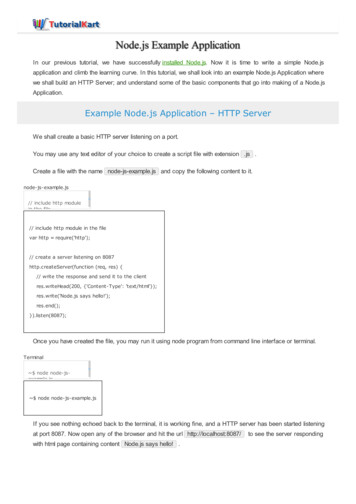
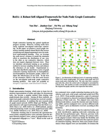
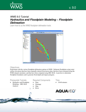
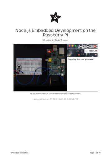
V. Grégoire et al. / Radiotherapy and Oncology 110 (2014) 172–181Within this framework, an initial set of recommendations forselection and delineation of the neck node target volumes waspublished in 2000 [2]. Three years later, a consensus was established among major stakeholders in H&N clinical research for thedelineation of node levels in the node-negative neck [3]. Theserecommendations were subsequently expanded for the post-operative situation and the node-positive neck [4]. Although it appearsthat these recommendations are well accepted and used amongthe radiation oncology community, they are also associated withsome shortcomings. Firstly, not all the neck node areas describedin the TNM atlas were included [5]. In particular, the nodal areasin the lower and posterior neck typically involved in nasopharyngeal carcinoma were not properly discussed; also the lymph noderegions draining the face, the scalp, and those nodal regions closeto the base of skull received inadequate attention. Secondly, it appears that the description of the anatomic boundaries of some ofthese levels (e.g. in the lower neck) was not sufficiently accurateand thus required some interpretation from the users. Thirdly, inthe guidelines for the node-positive neck, the proposed extensionaround positive nodes into normal structures to generate theClinical Target Volume (CTV) was both arbitrary and potentiallyimprecise. New data on tumor extra-capsular extension in lymphnodes have emerged permitting specific recommendations to beproposed [6,7]. Lastly, in the original publication, the illustrationsof the nodal levels were not available in DICOM or DICOM-RTformat, consequently limiting the easy use of the atlas, andpreventing its direct importation into treatment planning systems.In this context, in a continuing effort to standardize and simplify the H&N IMRT process for safe and effective global advancement of clinical practice, it was decided to update the previouslypublished guidelines on target delineation in the neck. A task forceassembling opinion leaders in the field of H&N radiation oncologyfrom various clinical research organizations and from variousgeographic regions was formed to review and update thepreviously published guidelines on nodal level delineation.Methodology used to reach consensus guidelines for thedelineation of the neck node levelsA multidisciplinary working party, including European individuals who already participated in the creation of the previous guidelines was created to update the previous recommendations forneck level delineation. The working party was enlarged to includerepresentatives of North American, Australia/New Zealand andAsian cooperative groups. All physicians who contributed to thecreation of these guidelines are listed as co-authors of this manuscript. The general principles guiding the activities of the workinggroup were (1) to integrate as accurately as possible anatomicknowledge and previously defined surgical and radiologicalguidelines into a set of recommendations based on axial CTsections useful to radiation oncologists, (2) to minimize differencesin interpretation of the guidelines by refining the description of theboundaries of the various levels, and (3) to extend the previouslypublished guidelines to cover all the head and neck lymph nodeareas, especially in the lower neck (e.g. supraclavicular nodes),the scalp (e.g. retroauricular and occipital nodes), and the face(e.g. buccal nodes, parotid nodes).Practically, with the help of an anatomist (BL) and a head andneck surgeon (MH), all the individual lymph node groups identifiedin the TNM atlas for lymph nodes of the neck [5] were reviewedand considered in the context of the anatomic description ofRouvière [8] and with the classification proposed by the AmericanHead and Neck Society and the American Academy of Otolaryngology-Head and Neck Surgery [9,10]. Finally, they were depictedspecifically on axial CT slices. When the existing classifications173were considered insufficiently detailed, the working party proposed subdivisions of certain groups into several clearly identifiedlevels; for example the caudal jugular nodes in the TNM atlas(group #5) were subdivided into the lower jugular group (levelIVa in the current proposal) and the medial supraclavicular group(level IVb in the current proposal) to better reflect the differencein the risk of nodal infiltration in these 2 levels according to primary tumor location and nodal stage; another example is that ofthe retropharyngeal nodes in the TNM atlas (group #9), whichthe working party proposed to subdivide into the retropharyngealnodes and the retro-styloid nodes, two distinct node levels according to Rouvière. Lastly, in the publications of Robbins from theAmerican Head and Neck Society and the American Academy ofOtolaryngology-Head and Neck Surgery, only 6 node levels weredescribed [9,10]. In the present consensus we propose to extendthe levels to 10 (some being divided into sub-levels) to correspondmore completely with the TNM atlas.The consensus guidelines for the delineation of the node levelsin the neckFig. 1 presents lateral views of the superficial and the deep nodegroups of the head and neck region [11]. For all these groups, Table 1 compares the terminology of the TNM atlas and the proposedterminology of the lymph node levels modified from Robbins. Inthe following sections, each of these levels will be briefly reviewedwith a short description of their main anatomic boundaries, thenormal structures juxtaposed to these nodes, and the main tumorsites at risk for harboring metastases in those levels [11]. Theboundaries refer to a patient lying supine with his/her head in a‘‘neutral’’ position. The terms ‘‘cranial’’ and ‘‘caudal’’ refer to structures closer to the cephalic and pedal ends, respectively. The terms‘‘anterior’’ and ‘‘posterior’’ were chosen to be less confusing thanthe terms ‘‘ventral’’ and ‘‘dorsal’’, respectively. A set of 6 transverseCT sections with the various delineated levels is presented in Fig. 2.Level IaLevel Ia (Table 2) is a median region located between the anterior belly of the digastric muscles, which contains the submentalnodes. The medial limit of level Ia is virtual, as the region is continuous with the contralateral level Ia.Nodes in level Ia drain the skin of the chin, the mid-lower lip,the tip of the tongue, and the anterior floor of the mouth. LevelIa is at greatest risk of harboring metastases from cancer arisingfrom the floor of the mouth, the anterior oral tongue, the anteriormandibular alveolar ridge, and the lower lip.Level IbLevel Ib (Table 2) contains the submandibular nodes located inthe space between the inner side of the mandible laterally and thedigastric muscle medially, from the symphysis menti anteriorly tothe submandibular gland posteriorly.Level Ib nodes receive efferent lymphatics from the submentallymph nodes (level Ia), the lower nasal cavity, the hard and softpalate, the maxillary and mandibular alveolar ridges, the cheek,the upper and lower lips, and most of the anterior tongue. Nodesin level Ib are at risk of developing metastases from cancers ofthe oral cavity, anterior nasal cavity, soft tissue structures of themid-face and the submandibular gland.Level IILevel II (Table 3) contains the upper jugular nodes locatedaround the upper one-third of the internal jugular vein (IJV) and
174Delineation of neck node levelsFig. 1. Superficial (top) and deep (bottom) lymphatic node groups of the head and neck. These groups are named according to the node levels modified from Robbinsclassification (see Table 1) [modified from Lengelé [11]. AJ: anterior jugular; B: buccal; diP: deep intraparotid; F: facial; iH: infrahyoid; M: malar; Mt: mastoid; pA:preauricular; pL: prelaryngeal; pT: pretracheal; R: recurrent or paratracheal; sA: subauricular; SAN: spinal accessory nerve; SEJ: superficial external jugular; siP: superficialintraparotid; sMb: submandibular; sMt: submental; sP: subparotid; TCA: transverse cervical artery.the upper spinal accessory nerve (SAN). The nodes lie in the spacebetween the deep (medial) surface of the sternocleidomastoid muscle laterally, and the medial edge of the internal carotid artery andthe scalenius muscle medially; this space extends from the posterior edge of the submandibular gland anteriorly to the posterior edgeof the sternocleidomastoid muscle posteriorly, and from the lateralprocess of the first cranial vertebra to the caudal edge of the hyoidbone. Level II can be further subdivided into level IIa and IIb by theposterior edge of the internal jugular vein. The usefulness of the distinction between these 2 sub-levels is a matter of discussion in theradiotherapy community.Level II receives efferent lymphatics from the face, the parotidgland, and the submandibular, submental and retropharyngealnodes. Level II also directly receives the collecting lymphatics fromthe nasal cavity, the pharynx, the larynx, the external auditory canal, the middle ear, and the sublingual and submandibular glands.The nodes in level II are therefore at greatest risk of harboringmetastases from cancers of the nasal cavity, oral cavity, nasopharynx, oropharynx, hypopharynx, larynx, and the major salivaryglands. Level IIb is more likely associated with primary tumors ofthe oropharynx or nasopharynx, and less frequently with tumorsof the oral cavity, larynx or hypopharynx.Level IIILevel III (Table 3) contains the middle jugular nodes locatedaround the middle third of the IJV. It is the caudal extension of levelII. It extends from the caudal edge of the body of the hyoid bone to
175V. Grégoire et al. / Radiotherapy and Oncology 110 (2014) 172–181Table 1Comparison between the TNM atlas for lymph nodes in the neck [5] and the guidelines of the lymph node levels modified from Robbins [9].TNM atlas for lymph nodes of the neckGroup numberNode levels modified from RobbinsTerminologyLevelTerminology12345Submental nodesSubmandibular nodesCranial jugular nodesMiddle jugular nodesCaudal jugular nodes6Dorsal cervical nodes along the spinal accessory nerve78Supraclavicular nodesPrelaryngeal and paratracheal nodes9Retropharyngeal IXXXaXbSubmental groupSubmandibular groupUpper jugular groupMiddle jugular groupLower jugular groupMedial supraclavicular groupPosterior triangle group- Upper posterior triangle nodes- Lower posterior triangle nodesLateral supraclavicular groupAnterior compartment group:- Anterior jugular nodes- Prelaryngeal, pretracheal, & paratracheal nodesPrevertebral compartment group:- Retropharyngeal nodes- Retro-styloid nodesParotid groupBucco-facial groupPosterior skull group:- Retroauricular & subauricular nodes- Occipital nodes101112Parotid nodesBuccal nodesRetroauricular and occipital nodesthe caudal edge of the cricoid cartilage. The anterior limit is theanterior edge of the sternocleidomastoid muscle or the posteriorthird of the thyro-hyoid muscle, and the posterior limit is the posterior edge of the sternocleidomastoid muscle. Laterally, level III islimited by the deep surface of the sternocleidomastoid muscle andmedially by the medial edge of the common carotid artery and thescalenius muscles.Level III receives efferent lymphatics from levels II and V, andsome efferent lymphatics from the retropharyngeal, pretrachealand recurrent laryngeal nodes. It collects the lymphatics from thebase of the tongue, tonsils, larynx, hypopharynx and thyroid gland.Nodes in level III are at greatest risk of harboring metastases fromcancers of the oral cavity, nasopharynx, oropharynx, hypopharynxand larynx.Level IVaLevel IVa (Table 3) contains the lower jugular lymph nodes located around the inferior third of the IJV from the caudal limit oflevel III to a limit set arbitrarily 2 cm cranial to the sternoclavicularjoint, caudally. This caudal limit was set after a critical examinationof level IVa neck node dissection, which typically does not go allthe way down to the clavicle and definitely never reaches the medial portion of the clavicle at the level of the sternoclavicular joint.The anterior limit is the anterior edge of the sternocleidomastoidmuscle cranially and the body of the sternocleidomastoid musclecaudally; the posterior limit is the posterior edge of the sternocleidomastoid muscle cranially and the scalenius muscles caudally.Laterally, level IVa is limited by the deep (medial) surface of thesternocleidomastoid muscle cranially and the lateral edge of thatmuscle caudally; the medial limit of level IVa is the medial edgeof the common carotid artery, the medial edge of the thyroid glandand the scalenius muscle cranially, and the medial edge of the sternocleidomastoid muscle caudally.Level IVa receives efferent lymphatics primarily from levels IIIand V, some efferent lymphatics from the retropharyngeal, pretracheal, and recurrent laryngeal nodes, and collecting lymphaticsfrom the hypopharynx, larynx, and thyroid gland. Level IVa nodesare at high risk for harboring metastases from cancers of the hypopharynx, larynx, thyroid and cervical esophagus. Rarely metastasesfrom the anterior oral cavity may manifest in this location withminimal or no proximal nodal disease.Level IVbLevel IVb (Table 3) contains the medial supraclavicular lymphnodes located in the continuation of level IVa down to the cranialedge of the sternal manubrium. The anterior limit is the deep surface of the sternocleidomastoid muscle; the posterior limit is theanterior edge of the scalenius muscle cranially, and the apex ofthe lung, the brachiocephalic vein, the brachiocephalic artery (rightside) and the common carotid artery and subclavian artery on theleft side, caudally; the lateral limit is the lateral edge of the scalenius muscle, while the medial limit abut level VI and the medialedge of the common carotid artery.Level IVb receives efferent lymphatics primarily from levels IVaand Vc, some efferent lymphatics from the pretracheal, and recurrent laryngeal nodes, and collecting lymphatics from the hypopharynx, esophagus, larynx, trachea and thyroid gland. Level IVbnodes are at high risk for harboring metastases from cancers ofthe hypopharynx, sub-glottic larynx, trachea, thyroid and cervicalesophagus.Level V (Va and Vb)Level Va and Vb (Table 4) contains the nodes of the posterior triangle group located posteriorly to the sternocleidomastoid musclearound the lower part of the spinal accessory nerve and the transverse cervical vessels. It extends from a plane crossing the cranialedge of the body of the hyoid bone to a plane crossing the cervicaltransverse vessels caudally. Originally, the cranial limit of level Vwas defined by the convergence of the sternocleidomastoid andthe trapezius muscles [9,10]. From an anatomic point of view,the uppermost part of level V includes lymph nodes belonging tothe occipital region (see level Xb) [12]. Thus, it was proposed touse the hyoid bone as a radiological landmark to define the craniallimit of level V. Laterally, level V is limited by the platysma muscleand the skin and medially by the levator scapulae (cranially) andthe posterior scalenius (caudally) muscles. Posteriorly level Vnodes extend to a limit set at the anterior border of the trapeziusmuscles. From a surgical point of view, level V is currently subdi-
176Delineation of neck node VIIIIIXbXbC203536D1719Ia28341IbIIVEIVaVIb 41415221621Ib30Xa11IX23386 26 1 41622 44 52442 46 45415165222320FIVbVIb VIa872026 16 1 4 10 922 41244443 46 45 20 23Fig. 2. Head and neck CT sections performed on a 32 year-old volunteer immobilized with a head-neck-shoulder thermoplatic mask. The head was set in a ‘‘neutral’’ position.Sixty ml of iodinated contrast medium (Omnipaque 350, HealthCare, Diegem, BE) was injected intravenously at a rate of 1 ml/s, then after a 3-min gap, another 50 ml wereinjected at a rate of 1.5 ml/s. The examination was performed on a Toshiba (Toshiba Aquilon LB, Toshiba Medical System corporation, Japan) helicoidal CT (300 mAs and120 keV) using a slice thickness of 2.0 mm, an interval reconstruction of 2.0 mm and a helicoidal pitch of 11. CT sections were reconstructed using a 512 512 matrix.Sections were taken at the level of the top edge of C1 (panel A), the bottom edge of C2 (panel B), mid C4 (panel C), the bottom edge of C6 (panel D), mid Th1 (panel E), and topedge of Th2 (panel F). Neck node levels were drawn on each CT slice using the radiological boundaries detailed in Table 1. Each node level corresponds to node groups, andthus does not include any security margin for organ motion or set-up inaccuracy. 1: common carotid artery; 2: internal carotid artery; 3: external carotid artery; 4: internaljugular vein; 5: external jugular vein; 6: anterior jugular vein; 7: right brachiocephalic trunc; 8: right brachiocephalic vein; 9: left susclavian artery; 10: left susclavian vein;11: facial vessels; 12: masseter m.; 13: pterygoid m.; 14: longus capitis m.; 15: longus colli m.; 16: sternocleidomastoid m.; 17: digastric (ant. belly) m.; 18: digastric (post.belly) m.; 19: platysma m. 20: trapezius m.; 21: splenius capitis m.; 22: scalenius m.; 23: levator scapulae m.; 24: serratus anterior m.; 25: thyro-hyoid m.; 26: sterno-hyoidm.; 27: parotid gland; 28: sub-mandibular gland; 29: thyroid gland; 30: mastoid; 31: styloid process; 32: mandible; 33: maxilla; 34: hyoid bone; 35: odontoid process; 36:2nd cervical vertebra; 37: 4th cervical vertebra; 38: 6th cervical vertebra; 39: thyroid cartilage; 40: cricoid cartilage; 41: clavicle; 42: 1st thoracic vertebra; 43: 2nd thoracicvertebra; 44: rib; 45: lung apex; 46: esophagus; 47: Bichat’s fat pad; 48: pre-styloid para-pharyngeal space.vided into levels Va and Vb using the caudal edge of the cricoid cartilage as an anatomic landmark.Level V receives efferent lymphatics from the occipital and retroauricular nodes as well as those from the occipital and parietal
177V. Grégoire et al. / Radiotherapy and Oncology 110 (2014) 172–181Table 2Node levels Ia and Ib.BoundariesLevel Ia (submental group)Level Ib (submandibular oid m.Platysma m. (caudal edge of the anteriorbelly of digastric mm)Symphysis mentiBody of hyoid bone/mylo-hyoid m.Medial edge of ant. belly of digastric m.Medialn.a.Cranial edge of submandibular gland; anteriorly, mylo-hyoid m.Plane through caudal edge of hyoid bone and caudal edge of mandible; alternatively caudal edge ofsubmandibular gland (whichever is more caudal)/platysma m.Symphysis mentiPosterior edge of submandibular gland (caudally)/posterior belly of digastric m. (cranially)Medial aspect (innerside) of mandible down to caudal edge/platysma m. (caudal)/medial pterygoid m.(posteriorly)Lateral edge of ant. belly of digastric m. (caudally)/posterior belly of digastric m. (cranially)Table 3Node levels II, III, IVa and II (upper jugulargroup)#Posterior edge of the Posterior edge ofCaudal edge of the Caudal edge of thesternocleidomastoid m.body of the hyoid bone submandibularlateral process ofgland/posterior edgeC1of posterior belly ofdigastric m.III (middle jugulargroup)Caudal edge of the Caudal edge of cricoidbody of the hyoid cartilageboneIVa (lower jugulargroup)Caudal edge ofcricoid cartilage2 cm cranial to sternalmanubriumIVb (medialsupraclaviculargroup)Caudal border oflevel IVa (2 cmcranial to sternalmanubrium)Cranial edge of sternalmanubriumAnterior edge ofsternocleidomastoidm./posterior third ofthyro-hyoid m.Anterior edge ofsternocleidomastoidm. (cranially)/bodyofsternocleidomastoidm. (caudally)Deep surface ofsternocleidomastoidm./deep aspect ofclaviclePosterior edge ofsternocleidomastoid m.Posterior edge ofsternocleidomastoid m.(cranially)/scalenius mm.(caudally)LateralMedialDeep (medial)surface ofsternocleidomastoidm./platysma m./parotid gland/posterior belly ofdigastric m.Deep (medial)surface ofsternocleidomastoidm.Deep (medial)surface ofsternocleidomastoidm. (cranially)/lateraledge ofsternocleidomastoidm. (caudally)Medial edge ofinternal carotidartery/scalenius m.Anterior edge of scalenius Lateral edge ofscalenius m.mm. (cranially)/apex oflung, the brachiocephalicvein, the brachiocephalictrunc (right side) and thecommon carotid arteryand subclavian artery onthe left side (caudally)Medial edge ofcommon carotidartery/scalenius mm.Medial edge ofcommon carotidartery/lateral edge ofthyroid gland/scalenius mm.(cranially)/medialedge ofsternocleidomastoidm. (caudally)Lateral border of levelVI (pre-trachealcomponent)/medialedge of commoncarotid arteryLevel II can be divided into level IIa and level IIb by drawing an artificial line at the posterior edge of the internal jugular vein.Table 4Levels V (Va and Vb) and Vc.aBoundariesLevel V (posterior triangle group)aLevel Vc (lateral supraclavicular ranial edge of the body of hyoid bonePlane just below transverse cervical vesselsPosterior edge of sternocleidomastoid m.Anterior border of trapezius m.Platysma m./skinLevator scapulae m./scalenius m. (caudally)Plane just below transverse cervical vessels (caudal border of level V)2 cm cranial to sternal manubrium, i.e. caudal border of level IVaSkinAnterior border of trapezius m. (cranially)/ 1 cm anterior to serratus anterior m. (caudally)Trapezius m (cranially)/clavicle (caudally)Scalenius m./lateral edge of sternocleidomastoid m, lateral edge of level IVaSurgically, level V is subdivided in two groups of upper (Va) and lower (Vb) nodes according to their respective relationships with the cricoid cartilage.scalp, the skin of the lateral and posterior neck and shoulder, thenasopharynx, the oropharynx and the thyroid gland. Level V lymphnodes are at high risk for harboring metastases from cancers of thenasopharynx, oropharynx and thyroid gland. Nodes in level V aremost often associated with primary cancers of the nasopharynx,the oropharynx, the cutaneous structures of the posterior scalp,and the thyroid gland (level Vb).Level VcLevel Vc (Table 4) contains the lateral supraclavicular nodes located in the continuation of the posterior triangle nodes (level Vaand Vb) from the cervical transverse vessels down to a limit setarbitrarily 2 cm cranial to the sternal manubrium, i.e. a similar limit than the caudal border of level IVa. It corresponds partly to thearea known as the ‘‘the supraclavicular fossa’’ also called the triangle of Ho, which was clinically defined in the mid-seventies beforethe era of CT for the neck staging of nasopharyngeal carcinoma[13]. Originally, this fossa also included the caudal portions of levelIV and V. Instead of proposing new boundaries of its own, weelected to subsume this fossa into other lower neck nodal levels.The anterior limit of level Vc is the skin. Its posterior limit is theanterior border of the trapezius muscles (cranially) or 1 cm fromthe anterior surface of the serratus anterior muscle (caudally).
178Delineation of neck node levelsTable 5level VIa and VIb.aBoundariesLevel VIa (anterior jugular nodes)Level VIb (pre-laryngeal, pre-tracheal, para-tracheal-recurrent laryngeal nerve nodes)CranialCaudal edge of the thyroid cartilageaCaudalAnteriorPosteriorCaudal edge of the hyoid bone or caudal edge of the submandibular gland, whichever is more caudalCranial edge of the sternal manubriumSkin/platysma m.Anterior aspect of the infrahyoid (strap) mm.LateralMedialAnterior edges of both sternocleidomastoid mm.n.a.Cranial edge of the sternal manubriumPosterior aspect of infrahyoid (strap) mm.Anterior aspect of larynx, thyroid gland and trachea (pre-laryngeal and pre-trachealnodes)/pre-vertebral m. (right side)/esophagus (left side)Common carotid artery on both sidesLateral aspect of trachea & esophagus (caudally)For tumors located on the anterior floor of month, tip of the tongue and lower lip, caudal edge of the body of the hyoid bone.Medially level Vc abuts level IVa; laterally it is limited by the trapezius muscle (cranially) and the clavicle (caudally).Level Vc receives efferent lymphatics from the posterior trianglenodes (level Va and Vb) and is more commonly associated withnasopharyngeal tumors.esophagus. These nodes are at high risk for harboring metastasesfrom cancers of the lower lip, the oral cavity (floor of mouth andtip of the tongue), the thyroid gland, the glottic and sub
In 2003, a panel of experts published a set of consensus guidelines for the delineation of the neck node levels in node negative patients (Radiother Oncol, 69: 227-36, 2003). In 2006, these guidelines were extended to include the characteristics of the node positive and the post-operative neck (Radiother Oncol, 79: 15-20, 2006).