Transcription
JOURNAL OF ORAL & MAXILLOFACIAL RESEARCHStarch-Jensen et al.Maxillary Sinus Floor Augmentation with Autogenous Bone GraftAlone Compared with Alternate Grafting Materials: a SystematicReview and Meta-Analysis Focusing on HistomorphometricOutcomeThomas Starch-Jensen1, Daniel Deluiz2, Niels Henrik Bruun3, Eduardo Muniz Barretto Tinoco2Department of Oral and Maxillofacial Surgery, Aalborg University Hospital, Aalborg, Denmark.Department of Periodontology, Rio de Janeiro State University, Rio de Janeiro, Brazil.3Unit of Clinical Biostatistics, Aalborg University Hospital, Aalborg, Denmark.12Corresponding Author:Thomas Starch-JensenDepartment of Oral and Maxillofacial SurgeryAalborg University Hospital18-22 Hobrovej, DK-9000 AalborgDenmarkPhone: 45 97 66 27 98Fax: 45 97 66 28 25E-mail: thomas.jensen@rn.dkABSTRACTObjectives: The objective of present systematic review was to test the hypothesis of no difference in histomorphometricoutcome after maxillary sinus floor augmentation with autogenous bone graft alone compared with alternate grafting materialsapplying the lateral window technique.Material and Methods: MEDLINE (PubMed), Embase and Cochrane library search in combination with hand-searchof relevant journals were conducted. Human studies published in English until the 25th of March, 2020 were included.Histomorphometric outcomes were evaluated by descriptive statistics and meta-analysis including 95% confidence interval (CI).Results: Electronic search and hand-searching resulted in 1902 entries. Sixteen randomized controlled trials with unclear riskof bias fulfilled the inclusion criteria. Descriptive statistics showed comparable or improved histomorphometric outcomeswith autogenous bone graft. Meta-analysis revealed a mean difference of -7.1% (CI -11.0 to -3.2) indicating a significanthigher amount of bone after maxillary sinus floor augmentation with autogenous bone graft compared with alternate graftingmaterials. Subgroup analysis demonstrated a non-significantly differences of -3.7% (CI -10.9 to 3.4), -11.5% (CI -25.9to 2.8), 2.2% (CI -16.9 to 21.3), and -4.6% (CI -14.4 to 5.2), when autogenous bone graft was compared with allogeneicbone graft, xenograft, composite grafting materials involving xenograft or synthetic biomaterial mixed with autogenous bonegraft, respectively.Conclusions: Maxillary sinus floor augmentation with autogenous bone graft seems to facilitate improved histomorphometricoutcomes compared with alternate grafting materials. However, the included studies were characterised by an unclear risk ofbias and various methodological confounding factors. Hence, the conclusions drawn from the results of present study shouldbe interpreted with caution.Keywords: alveolar ridge augmentation; dental implants; oral surgical procedures; review; sinus floor augmentation.Accepted for publication: 10 September 2020To cite this article:Starch-Jensen T, Deluiz D, Bruun NH, Tinoco EMB.Maxillary Sinus Floor Augmentation with Autogenous Bone Graft Alone Compared with Alternate Grafting Materials: aSystematic Review and Meta-Analysis Focusing on Histomorphometric OutcomeJ Oral Maxillofac Res 2020;11(3):e2URL: e2.pdfdoi: rchives/2020/3/e2/v11n3e2ht.htmJ Oral Maxillofac Res 2020 (Jul-Sep) vol. 11 No 3 e2 p.1(page number not for citation purposes)
JOURNAL OF ORAL & MAXILLOFACIAL RESEARCHINTRODUCTIONMaxillary sinus floor augmentation (MSFA) wasoriginally developed by Tatum in the mid-seventiesand afterwards described by Boyne and James in 1980[1,2]. Nowadays, MSFA applying the lateral windowtechnique is the most commonly used surgicalprocedure to increase the vertical alveolar bone heightof the posterior maxilla before or in conjunctionwith placement of dental implants, and the treatmentoutcome involving various types of grafting materialshave been documented in systematic reviews andmeta-analyses [3-10]. Autogenous bone graft isgenerally considered the preferred grafting materialfor MSFA due to its osteoinductive, osteogenic andosteoconductive characteristics [11]. Long-termstudies have demonstrated high survival rates ofsuprastructures and implants, limited peri-implantmarginal bone loss and few complications afterMSFA with autogenous bone graft [12,13]. However,harvesting of autogenous bone graft is associatedwith risk of donor site morbidity and prolongedtreatment time [14,15]. Various bone substitutesof biologic or synthetic origin are therefore usedincreasingly to simplify the surgical procedureby diminishing the need for bone harvesting [4].On the contrary, allogeneic bone graft, syntheticbiomaterials or bone substitutes of natural transplantsinvolves a risk of contamination, activation of hostimmune system and disease transmission as wellas they can be refused by patients for religiousreasons or because they are in contrast with chosenlifestyle. Thus, selection of the most appropriategrafting material for MSFA sustains a matter ofcontroversy.Impaired bone quality and quantity is detrimental toobtaining adequate osseointegration of implants inthe posterior part of the maxilla [16]. MSFA with theuse of a grafting material facilitates bone regenerationand increases bone volume, which enhances thepotential for achieving sufficient bone-to-implantcontact (BIC) and long-term implant survival [17].Histomorphometric analysis of tissue specimens ormicro-computed tomography are the most commonlyapplied methods for assessing percentage of newlyformed bone, non-mineralized tissue, residualgrafting material and BIC. Histomorphometricparameters are frequently presented as total bonevolume (TBV), bone area fraction (BAF) or newlyformed bone, non-mineralized tissue and residualgraft material in a clearly specified region of interest.Experimental studies evaluating histomorphometricvariables and BIC after MSFA with 3/e2/v11n3e2ht.htmStarch-Jensen et al.bone graft compared with various grafting materialshave revealed disputing results, without sticswith any of the used grafting materials [17-25].A recent published systematic review and metaanalysis assessing MSFA in humans concluded thatautogenous bone graft enables the highest amount ofnewly formed bone compared with the use of bonesubstitutes alone or different compositions of bonesubstitutes and autogenous bone graft [17]. Thisis in accordance with a previous published metaanalysis demonstrating that MSFA with autogenousbone graft generates a significantly higher TBVin the early healing period compared with bonesubstitutes alone or different compositions ofautogenous bone graft and bone substitutes, whilea non-significant difference was reported aftera longer observation period [26]. Consequently,type and composition of the grafting material aswell as length of healing period seems to influencethe histomorphometric outcome. Therefore, theobjective of the present systematic review andmeta-analysis was to test the hypothesis of nodifferences in histomorphometric outcomes aftermaxillary sinus floor augmentation with autogenousbone graft alone compared with alternate graftingmaterials.MATERIAL AND METHODSProtocol and registrationThe present systematic review was conducted inaccordance with the Preferred Reporting Items forSystematic Reviews and Meta-Analyses (PRISMA)statement for reporting systematic reviews [27].Methods of the analysis and inclusion criteria werespecified in advance and documented in a protocoland registered in PROSPERO, an internationalprospective register of systematic reviews.Registration number: CRD42020177003.The protocol can be accessed at:https://www.crd.york.ac.uk/prospero/display record.php?ID CRD42020177003.Focus questionThe focus question was developed according to thePatient, Intervention, Comparison and Outcome(PICO) framework as described in Table 1:Are there any differences in the histomorphometricoutcomes after MSFA with autogenous bonegraft alone compared with alternate graftingmaterials?J Oral Maxillofac Res 2020 (Jul-Sep) vol. 11 No 3 e2 p.2(page number not for citation purposes)
JOURNAL OF ORAL & MAXILLOFACIAL RESEARCHStarch-Jensen et al.Table 1. PICOS guidelinesPatient and population (P)Intervention (I)Comparator or control group (C)Outcomes (O)Study design (S)Focused questionHealthy adult patients with vertical atrophy of the posterior part of the maxilla receiving maxillarysinus floor augmentation applying the lateral window technique.Autogenous bone graft alone.Alternate grafting materials.Bone-to-implant contact, total bone volume, bone area fraction, newly formed bone, nonmineralized tissue and residual graft material.Randomized controlled trials.Are there any differences in histomorphometric outcomes after maxillary sinus floor augmentationwith autogenous bone graft alone compared with alternate grafting materials?Types of studies and publicationsRandomized controlled trials in humans.PopulationIncluded studies involved healthy adult partiallyor totally edentulous patients with atrophy of theposterior part of the maxilla receiving MSFA applyingthe lateral window technique with autogenousbone graft alone compared with alternate graftingmaterials.Types of outcome measures BIC defined as percentage of bone area in directcontact with the implant surface.TBV defined as percentage of bone volume of theexamined tissue sample.BAF defined as percentage of bone area in aclearly specified region of interest.Percentage of newly formed bone, nonmineralized tissue and residual graft materialas evaluated by histomorphometric analysis ormicro-computed tomography.Periodontology”, “Journal of Prosthetic Dentistry”,“Journal of Craniofacial Surgery”, “Journal ofCranio-Maxillo-Facial Surgery”, “Journal of Oraland Maxillofacial Surgery”, “Periodontology 2000”,“Oral and Maxillofacial Surgery” and “Oral SurgeryOral Medicine Oral Pathology Oral Radiology”. Themanual search also included the bibliographies ofall articles selected for full-text screening as well aspreviously published reviews relevant for the presentsystematic review.SearchA MEDLINE (PubMed), Embase, and CochraneLibrary search was conducted. Human studiespublished in English until the 25th of March, 2020were included. Grey literature, unpublished literatureas well as other databases like Scopus, GoogleScholar, or Research Gate were not included in thesearch strategy of the present systematic review.The search strategy was performed in collaborationwith a librarian and utilized a combination ofMedical subject heading (MeSH) and free text terms.A detailed description of the search strategy isoutlined in Appendices 1, 2 and 3.Information sourcesSelection of studiesThe search strategy incorporated examinations ofelectronic databases, supplemented by a thoroughhand-search page by page of relevant journalsincluding “British Journal of Oral and MaxillofacialSurgery”, “Clinical Implant Dentistry and RelatedResearch”, “Clinical Oral Implants Research”,“European Journal of Oral Implantology”, “ImplantDentistry”, “International Journal of Oral andMaxillofacial Implants”, “International Journal ofOral and Maxillofacial Surgery”, “InternationalJournal of Periodontics and Restorative Dentistry”,“International Journal of Prosthodontics”, “Journalof Clinical Periodontology”, “Journal of DentalResearch”, “Journal of Oral Implantology”, “Journalof Oral & Maxillofacial Research”, “Journal ofPRISMA flow diagram presents an overview ofthe selection process (Figure 1). Titles of identifiedreports were initially screened with duplicatesremoved. Abstracts were assessed when the titleindicated that the study was relevant. Full-textanalysis was obtained for those with apparentrelevance or when the abstract was unavailable.References of papers identified and previouslypublished systematic reviews were cross-checked forunidentified articles. Study selection was performedby one reviewer /e2/v11n3e2ht.htmInclusion criteriaStudies assessing MSFA with particulated autogenousJ Oral Maxillofac Res 2020 (Jul-Sep) vol. 11 No 3 e2 p.3(page number not for citation purposes)
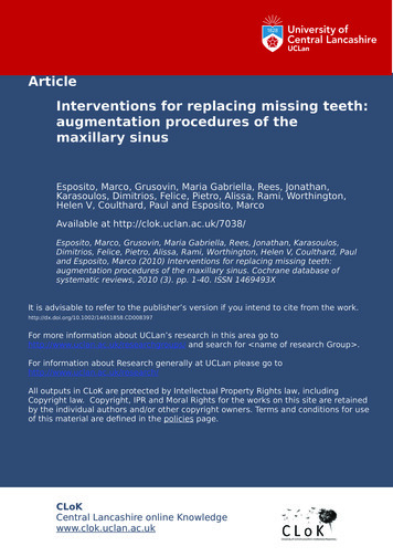
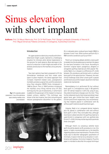
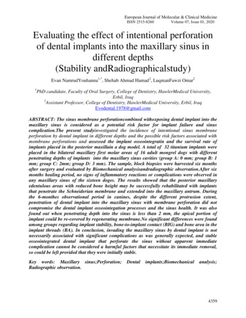
IncludedEligibilityScreeningIdentificationJOURNAL OF ORAL & MAXILLOFACIAL RESEARCHTitles identified throughdatabase searchingn 1902Starch-Jensen et al.Additional titles identifiedthrough hand-searchingn 0Titles after duplicates removedn 1032Abstracts screenedn 176Abstracts excludedn 148Full-text articlesassessed for eligibilityn 28Full-text articlesexcluded, with reasonsn 12Studies included inqualitative synthesisn 16Figure 1. PRISMA (Preferred Reporting Items for Systematic Reviews and Meta-Analyses) flow diagram demonstratingthe results of the systematic literature search.bone graft alone compared with alternate graftingmaterial were included by addressing the previousdescribed outcome measures. The review exclusivelyfocused on studies presenting histomorphometricoutcome. In addition, at least five patients involvingfive sinus augmentation procedures in each groupshould be included and the used grafting material aswell as number of sinus augmentation procedures hadto be clearly specified.Exclusion criteriaFollowing exclusion criteria were applied: MSFAwith autogenous bone block graft, unspecified lengthof observation period as well as studies involvingmedically compromised patients. Moreover, letters,editorials, PhD theses, letters to the editor, casereports, abstracts, technical reports, conferenceproceedings, animal or in vitro studies and literaturereview papers were also excluded.Data extractionData were extracted by one reviewer (T.S-J.) accordingto a data-collection form ensuring systematicrecording of the outcome measures. In /e2/v11n3e2ht.htmrelevant characteristics of the study were recorded.Corresponding authors were contacted by e-mail in theabsence of important information or ambiguities.Data itemsFollowing items were collected and arranged in thefollowing fields: author, year of publication, studydesign, number of patients, MSFA procedures,residual alveolar bone height, type of graftingmaterial, length of observation period, number ofbiopsies, BIC, bone regeneration including percentageof TBV, BAF, newly formed bone, non-mineralizedtissue and residual graft material.Assessment of methodological qualityQuality assessment was undertaken by one reviewauthor (T.S-J.) as part of the data extraction process.Cochrane Collaboration’s tool for assessing the riskof bias suggested in the Cochrane Handbook forSystematic Reviews of Interventions was used forincluded randomized controlled trials (version 5.1.0)[28]. Following items were evaluated: Random sequence generation; Allocation concealment;J Oral Maxillofac Res 2020 (Jul-Sep) vol. 11 No 3 e2 p.4(page number not for citation purposes)
JOURNAL OF ORAL & MAXILLOFACIAL RESEARCH Patient blinding;Outcome blinding;Incomplete outcome data addressed;Selective reporting.Publications were grouped into the followingcategories [29]:Low risk of bias (possible bias not seriouslyaffecting results) if all criteria were met.High risk of bias (possible bias seriouslyweakening reliability of results) if one or morecriteria were not met.Unclear risk of bias when too few details wereavailable for classification as high or low risk.Statistical analysisMeta-analysis (with random effect) where conductedto combine data from multiple studies where similareffects were measured. Results were expressed as themean difference (MD) between autogenous bone graftalone compared with alternate grafting materials. The95% confidence interval (CI) was also calculated.Parametric data are expressed as mean and standarddeviation (M [SD]). Statistical significance level wasdefined at P 0.05.Assessment of heterogeneityThe significance of any discrepancies in the estimatesof the treatment effects of the different studies wasassessed by means of Cochran’s test for heterogeneityand the I2 statistic, which describes the percentageof total variation across studies that is due toheterogeneity rather than chance. Heterogeneity byCochran’s test was considered statistically significantif P 0.1. A rough guide to the interpretation of I2given in the Cochrane Handbook for SystematicReviews of Interventions is as follows [28]: 0 - 40% the heterogeneity might not be important; 30 - 60% may represent moderate heterogeneity; 50 - 90% may represent substantial heterogeneity; 75 - 100% may represent considerable heterogeneity.RESULTSStudy selectionArticle review and data extraction were performedaccording to the PRISMA flow diagram (Figure 1).The systematic electronic literature search resultedin 1902 entries. No additional articles were identifiedthrough hand-searching. Of these 1902 articles, 870were excluded because they had been retrieved inmore than one search. A total of 176 abstracts 11n3e2ht.htmStarch-Jensen et al.reviewed and full-text analysis included 28 articles.Finally, 16 studies were included [30-45].Exclusion of studiesReasons for excluding studies after full-textassessment were as follows: less than five patientsincluded [46], less than five sinus augmentationprocedures in each group [47], controlled clinicaltrials without randomization [48-52], MSFA inconjunction with additional augmentation procedures[53], alveolar ridge augmentation not involvingMSFA [54], autogenous bone block graft was used asgrafting material [55], histomorphometric outcomewas not expressed in percentage [56], and MSFA wasconducted with the use of space-maintaining mesh [57].Study characteristicsIncluded studies consisted of 16 randomizedcontrolled trials with an unclear risk of bias [3045]. Partially and totally edentulous patients withatrophy of the posterior maxilla were enrolled in theincluded studies. Power calculation was performedin three studies [42,44,45]. A split-mouth studydesign was used in seven studies [30,31,33-35,40,41].No significant difference in patient demographicswas reported in one study [35]. Residual boneheight as well as inclusion and exclusion criteriawere clearly specified in all included studies[30-45]. Randomization was conducted using arandomization table [32,35,40], computer-generatedrandomization code [41], allocation concealmentprocess [37], drawing lots [42-45], coin-toss method[34], or no information was provided about therandomization procedure [30,31,33,36,38,39]. Thesurgical procedure was performed by one surgeon[36,39,40,44] or no information was provided aboutthe number of surgeons involved [30-35,37,38,4143,45]. Autogenous bone graft was harvested fromthe ascending mandibular ramus [31,37,38,40,42-45],mandibular symphysis [32,37,42-45], lateral sinuswall [41], zygomatic buttress [36,41], tuberosity area[39,41] or iliac crest [30,32,33,35]. Autogenous bonegraft was compared with a synthetic biomaterialalone involving BioGran (3i Implant Innovations,Inc.; Palm Beach Gardens, FL, USA) [32,45],BoneCeramic (Straumann AG; Basal, Switzerland)[38,39,41], Cerasorb (Curasan AG; Kleinostheim,Deutschland) [34], and chronOS (Synthes Inc.; WestChester, PA, USA) [43] or composite graft consistingof 50% BioGran and 50% autogenous bone graft[30,42,44,45] or 50% chronOs and 50% autogenousbone graft [43]. Autogenous bone graft was comparedJ Oral Maxillofac Res 2020 (Jul-Sep) vol. 11 No 3 e2 p.5(page number not for citation purposes)
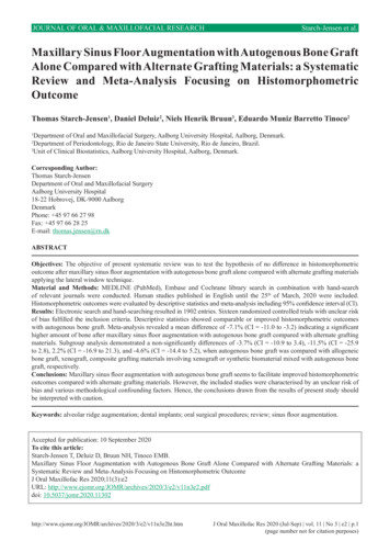
JOURNAL OF ORAL & MAXILLOFACIAL RESEARCHwith xenograft alone involving Bio-Oss (GeistlichPharma AG; Wolhusen, Switzerland) [38], OsteoBiol (Tecnoss Dental s.r.l.; Turin, Italy) [33] or a compositexenograft involving 20% Bio-Oss mixed with 80%autogenous bone graft [31]. Autogenous bone graftwas compared with allogeneic bone graft involvingPuros (Zimmer Dental GmbH; Munich, Germany)[38] or fresh frozen bone [40]. Autogenous bonegraft was also compared with platelet-rich plasmamixed with autogenous bone graft [35,37] or bloodcoagulum including replacement of the lateral bonewindow or barrier membrane coverage of the lateralwindow [36]. Different collagen barrier membraneswere used to cover the lateral window involving BioGide (Geistlich Pharma AG; Wolhusen, Switzerland)[31,38-40], Tecnoss (Tecnoss Dental s.r.l.; Torino,Italy) [33], Spongostan film (Johnson & JohnsonMedical N.V.; Courcelles, Belgium) [36], type ofmembrane was not specified [37,41], no informationwas provided about the use of a membrane [30,35,4245] or no membrane was used [32,34]. None of themembranes were fixed with tacks. Experimentalmicro-implants were retrieved for assessment of BICafter six [31] and seven months [36]. MSFA withsimultaneous implant placement was performed inone study [36], while delayed implant placement wasconducted after four months [30,37], five months[30,33,38], six months [30-32,34,35,37,40-45],nine months [39] or 16 months [30], respectively.Different implant systems were used includingAnkylos (Dentsply Implants Manufacturing GmbH;Mannheim, Germany) [34], Conexão System (Gestão de TI e Impressão; São Paulo, Brazil) [40],Nobel Biocare (Nobel Biocare AB; Gothenburg,Sweden) [31], Premium TG (Sweden & MartinaSpA; Padova, Italy) [33], Protetim (Protetim OrvosiMűszergyártó Kft.; Hódmezővásárhely, Hungary)[34], Spectra-System (Core-Vent Corporation, LasVegas, NV, USA) [37], Straumann (Straumann AG;Basal, Switzerland) [32,36,38,39,41], whereas theused implant system was not specified in six volvingmicro-computedtomographywasperformed after six months [45] and seven months[36], respectively. Tissue specimens were evaluatedafter four months [30,37], five months [30,33,38],six months [30-32,34,35,37,40-45], nine months[39] and 16 months [30], respectively. Number ofbiopsies were clearly specified in eight studies [3036,40], while no information was provided in sevenstudies [37,39,41-45]. Bone cores were retrieved withtrephine drill through the previous lateral window[31,32,40,41], at planed implant site [30,33,34,38,39],or not specified [35-37,42-45]. Percentage of BIC 1n3e2ht.htmStarch-Jensen et al.evaluated in two studies [31,36]. Percentage of TBVwas assessed in seven studies [30,32-35,37,45], BAFin three studies [31,38,39], and percentage of newlyformed bone was estimated in five studies [40-44].None of the included studies provided informationabout blinding, training or calibration of surgeons orthe examiners assessing the clinical and radiographicoutcome. Numbers of drop-outs were reported in threestudies [43-45].Outcome measuresBIC, TBV, BAF and newly formed bone areconsidered the primary outcome measures, whilenon-mineralized tissue and residual graft materiel aresurrogate outcome. Percentage of non-mineralizedtissue and residual graft materiel are therefore solelypresented in Table 2. Results of each primary outcomeare described below including a short summary andoutlined in Table 2.Bone-to-implant contactPercentage of BIC with autogenous bone graftcompared with mixture of 80% Bio-Oss and 20%autogenous bone graft were 34.6 (9.5) and 54.3 (33.1)after six to seven months, respectively [31]. There wasno significant difference (P 0.05) [31].Percentage of BIC with autogenous bone graftcompared with blood coagulum including eitherreplacement of the lateral bone window or barriermembrane coverage were 93.5 (3.3), 93.5 (3.3) and 92(4.1) after seven months, respectively [36]. There wasno significant difference (P 0.502) [36].SummaryBIC after MSFA with autogenous bone graft wascomparable to alternate grafting material consistentof 80% xenograft mixed with 20% autogenous bonegraft or blood coagulum.Total bone volumePercentage of TBV with autogenous bone graft was40.9 (3.3), 42.2 (4.5), 43.7 (2.4), and 45.1 (38.1)after four, five, six, and 16 months, respectively [30].Corresponding measurements for 50% BioGran and50% autogenous bone graft were 28.5 (1.4), 34.5(1.6), 38.1 (5.7), and 44.5. No statistically analysiswas performed [30].Percentage of TBV with autogenous bone graftcompared with Cerasorb was 41 (10) and17.5 (5) after six months, respectively [32].J Oral Maxillofac Res 2020 (Jul-Sep) vol. 11 No 3 e2 p.6(page number not for citation purposes)
JOURNAL OF ORAL & MAXILLOFACIAL RESEARCHStarch-Jensen et al.Table 2. Histomorphometric outcomes after maxillary sinus floor augmentation with autogenous bone graft compared with alternate grafting materialsMaterials and methodsStudyTadjoedin et al. [30]Year ofpublication2000Numberofpatients10Hallman et al. [31]200221Zerbo et al. [32]20049Barone et al. [33]200518Szabó et al. [34]200520Bettega et al. [35]200918Johansson et al. [36]201324Khairy et al. [37]201315Maxillarysinus m) 3 54-8 3 533.54.33.54.3 55Schmitt et al. [38]Tosta et al. [39]201320133030Xavier et al. [40]201515Danesh-Sani et al. [41]201610451515151510102.32.52.62.24.1 3 510Pereira et al. [42]2017301010Menezes et al. [44]Pereira et al. [45]201722201821201810710121214131291250% Biogran ; 50% autogenousAutogenous20% Bio-Oss ; 80% autogenousAutogenousCerasorb AutogenousOsteoBiol AutogenousCerasorb AutogenousAutogenous mixed with PRPAutogenousBloodclotBloodclot and membraneAutogenousLength ofobservation period(months)4 - 166.565667Autogenous mixed with PRP4-6AutogenousBoneCeramic Bio-Oss Puros Autogenous6BoneCeramic AutogenousFresh frozen boneAutogenousBoneCeramic Autogneous5966-8Numberofbiopsies3636883218NRTBV, BAF or NFBNon-mineralized tissueMean (SD)Mean (SD)Mean (SD)ResidualgraftMean (SD)NRNRNR12.3 (8.5)NRNRNRNRNRNRNRNRNRNRNRNRNR55.2 (5.6)53.7 (8.1)64.6 (2.8)57.3 (2.1)IntermediateApical33.4 (8)39.6 (11.1)59.2 (4.7)f60.6 (7.2)g56.8 (7.3)55.6 (14.5)38.9 (14.9)58.4 (10)iIntermediateApical56.6 (15.3)47.9 (12.1)62.5 (14.7)48.8 (17.7)61.2 (15.7)57.8 (16.2)IntermediateApical52.3 (9.9)55.1 (22.1)60.1 (13.9)55.5 (9.2)65 (10.2)50.2 (14.5)15.2 (2.1)21.4 (4.8)NR54.3 (33.1)34.6 (9.5)NRNR4040991091093.5 (3.3)92 (4.1)93.5 (3.3)NRNR14151212NR15151010NRNR 550% Biogran ; 50% autogenousAutogenousNR 5 550% chronOS ; 50% autogenousAutogenous50% Biogran ;50% autogenousAutogenousBiogran 50% Biogran ; 50% autogenousAutogenous bone5 month6 month34.5 (1.6) 38.1 (5.7)42.2 (4.5) 43.7 (2.4)39.9 (8)37.7 (31.3)17 (5)41 (10)a67 (14.9)70 (19.9)36.5 (6.9)38.3 (7.4)43.2504 month27.3 (6.3)NRNRNR6NRNR6NRNR6NRNR6 month28 (4.1)39.5 (7.4)b30.3 (2.2)24.9 (5.7)35.4 (2.8)42.7 (2.1)cIntermediate33.7 (8.1)41 (4.6)dApical26.7 (3.9)38.6 (7.5)e8.3 (3)8.3 (3.4)28.2 (8.4)36.8 (11.5)hNR616 month44.545.1NRNRchronOS 54 month28.5 (1.4)40.9 (3.3)NRBiogran 12Pereira et al. [43]Type of grafting materialOutcome measuresBone 0.7 (14)33.2 (13.3)35.3 (14.7)Intermediate47.6 (9.9)32.5 (13.7)31 (13)Intermediate33.2 (13.3)35 (13.9)Apical45.6 (13.5)45.8 (13.9)39.9 (15.8)Apical44.8 (22.1)32.8 (16)46.1 (16.3)Apical45.8 (13.8)42 (16.6)52.157.457.2NRNR34.9 (6.4)36.1 (12.8)32.9 (8.1)4.8 (2.4)jNRNRNRNRNRNRP 0.05 (Independent t-test); bP 0.003 (Paired t-test); cP 0.0001 (Kruskal-Wallis test); dP 0.008 (Mann-Whitney test); eP 0.0001 (Mann-Whitney test); fP 0.0001 (Mann-Whitney test); gP 0.0001 (Mann-Whitney test); hP 0.0032 (Paired t-test); iP 0.0001 (Paired t-test); jP 0.0001 (Paired t-test).BAF bone area fraction; NFB newly formed bone; NMT non-mineralized tissue; PRP platelet rich plasma; RBH residual bone height; RCT randomized controlled trial; SD standard deviation; TBV total bone e1/v11n3e1ht.htmJ Oral Maxillofac Res 2020 (Jul-Sep) vol. 11 No 3 e1 p.7(page number not for citation purposes)
JOURNAL OF ORAL & MAXILLOFACIAL RESEARCHThe difference was significant demonstrating higherTBV with autogenous bone graft (P 0.05) [32].Percentage of TBV with autogenous bone graftcompared with OsteoBiol was 70 (19.9) and 67(14.9) after five months, respectively [33]. There wasno significant difference (P 0.05) [33].Percentage of TBV with autogenous bone graftcompared with Cerasorb were 38.3 (7.4) and 36.5(6.9) after six months, respectively [34]. There was nosignificant difference (P 0.25) [34].Percentage of TBV with autogenous bone graftcompared with platelet-rich plasma mixed withautogenous bone graft was 50 (range 38 to 70) and43.2 (range 40 to 54.8) after six months, respectively[35]. There was no significant difference (P 0.474)[35].Percentage of TBV with autogenous bone graft was39.5 (7.4) after six months [37]. Correspondingmeasurements for platelet-rich plasma mixed withautogenous bone graft were 27.3 (6.3) and 28 (4.1)after four and six months, respectively. The differencewas significant demonstrating higher TBV withautogenous bone graft (P 0.003) [37].Percentage of TBV with autogenous bone graftcompared with mixture of 50% Biogran and 50%autogenous bone graft or Biogran alone were 52.1,57, and 57.2 after six months, respectively [41]. Therewas no significant difference (P 0.05) [45].SummaryTBV after MSFA with autogenous bone graftwas comparable with alternate grafting materialsconsistent of synthetic bone graft or xenograft aloneas well as autogenous bone graft mixed with syntheticbone graft or platelet-rich plasma. However, TBVwas statistically significant higher with autogenousbone graft compared with synthetic bone graft aloneor platelet-rich plasma mixed with autogenous bonegraft in other studies with similar observation period.Thus, percentage of TBV seems to be equivalent orenhanced after MSFA with autogenous bone graftcompared with alternate grafting materials.Bone area fractionPercentage of BAF with autogenous bone graftcompared with mixture of 80% Bio-Oss and 20%autogenous bone graft was 37.7 (31.3) and 39.9 (8)a
suprastructures and implants, limited peri-implant marginal bone loss and few complications after MSFA with autogenous bone graft [12,13]. However, harvesting of autogenous bone graft is associated with risk of donor site morbidity and prolonged treatment time [14,15]. Various bone substitutes of biologic or synthetic origin are therefore used