Transcription
Tra u m a t i c C e re b ro s p i n a lF l u id L e a k sJ. Drew Prosser, MDa, John R. Vender,C. Arturo Solares, MDa,*MDb,KEYWORDS Cerebrospinal fluid leak Skull base trauma Skull base surgery CSF rhinorrheaKey Points: CSF LEAKS Eighty percent of cerebrospinal fluid (CSF) leaks occur following nonsurgical trauma andcomplicate 2% of all head traumas, and 12% to 30% of all basilar skull fractures The most common presentation is unilateral watery rhinorrhea, and b2-transferrin is now thepreferred method of confirming a fluid as CSF High-resolution computed tomography (CT) is the preferred method of localizing the site ofskull base defect following craniomaxillofacial trauma, but can be coupled with magneticresonance imaging (MRI) or cisternography Conservative management of CSF leaks includes strict bed rest, elevation of the head, andavoidance of straining, retching, or nose blowing, and results in resolution of the majority oftraumatic CSF leaks over a 7-day period CSF diversion improves the rates of resolution when added to conservative therapy Endoscopic endonasal approaches should be the preferred method of repair with greaterthan 90% success rates; however, open transcranial and extracranial repairs still have theirplace in operative management Small postsurgical defects can be repaired endoscopically with mucosal grafts, while thepedicled nasal septal flap has emerged as the preferred method of repair for large defects Prophylactic antibiotics have not been shown to reduce the risk of meningitis and may selectfor more virulent organisms Data suggest that early surgical repair ( 7 days) can reduce the risk of meningitis.The authors have no relevant disclosures.aDepartment of Otolaryngology, Medical College of Georgia, 1120 15th Street, BP 4109,Augusta, GA 30912, USAbMedical Director of Southeast Gamma Knife Center, Department of Neurosurgery, MedicalCollege of Georgia, 1120 15th Street, BI 3088, Augusta, GA 30912, USA* Corresponding author.E-mail address: csolares@mcg.eduOtolaryngol Clin N Am 44 (2011) / – see front matter Ó 2011 Elsevier Inc. All rights reserved.oto.theclinics.com
858Prosser et alEBM QuestionDo spontaneous CSF leaks represent a distinct clinicalentity and a variant of idiopathic intracranialhypertension?Does a reduction in CSF pressure alone lead to resolutionof CSF leaks?Does decreasing ICP, transiently or long-term, improveoutcomes of endoscopic closure of spontaneous CSFleaks?Level ofEvidenceGrade ofRecommendation2bB5D4CCSF is an ultrafiltrate of plasma or serum, which contains electrolytes, glucose, andproteins. Located in the cerebral ventricles and cranial/spinal subarachnoid space,this fluid serves in physical support and provides buoyancy for the brain and spinalelements. CSF also serves to maintain homeostasis of neural tissues by removingby-products of metabolism and regulating the chemical environment of the brain.1CSF is produced by the choroid plexus in the lateral, third, and fourth ventricles. Itcirculates from the lateral ventricles through the foramina of Monro into the thirdventricle and then to the fourth ventricle through the aqueduct of Sylvius, then communicates with the subarachnoid space through the foramen of Magendie and foraminaof Luschka. CSF circulates throughout the meninges between the arachnoid and isreabsorbed into the venous system via arachnoid villi, which project into the duralvenous sinuses. Adults average 140 mL of CSF volume, and the body produces0.33 mL of CSF per minute and thus regenerates its CSF volume 3 times per day.1As CSF is an ultrafiltrate of serum, serum abnormalities will be reflected in CSF(ie, hyperglycemia). Fluctuations in the cerebral blood flow related to the cardiacoutput result in generalized brain volume fluctuations, which gives rise to the vascularpulsations seen in CSF. In addition, major branches of the circle of Willis are located inthe subarachnoid space and are likely contribute to this phenomenon.CSF leaks are rare but are associated with morbidity such as general malaise andheadache. More importantly, they can lead to potentially life-threatening complications such as meningitis. As such, they require thorough and timely evaluation andtreatment. CSF leaks occur when the bony cranial vault and its underlying dura arebreached. Such leaks may be broadly categorized as traumatic or nontraumatic inorigin. Traumatic causes can be further subclassified into surgical and nonsurgical,with surgical causes divided into planned (as in failure of reconstruction of a planneddural resection) or unplanned (as a complication following an ethmoidectomy).2 Nontraumatic CSF leaks may be subclassified into high or normal pressure leaks, with therecognition that tumors can occupy either subclass by mass effect in the highpressure group or by direct erosive effect on the skull base in the normal-pressuregroup. If no cause can be found the leak may be classified as idiopathic; however,with careful history taking, physical examination (including nasal endoscopy), andradiologic evaluation, true idiopathic leaks are rare.2EPIDEMIOLOGY OF CEREBROSPINAL FLUID LEAKSApproximately 80% of CSF leaks result from nonsurgical trauma, 16% from surgicalprocedures (although this number is rising), and the remaining 4% are nontraumatic.Of the traumatic leaks, more than 50% are evident within the first 2 days, 70% withinthe first week, and almost all present within the first 3 months.3,4 Delayed presentationmay result from wound contraction or scar formation, necrosis of bony edges or soft
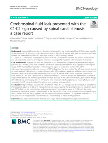
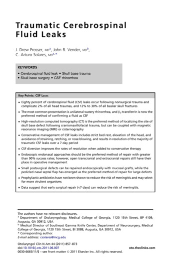
Traumatic Cerebrospinal Fluid Leakstissue, slow resolution of edema, devascularization of tissues, posttreatment tumorretraction, or progressive increases in intracranial pressure (secondary to brain edemaor other process). As with most maxillofacial trauma, traumatic CSF leaks occur mostcommonly in young males and complicate 2% of all head traumas, and 12% to 30% ofall basilar skull fractures.5Anterior skull base leaks are more common than middle or posterior leaks, due tothe firm adherence of the dura to the anterior basilar skull. The most common sitesof CSF rhinorrhea following accidental trauma are the sphenoid sinus (30%), frontalsinus (30%), and ethmoid/cribriform (23%) (Fig. 1).6 Temporal bone fractures withresultant CSF leak can present with CSF otorrhea or rhinorrhea via egress throughthe Eustachian tube with an intact tympanic membrane. Although a rare complicationof functional endoscopic sinus surgery (FESS), the frequency in which this procedureis performed makes it a significant cause of CSF leaks (Fig. 2). When looking atsurgical trauma, the most common sites of CSF leak following FESS are ethmoid/cribiform (80%), followed the frontal sinus (8%) and sphenoid sinus (4%). After neurosurgical procedures the most common site of CSF leak is the sphenoid sinus (67%)because of the high number of pituitary tumors that are addressed via transsphenoidalapproach.6Regarding nontraumatic leaks, high-pressure leaks comprise approximately half ofthese, with more than 80% resulting from tumor obstruction.7 The remainder is causedby either benign intracranial hypertension (BIH) or hydrocephalus; however, recentpublications suggest an increased role of BIH in idiopathic or “spontaneous” CSFleaks.8 One hypothesis is that in patients with occult elevated CSF pressure, an intermittent CSF leak may serve as a release valve that decompresses the elevated pressure. Once the leak resolves, either by the normalization of CSF pressure with anintermittent leak or by surgical repair, the CSF pressure will slowly increase and resultin recurrence of the leak. It is not surprising that the association between idiopathicFig. 1. Magnified view of the ethmoid bone in the coronal plane with the most commonsites of fracture labeled. CN, cranial nerve.859
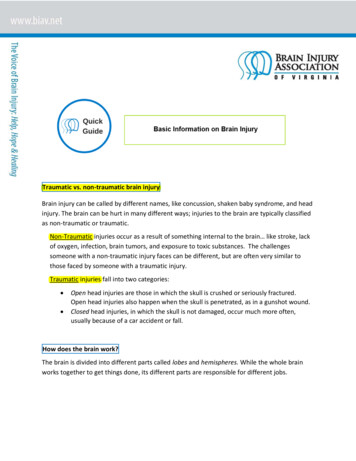
860Prosser et alFig. 2. Coronal CT scan demonstrating a defect in the roof of the left ethmoid sinus (A), anda defect in a separate patient with displacement of bony fragments intracranially (B). Bothare the result of surgical trauma, sustained at another institution.CSF leaks and BIH has been made, as the demographics of the populations are quitesimilar.9 Furthermore, the clinical manifestations and demographic profile of patientswith empty sella syndrome are highly similar to those for patients with BIH and patientswith nontraumatic CSF leaks.10 These associations have important clinical implications in the management of idiopathic CSF leaks as well as surgical failures after traumatic CSF leak repair.DETECTION OF CEREBROSPINAL FLUID LEAKSPresentationPresentation of traumatic CSF leaks can be subtle, and diligence is required when oneis suspected. Fain and colleagues11 presented an analysis of 80 cases of trauma to thecranial base. From these cases they determined that there are 5 types of frontobasaltrauma. Type I involves only the anterior wall of the frontal sinus. Type II involves theface (craniofacial disjunction of the Lefort II type or crush face), extending upward tothe cranial base and to the anterior wall of the frontal sinus, because of the facial retrusion. Type III involves the frontal part of the skull and extends down to the cranialbase. Type IV is a combination of types II and III, whereas type V involves only ethmoidor sphenoid bones. In this study CSF leaks were infrequent in types I and II, butoccurred more frequently in types III, IV, and V, which included a dural tear in eachcase. Therefore, when a patient presents with one of these fractures a CSF leak shouldbe suspected.The most common presenting sign is unilateral watery rhinorrhea following skull basetrauma. Patients may complain of a salty or even sweet taste in their mouth; however,with severe trauma to the skull, history may be unobtainable secondary to the patient’sneurologic status. Rhinorrhea is classically positional in nature and is most commonlyassociated with standing or leaning forward. Although this presentation seemsstraightforward, establishment of the diagnosis and precise localization of the leakcan present major challenges. Other rhinologic pathology, including seasonal allergicrhinitis, perennial nonallergic rhinitis, and vasomotor rhinitis, are relatively common,and may mimic some of the signs and symptoms of CSF rhinorrhea or may occur simultaneously with a CSF leak. Furthermore, CSF rhinorrhea is often intermittent, even after
Traumatic Cerebrospinal Fluid Leakstrauma, which may lead to false-negative results on diagnostic testing if testing isperformed during the quiescent phase. Lastly, the subarachnoid cistern is a relativelylow-pressure system. Thus, leaks may be of low volume, which can lead to falsenegative testing or failure to recognize that a leak even exists. In cases of high clinicalsuspicion and initially negative diagnostic testing, further follow-up with repeat testingis warranted.IdentificationTraditionally the presence of a halo sign (clear ring surrounding a central bloody spot)on gauze, tissue, or linen has been used to predict CSF leak following trauma. Thishalo forms as blood and CSF separate; however, this test should only be usedto arouse suspicion as tears, saliva, and other non-CSF rhinorrhea can give falsepositive results. Historically the components of the rhinorrhea (including glucose,protein, and electrolytes) have been measured to confirm the diagnosis of CSF. Thesetests, however, should not be relied on, as their sensitivity and specificity are unacceptably low.7,12,13b2-Transferrin has emerged as a highly sensitive and specific way of identifying CSF,and is now the preferred method of confirming a fluid as CSF. The method was initiallydiscovered in 1979 by Meurman and colleagues14 who, when performing protein electrophoresis of CSF, tears, nasal secretions, and serum, noted a b2-transferrin fractiononly in the CSF samples. Techniques for isolating this marker have been subsequentlysimplified and refined, leading to enhanced sensitivity and specificity.15,16 As with anytest, a reliable result requires an adequate sample. Of note, although quite specific,there are reports of b2-transferrin being detected in aqueous humor17 and in the serumof patients with alcohol-related chronic liver disease.18Beta-trace protein (bTP) is another marker that has been used for the detection ofCSF. This protein is produced by the meninges and choroid plexus and is releasedinto CSF. It is present in other body fluids, including serum, but at much lower concentrations than in CSF. Detection of bTP has 100% sensitivity and specificity in cases ofconfirmed CSF rhinorrhea, but cannot be reliably used in patients with renal insufficiency or bacterial meningitis, because serum and CSF levels of bTP substantially increase with reduced glomerular filtration rate and decrease with bacterialmeningitis.19,20LOCALIZATIONOnce confirmed, localization of the dural defect is critical to management of CSFleaks, particularly if operative management is considered. Following identification ofa traumatic CSF leak, nasal endoscopy should be performed. This procedure maynarrow the side/site of the leak. Findings commonly are nonspecific, including glistening of nasal mucosa, but occasionally active leaks can be identified. Althoughdirect visualization plays an important role, imaging of the skull base is critical to localization of CSF leaks, particularly traumatic leaks.High-Resolution CT ScanMultiple imaging studies have been used to localize defects, but the most common ishigh-resolution CT (HRCT) scanning. This technology uses 1- to 2-mm sections in boththe coronal and axial planes with bone algorithm (see Fig. 2), resulting in localization ofthe majority of skull base defects that result in CSF leak. However, it is important torecognize that congenital or acquired thinning or absence of portions of the bony skullbase may be identified and may not necessarily correspond to the site of CSF leak.21861
862Prosser et alThis technology can be used with most surgical image guidance systems. Due to therelative ease of obtaining this study and high degree of accuracy, this method shouldbe used as the primary imaging modality for traumatic CSF leaks. Plain CT scans maylead to false-positive results secondary to volume averaging, and their use should belimited. The use of intrathecal fluorescein in combination with HRCT allows for theidentification of most CSF leaks.Intrathecal FluoresceinIntrathecal agents have been used both to confirm the presence of and to attempt tolocalize CSF leaks. These agents are administered via lumbar puncture into thesubarachnoid space and, as such, complications can be severe. Visible dyes, radiopaque dyes, and radioactive markers have been used with a positive result beingvisualization, either directly or radiographically, of the agent within the nose and paranasal sinuses.Intrathecal fluorescein is the most popular visible agent. Popularized byMesserklinger,22 intrathecal fluorescein has been associated with multiple complications, including grand mal seizures and even death. However, in a study of 420 administrations low-dose (50 mg or less) intrathecal fluorescein was found to be useful inlocalizing CSF fistulas and was deemed unlikely to be associated with adverse events,as most complications were dose-related.23 The current recommended dilution is 0.1mL of 10% intravenous fluorescein (not ophthalmic preparation) in 10 mL of thepatient’s own CSF, which is infused slowly over 30 minutes. Patients should be extensively counseled about the risks, as this use is not approved by the US Food and DrugAdministration (ie, off-label use).CT CisternogramsCT cisternography involves the intrathecal administration of radiopaque contrast (metrizamide, iohexol, or iopamidol) followed by CT scanning. Studies have shown thatapproximately 80% of CSF leaks can be confirmed through this technology.20 Weaknesses of this technology include its invasive nature, which can limit its use particularlyin the pediatric population, as well as its low sensitivity in intermittent leaks.24 Positivefindings usually reveal pooling of contrast in the frontal or sphenoid sinuses, but maynot necessarily locate the actual defect. Furthermore, the density of the dye mayobscure bony anatomy, leading to more difficulty in locating the bony defect.Radionuclide CisternogramsA variety of radioactive markers have been used to detect CSF leaks, including radioactive iodine (131I)-labeled serum albumin, technetium (99mTc)-labeled serum albuminor diethylenetriamine penta-acetic acid (DTPA), and radioactive indium (111In)-labeledDTPA. This technique is similar to intrathecal fluorescein and involves administration ofthe tracer via a lumbar puncture. Intranasal pledgets are placed in defined locationsunder endoscopic guidance and analyzed for tracer uptake approximately 12 to24 hours later. A scintillation camera is also used, but has poor resolution and difficultyprecisely localizing the leak. Overpressure radionuclide cisternography increases theintrathecal pressure with a constant infusion to improve the sensitivity of radionuclidecisternography25; however, this modality in clinical practice results in a high degree offalse-positive findings with sensitivities from 62% to 76%, limiting its utility.24MRI and MR CisternogramsIn contrast to the previously discussed cisternograms, MR cisternography is a noninvasive method for assessing the presence of intranasal CSF. This technique uses
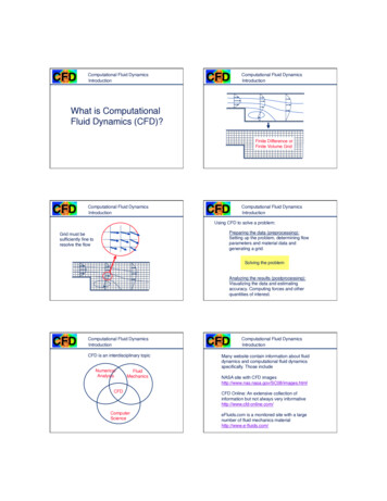
Traumatic Cerebrospinal Fluid LeaksT2-weighted images with fat suppression and image reversal to highlight CSF. Thecharacteristic signal tracking from the intracranial space to the paranasal sinusesrepresents a CSF leak. The sensitivity of this test is reported to be 85% to 92%,with 100% specificity.26 MRI and MR cisternography are able to distinguish inflammatory tissue from meningoencephaloceles; however, bony detail is poor (Fig. 3).Cisternography warrants discussion however, because of the specificity and noninvasive nature of b2-transferrin testing, the role of various cisternography studies hasbeen significantly altered. In the authors’ practice, after the diagnosis of a CSF leakhas been confirmed through b2-transferrin testing, high-resolution CT and MRI ofthe skull base are used to detail the anatomic integrity of the skull base. Endoscopicexploration confirms the location and is used for repair of the defect (discussed later)in the majority of cases. This philosophy has been adopted by other investigators aswell.27 Using this approach CT and MRI are complementary studies, with CT providingdetailed bony anatomy, particularly skull base dehiscence/fractures, and MRI providing soft-tissue detail, including coincident meningoencephaloceles and incidentalintracranial pathology. Modern image-guidance software enables the application ofCT-MRI fusion for surgical navigation, and results in accurately identifying and localizing the site of CSF leakage in 90% of cases.28CONSERVATIVE MANAGEMENTOnce a CSF leak has been confirmed and localized, the optimal management decision depends on a variety of factors. Even if the otolaryngologist is primarilymanaging the CSF leak, direct input should be obtained from the trauma service,neurosurgical team and, particularly if meningitis is suspected, infectious diseasecolleagues. Conservative treatment consists of strict bed rest and elevation of thehead at least 30 . In addition, patients should be advised to refrain from coughing,sneezing, nose blowing, and straining or Valsalva maneuvers. Stool softeners arerecommended, as well as antiemetics to avoid emesis or retching, antitussives toFig. 3. Axial MRI scan post gadolinium following skull base trauma showing an enhancinglesion in the inferior-medial frontal lobe (A) with follow-up scan showing resolution of thelesion in 3 months (B).863
864Prosser et alavoid coughing, and strict blood pressure management. The goal of these measuresis to reduce active flow through the leak, reduce CSF pressure, and allow healing ofthe defect to seal the leak, avoiding surgical intervention. In a series of 81 cases oftraumatic CSF fistula, the overall rate of cessation with conservative treatment was39.5% when used for 3 days. Resolution with conservative treatment of CSF fistulasinvolving temporal bone origin was 60%, whereas anterior skull base defectsresolved 26.4% of the time with conservative treatment.29 If conservative management is extended to 7 days, resolution rates improve to 85%, again with leaks oftemporal bone origin healing with a significantly higher rate than those of anteriorskull base origin.30 The main reason for this discrepancy may be anatomic differences in the skull base bone and dural structures that are damaged with traumato these subsites (ie, thin bone of the anterior skull base is more likely to causesignificant dural lacerations than the thicker temporal bone).Cerebrospinal Fluid DiversionIf there is persistence of the leak with conservative treatment, CSF diversion (mostcommonly with a lumber drain but occasionally serial lumbar punctures) is pursued.Lumbar drains are passive devices yet they require active management, as CSFcell count, protein and glucose measurements, and cultures should be collectedfrequently to monitor for meningitis, particularly if systemic signs exist. Averagedrainage rates are around 10 mL per hour. Optimal drainage lowers CSF pressureto decompress the leak; however, if drainage is too high severe headaches and pneumocephalus may result from drawing of air through the skull base defect into thecranial vault. There is also the added risk of meningitis. The benefits are that the addition of CSF diversion to conservative measures raises success rates to 70% to 90%with the average duration of drainage being 6.5 days.30,31 Another benefit of this treatment is that it can be performed at the bedside, even if patients are not stable enoughto go to the operating room. Lumbar drains can also be used as an adjunctive treatment to increase the success rates following a variety of surgical repairs.31SURGICAL MANAGEMENTTranscranial ApproachAlthough CSF rhinorrhea was initially described in the seventeenth century, it was notuntil 1926 that Dandy32 reported the first successful repair by using a bifrontal craniotomy for access and a fascia lata graft for repair. With this approach, access to thecribriform plate region and roof of the ethmoid is obtained via a frontal craniotomy. Anextended craniotomy and skull base dissection are required to access defects in thesphenoid sinus. After craniotomy the brain is retracted and the site of the defect isidentified. Multiple tissues can be used for repair including fascia lata grafts, muscleplugs, and pedicled galeal or pericranial flaps. Tissue sealants, such as fibrin glue,can be used to hold the graft in position; however, this will only last a few weeks,leading the authors to prefer suture closure to the dura distal to the defect, whichmore securely holds the graft in place. Reported success rates vary; however, recurrence rates as high as 27% have been reported.33 The clear advantage of thisapproach is that it provides direct access to the defect and allows for repair of multiplesites; however, with high reported failure rates, the morbidity of a craniotomy, andbrain retraction (including potential hematoma, seizures, and anosmia), extracranialtechniques are now preferred in most circumstances. At present these techniquesare mostly used in patients who require a craniotomy and exposure of the skullbase to treat associated intracranial pathology.
Traumatic Cerebrospinal Fluid LeaksExtracranial ApproachThe first extracranial approach was described by Dohlman34 in 1948, when he useda naso-orbital incision to repair a CSF leak. Dissection is then carried into the sinuscavities through the external incision for trans-sinus access to the skull base defect.The defect is identified and repaired directly using tissue grafts. Success rates withthis approach range from 86% to 97%.35,36 The benefits of this approach includeimproved success rates with decreased morbidity, including avoiding anosmia andbrain retraction with improved exposure of the posterior wall of the frontal sinus, foveaethmoidalis, cribriform plate, posterior ethmoids, sphenoid, and parasellar regions.Drawbacks include the necessity of a facial scar, facial numbness, orbital injury,and relative difficulty of dissection. Further intracranial pathology as well as leaks originating from the lateral aspects of the frontal and sphenoid sinuses cannot beaddressed.Transnasal ApproachFurther advancements were made in 1952, when Hirsch37 reported the successfulclosure of two sphenoid sinus CSF leaks via a transnasal approach. In 1964, Vrabecand Hallberg38 repaired a cribriform defect using this approach. In an attempt toimprove visualization, Lehrer and Deutsch39 used a microscope; however, accessand visualization of the lateral and superior walls of the sphenoid sinus were limited.Inherent to this approach is the risk of facial numbness and septal perforation, andwith the advent of endoscopic techniques this approach became rarely used.Endoscopic Endonasal ApproachEndoscopic techniques have emerged as the preferred approach to the repair of skullbase defects since their initial description by Wigand40 in 1981. This initial reportdescribed repair of a defect encountered during sinus surgery. In 1989 the first reportof the use of rigid transnasal endoscopy for the endonasal repair of CSF rhinorrheawas described,41 followed by a series of cases presented by Mattox and Kennedy42addressing CSF rhinorrhea with the aid of endoscopic visualization. Following identification and localization of the skull base defect, standard endoscopic techniques areused to expose the defect site. This approach provides excellent exposure of theethmoid roof, cribriform plate, and the sphenoid sinus.When lateral exposure in the sphenoid is necessary, endoscopic dissection of thepterygomaxillary space can be performed. Exposure of the posterior aspect of thefrontal sinus may require an osteoplastic flap or simple trephine. If needed, intrathecalfluorescein can be used to aid in identification of the skull base defect. Associatedmeningoencephaloceles are then addressed with bipolar electrocautery.Mucosa that is adherent to dura must be dissected away. It is critical that themucosa surrounding the skull base defect be removed for approximately half a centimeter. This technique both stimulates osteogenesis (thickening the bone surroundingthe defect) and improves graft incorporation. Following site preparation, the decisionfor graft type can be made.The choice of graft material has been a source of debate for some time; however,based on a recent meta-analysis it appears graft material does not affect successrate as long as sound surgical technique is used.43 Graft choices include temporalisfascia, fascia lata, muscle plugs, mucosal grafts (with or without bone), autogenousfat, free cartilage grafts (from the nasal septum or auricle), and free bone grafts(from the nasal septum, calvarium, or iliac crest). For small defects, free mucosal orfree fascial grafts can be placed in an overlay fashion. Significant overcorrection865
866Prosser et alshould be performed when forming the graft, as one can expect approximately 20%shrinkage in free mucosal graft size, starting in the first few postoperative days. Largedefects at risk for secondary encephalocele are sometimes treated by placement ofa bone or cartilaginous graft in an underlay fashion, with a fascial or mucosal graftplaced as an overlay to form a watertight seal. In the authors’ experience, this isusually not necessary. For large skull base defects the use of vascularized pedicledmucosal flaps are preferred, and this method is discussed in the following section.In general, the authors’ technique involves the placement of an inlay dural substitutegraft (ie, Duragen; Integra, Plainsboro, NJ, USA) followed by a mucosal graft in thecase of small defects or a vascularized pedicle flap for larger defects. Following graftplacement, a sealant (such as fibrin glue) can be used to hold the graft in place.Absorbable nasal packing (gel-foam) is usually placed directly against the mucosalsurface of the graft for additional support, followed by placement of nonabsorbablepacking for further support and hemostasis. Packs are usually removed in 5 to 7days, and antistaphylococcal antibiotic coverage should be used while nonabsorbable packs are in place.Postoperatively the patient should be placed on conservative measures (as previously discussed). As dissection occurs through a contaminated operative field,most surgeons elect to use perioperative antibiotics. Antibiotic choice should havegood CSF penetration (eg, ceftriaxone). At a minimum an antistaphylococcal antibioticshould be given postoperatively if nonabsorbable nasal packing is used. Admission tothe intensive care unit for monitoring and frequent neurologic status checks should beconsidered for the first 24 hours to monitor for complications such as hematoma orcerebral edema, although this is institution dependent. If a lumbar drain is in placefrom prior attempts to slow CSF drainage, or if one was placed intraoperatively, itshould be actively managed as discussed previously. Following removal of the nasalpacking, serial nasal endoscopies should be performed for debridement of any crusting and to inspect the operative site. Care should be taken not to disrupt the graftduring debridements, and patients should avoid strenuous activity or straining forapproximately 6 weeks following the repair.The endonasal endoscopic technique offers several advantages, including excellentvisualization and identification of the defect as well as graft placement. Reportedoutcomes are excellent, with success rates of 90% for primary procedures and 96%for sec
trauma, which may lead to false-negative results on diagnostic testing if testing is performed during the quiescent phase. Lastly, the subarachnoid cistern is a relatively