Transcription
Updated: June 2021I’veLab 4: Gel ElectrophoresisStandardProjectGuide
Table of ContentsPage3ContentsIntroduction4Key Elements for Gel Electrophoresis5How to Read a Gel6How to Interpret Gel ElectrophoresisResultsGel Electrophoresis Lab7Pre-Lab Questions8Getting Started9Visual Equipment and ReagentsChecklist12Gel Electrophoresis Protocol:Standard14Results15Post-Lab Questions16Troubleshooting19Database Checklist20GlossaryContent is made available under the CreativeCommons Attribution-NonCommercial-NoDerivatives International License. Contact(wolbachiaproject@vanderbilt.edu) if youwould like to make adaptations fordistribution beyond the classroom.The Wolbachia Project: Discover the MicrobesWithin! was developed by a collaboration ofscientists, educators, and outreach specialists.It is directed by the Bordenstein Lab atVanderbilt jectSome figures created with BioRender.comLAB 4: GEL ELECTROPHORESIS2
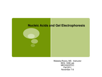
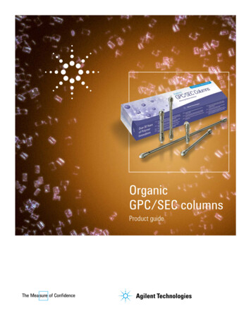
IntroductionThis lab will determine the presence or absence of amplified DNA in your samples by visualizationon an agarose gel. Arthropod and Wolbachia DNA, if present, will be distinguishable based on thesize, or base pair (bp) length, of the DNA molecule.Gel electrophoresisGel electrophoresis is a method of separating DNA fragments by movement through a Jello-likesubstance called agarose. Derived from a seaweed polysaccharide, agarose gels form small poresthat act as sieves to separate DNA based on size; whereby smaller DNA molecules move throughthe pores faster and easier than larger molecules. Loading wells are oriented at the top of the gelto allow for precise insertion of PCR products into the gel. An electrical current is applied to movenegatively charged DNA molecules away from a negative electrode (-) and toward a positiveelectrode ( ). DNA migrates through the gel in a single, vertical lane. Three factors influence thespeed of movement: (i) the voltage of the electrical field, (ii) the concentration of agarose, and(iii) most importantly, the size of the DNA molecule.DNA VisualizationDNA itself is not visible within an agarose gel. Therefore, a fluorescent stain is added to the gelthat binds DNA and fluoresces under UV or blue light. DNA will appear as a horizontal line, orband, on the agarose gel.Figure 4.1 Gel electrophoresis allows for differentiation of DNA by size. Larger fragmentsdo not move as fast as smaller fragments, and there is a nonlinear relationship betweenthe size of the DNA fragments and the distance migrated. A DNA ladder, a sample withknown fragment sizes, should always be run to identify the size of experimental bands.LAB 4: GEL ELECTROPHORESIS3
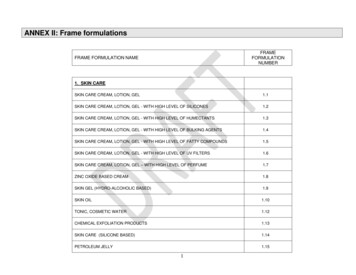
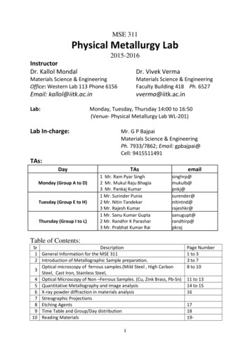
Key Elements for Gel ElectrophoresisPCR Products (DNA)The purpose of this lab is to visualize the PCR products, or amplified DNA, from your arthropodsamples.DNA LadderA DNA ladder is a cocktail of DNA fragments with pre-determined sizes. The ladder, also called aDNA marker, is loaded alongside experimental samples as a reference tool for estimating bandsize.Agarose GelAgarose powder is dissolved into running buffer and boiled until the solution becomes clear.After slightly cooling, it is poured into a casting tray with combs to solidify and form the agarosegel. DNA will migrate through the gel and form separate bands based on size (correlating tolength in bp).DNA StainA DNA stain is added to the agarose gel to visualize DNA under a UV or blue light. There are twoprimary methods of DNA Staining:- Pre-stain: DNA stain is added to the agarose gel mixture after melting, but beforepouring, the gel.- Post-stain: The gel is incubated in a stain solution after gel electrophoresis.Running BufferRunning buffer is a conductive liquid that allows the DNA to migrate through the agarose gel. Itis important that the agarose gel be made using the same buffer. Tris/Borate/ETDA (TBE) orTris/Acetic Acid/EDTA (TAE) are commonly used.Loading DyeLoading dye has two primary components: (i) a visible dye indicates how far the DNA has run onthe gel and (ii) glycerol, which is denser than the buffer, ensures that samples fall into the loadingwells rather than float back out. Some Taq Master Mixes (e.g., Promega GoTaq) already contain apre-mixed loading dye.Electrophoresis SystemRunning buffer and the agarose gel will be placed into the chamber of an electrophoresis system.After loading the samples, an electric current is applied to move the negatively charged DNAtowards the positive end of the system. Without this electric field, the DNA will not migratethrough the agarose gel. If the electric field is reversed, the DNA will run in the opposite direction,towards the top of the gel, and eventually exit the gel.Figure 3.4. Pictograph of all necessary components for gel electrophoresis.LAB 4: GEL ELECTROPHORESIS4
How to Read a GelLanesBandsDNA that was loaded into each well will migrate in a single, vertical lane towards the ( ) charge.When DNA becomes separated by size due to gel electrophoresis, they appear as bands in the gel.These are clearly defined, bright lines in the gel.DNA LadderThe DNA ladder will contain multiple bands in one lane. Each band represents a pre-determinedlength of DNA and can be used as a reference tool to estimate DNA size for each of theexperimental samples. Refer to the product information for specific band sizes.Primer DimersPCR reactions are set up with an excess of primers. In addition, some primers bind to each otherinstead of binding to the DNA, creating primer dimers. Primers are 25bp long, so excess primersappear as fuzzy bands on the bottom of the gel 25-50bp. This is normal and to be expected.Reading a Single PCR vs. Duplex PCR GelA single PCR gel will contain only one amplified PCR product, either Wolbachia or arthropod, ineach lane. A separate gel will need to be run for each DNA type.A duplex PCR means that both the arthropod barcoding gene and the 16S rRNA fragment fromWolbachia were amplified in the same PCR reaction. When visualizing this PCR reaction, twobands should appear in the same lane if Wolbachia is present, and only one band will appear ifthe arthropod is uninfected.Expected Band SizesArthropod Barcoding Gene (CO1): 708bpWolbachia Specific Gene (16S rRNA): 438bpLAB 4: GEL ELECTROPHORESIS5
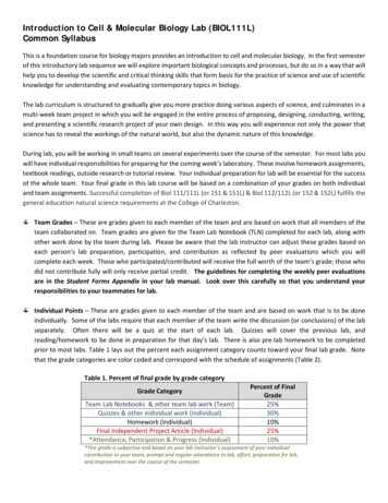
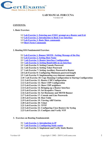
How to Interpret Gel Electrophoresis ResultsTo interpret gel electrophoresis results, first ensure that all controls are correct. The DNA ladder, ( )Arthropod control, (-) Arthropod control, and ( ) DNA control should produce bands of expected size,whereas the water lane should be empty.1. DNA LadderTo accurately read the gel, confirm the band size of experimental samples by comparing theirlocation in the gel to reference bands in the DNA Ladder. Refer to the information sheetaccompanying your DNA ladder for specific band sizes as the bands, or rungs, vary by product.Below is the DNA Marker (M3104) from MiniOne.2. Positive ControlsThe ( ) Arthropod control and ( ) DNA control should have both the CO1 and Wolbachia bandpresent. In a duplex PCR, as shown below, these will appear in the same lane. In a standard (single)PCR, these will be loaded into separate gels. The DNA ladder bands should be clearly present andseparated.3. Negative ControlsThe (-) Arthropod control should have a CO1 band, but no Wolbachia band. The negative watercontrol should not have any band or smudge.If all controls worked, the results of your experiment are valid, and the experimental bands can beanalyzed. If the controls have unexpected results, or if there is a band in the water lane, refer to theTroubleshooting guide on page 16.Lane1 2Loading Well34Band5Loading KeyLane123452000bpLadder1000bp500bp250bpSampleDNA Ladder( ) Arthropod(-) Arthropod( ) DNAWater100bpPrimer DimersFigure 4.3. Schematic of a duplex PCR gel where both Arthropod andWolbachia PCR products are loaded on the same gel. Common vocabularyterms are labeled on the gel, and the loading key is labeled according to eachlane.LAB 4: GEL ELECTROPHORESIS6
Pre-Lab QuestionsRead through the entire protocol and answer the questions below.1. Assume that single PCR reactions were loaded into two separate gels for arthropod andWolbachia DNA analysis. Fill in the expected bands for lanes 2-7 using the table below.Wolbachia (16S rRNA) gelArthropod (COI) gelLane1234567SampleDNA Ladder (bands already shown)An arthropod sample positive for WolbachiaAn arthropod sample negative for Wolbachia( ) Arthropod Control(-) Arthropod Control( ) DNA ControlWater2. This experiment included five controls. In the table below, list the following for each lab activity: ( ) for positive control (-) for negative control N/A for not applicableControlDNA ExtractionPCRGel ElectrophoresisDNA Ladder( ) Arthropod Control(-) Arthropod Control( ) DNA ControlWaterLAB 4: GEL ELECTROPHORESIS7
Getting StartedIntroductionIn this activity, you will use agarose gel electrophoresis to determine the presence and size of twodifferent gene fragments (arthropod COI, and Wolbachia 16S rRNA) previously amplified by PCR.If you ran two separate PCR reactions, arthropod and Wolbachia, you should prepare and run twogels. If you set up a duplex reaction (both primer sets in one PCR tube), you will only need onegel.Standard Gel Electrophoresis SystemThis protocol uses a standard electrophoresis system. The agarose gel will be made by addingagarose powder (or tablets) to running buffer, boiling the mixture, then letting it cool into agelatin-like slab. The agarose gel is run in a standard electrophoresis system, then visualized witha transilluminator.Pre-Lab PreparationMake sure your running buffer is made up and diluted to 1X. Running buffer (commonly TBE orTAE) is usually concentrated in a 10X or 20X solution. Dilute the buffer following manufacturer’sspecifications using deionized (DI) or distilled water, not tap water.Refer to your specific equipment manuals and reagent instructions before moving on.LAB 4: GEL ELECTROPHORESIS8
Visual Supplies ChecklistüNamePicturePurposeElectrophoresis tank& power supplyEquipmentA standard electrophoresis systemincludes a tank to hold buffer, and(-) and ( ) electrodes that areplugged into a power supply.Standardelectrophoresiscasting tray andcombEquipmentUsed to cast gels. Before pouringthe gel, the ends should be leakproof, and a comb should beadded.EquipmentThe microwave is used to melt theagar solution before casting into agel.EquipmentA transilluminator, geldoc, or otherimaging system to image andphotograph the gel.Microwave or hotwater bathImagerElectronic balanceEquipmentA balance is needed to weigh theamount of agarose powder tomake a 1% gel.Squirt bottle or spraybottle with 70%ethanolCleaning70% ethanol is used to clean theworkspace before and afterexperiments.GlovesPersonal Protective Equipment(PPE)Gloves are used to protect boththe scientist and sample fromcontamination.Oven mittPersonal Protective Equipment(PPE)A waterproof, heat-proof ovenmitt is used to protect the scientistfrom hot glass and steam aftermicrowaving the agarose.LAB 4: GEL ELECTROPHORESIS9
Eye protectionPersonal Protective Equipment(PPE)If you are using a UV light toobserve and image the gel, eyeproteciton is REQUIRED. UV lightcan damage the scientist’s eyes.Weigh DishSuppliesFor weighing agarose powder onthe electronic balance.1X Running Buffer(TAE or TBE)SuppliesRunning Buffer allows the gelfragments to migrate through thegel. It is a concentrated solutionand needs to be diluted prior touse.500mL flaskSuppliesUsed to boil the agarose powderand running buffer in themicrowave to make moltenagarose gel.Graduated cylinderSuppliesA graduated cylinder is used todilute the running buffer, and totransfer buffer to theelectrophoresis tank.Rack for 0.2mL PCRtubesOrganizationPCR tubes are small, it is necessaryto have a tube rack so they are notlost.20 μL pipetteLiquid ManagementPipettes are used to moveaccurate and precise amounts ofliquid from one place to another.20 μl pipette tipsLiquid Management20 μL tips are used to move 2-20μL of liquid. Tips should bechanged between each sample toavoid contamination.LAB 4: GEL ELECTROPHORESIS10
Waste cup for tipsDisposalKeeping all waste in one area untilthe end of the experimentincreases efficiency.SharpieOrganizationIt is extremely important to labelall tubes and samples.Parafilm (optional)SuppliesCan be used to cover the agarosemixture in the flask while meltingto prevent evaporation.It can alsobe used to mix PCR products withloading dye.Lab tape (optional)SuppliesCan be used to seal off the ends ofa casting tray before adding warmagarose.Visual Reagents ChecklistAgarose powderSuppliesAgarose powder is measured andcombined with running buffer tomake an agarose gel. The powderdissolves into solution afterboiling, or 80 seconds in themicrowave.DNA stainReagentDNA stain binds to DNA, making itvisible in UV or blue light.PCR products fromLab 3SamplesThese contain amplified DNA fromthe collected arthropod samples,control arthropods, control DNA,and water.ReagentThe DNA ladder is made up of predetermined fragment sizes of DNAand serves as a reference for bandsize determination.ReagentIf PCR products are colorless (i.e.,no loading dye in the Taq MasterMix), this should be added to eachsample prior to loading the gel. Ithelps to visualize movementthrough the agarose.DNA ladderLoading dye(optional)LAB 4: GEL ELECTROPHORESIS11
Gel Electrophoresis ProtocolStandardClass Preparation: Running Buffer Working Solution1. Together with your class, prepare a working solution of 1X electrophoresis running buffer.Prepare Lab Space2. Remove all unnecessary items from your lab station.3. Put on nitrile gloves and clean all surfaces by wiping down with 70% Ethanol.Prepare the Gel with DNA Stain4. Measure 1g agarose powder and add it to a 500mL flask.5. Add 100mL running buffer to the flask. (1% solution; note the total gel volume will varydepending on the size of the casting tray; refer to manufacturer instructions)6. Melt the agarose in a microwave or hot water bath until the solution becomes clear. Look for“lenses” in the liquid by holding the flask up to the light. If using a microwave, place a ball ofKimwipe tissue in the flask opening, or cover with parafilm and poke a hole for venting. Heatthe solution for several short ( 20 second) intervals, do not let the solution boil over.7. Let the solution cool to about 50-55 C, swirling the flask occasionally so it cools evenly. Theflask should be warm, but not too hot to touch.8. Seal the ends of the casting tray by placing it into the gel electrophoresis tank, or by sealingthe ends with wide lab tape. Refer to the manual of your electrophoresis system for moreinformation.9. Add 10uL GelRed (Biotium), or comparable DNA stain, to the agarose. Swirl to mix.10. Select a comb that will accommodate all samples. Place the comb(s) in the gel casting tray.11. Slowly pour the melted agarose solution into the casting tray. Gently pop any bubbles with apipette tip. Note: Fill the gel in the tray so the teeth of the comb are immersed under the gel,but the base of the comb is above the gel. You do not have to use all the melted agarose.12. Let gel cool undisturbed on a solid, flat surface until it is opaque and solid. This may take atleast 15-30 minutes. Moving the tray or not waiting until the gel is fully set may affect yourresults.13. Carefully pull out the combs and remove the tape or lift casting tray out of the electrophoresischamber.14. Place the gel in the electrophoresis chamber with the wells oriented near the (-) electrode.15. Add enough running buffer so there is 2-3mm of buffer over the gel.Prepare to Load the Gel16. Fill out the Loading Key on the next page.17. If your PCR Master Mix has no loading dye (your PCR products are clear), follow 17a. If yourPCR products are colored, move to Load the Gel.(a) Pipette 2uL drops of 5X loading buffer onto a piece of Parafilm or wax paper. Add10uL from each of your PCR reactions to a drop of loading buffer. Then mix well bygently pipetting up and down several times until the color of the liquid ishomogenous.Load the Gel18. Pipette 10uL of the DNA ladder in the first well. Hover your pipette above the well, and slowlyempty your pipette. Do not press to the second stop.LAB 4: GEL ELECTROPHORESIS12
19. Continue in this manner, carefully pipetting 10uL of each sample/sample loading buffermixture into separate wells in the gel. Change tips between each sample and store remainingPCR products in the freezer.Gel #1 Loading Key: ArthropodRun the Gel20. Place the lid on the gel box, connectingLane Samplethe electrodes appropriately (positive isDNA Ladder1red, negative is black). The negativeelectrode should be near the wells of2the gel, your DNA should “run to red”.3Also connect your electrodes to your( ) Arthropod Control4power supply.(-) Arthropod Control521. Turn on the power supply to about 100( ) DNA Control6volts. Maximum allowed voltage willWater7vary depending on size of theelectrophoresis chamber, and will beprinted on the label.22. Check to make sure current is running through the buffer by looking for bubbles that form oneach electrode.23. Check to make sure the current is moving in the correct direction by observing the movementof the loading dye. This may take a few minutes.24. Let the power run until the yellow (or bottom) band in the loading dye is ¾ down the gel.Then, turn off the power, disconnect the electrodes, and remove the lid and the gel usinggloves.Obtain an Image of the GelCAUTION; UV LIGHT CAN DAMAGE EYES!!! EYE PROTECTION REQUIRED!25. Place the gel on the transilluminator or other imaging equipment. Put on eye protectionbefore turning on the transilluminator. Use the equipment as directed, following the manual.26. Note the presence or absence of bands in each lane. Use the DNA ladder, with fragments ofa known size, to determine the size of each of the PCR products.27. Document your results on the next page.Repeat Steps 4-27 for the Wolbachia Gel ElectrophoresisClean your Work Station28. Discard used tips and wipe down thebench with 70% ethanol.29. Refer to your equipment’s manual toclean, dry, and store the electrophoresissystem.Label the Gel(s)30. Transfer the gel images to a computerand use a program such as Powerpoint,Google Slides or Preview to label each gel(Arthropod/Wolbachia)andcorresponding lanes.Gel #2 Loading Key: WolbachiaLane1234567LAB 4: GEL ELECTROPHORESISSampleDNA Ladder( ) Arthropod Control(-) Arthropod Control( ) DNA ControlWater13
ResultsUse the table below to record presence ( ) or absence (-) of bands.LaneDNA Source1DNA LadderArthropodCO1 Band?N/AWolbachia 16SBand?N/A234( ) Arthropod Control5(-) Arthropod Control6( ) DNA Control7WaterDo your control bands match the expected results you drew in Pre-Lab Question 1?Based on your answer, how confident are you that the experimental results are valid? Why?Complete the table below:Tube labelArthropod IDWolbachia-infected?(Yes, No, Unknown)LAB 4: GEL ELECTROPHORESISConfidence (High, Low)14
Post-Lab Questions1. In gel electrophoresis, pore size depends on agarose content. Higher % gels have smaller poreswhereas lower % gels have larger pores. In this lab, you used a 1% agarose gel. What wouldhappen if you used a 2% agarose gel, but ran it for the same amount of time?2. (a) Based on these results, which step of the experiment likely went wrong? Explain. (b) Are thearthropods tested here confidently infected with Wolbachia?Lane Sample1234567DNA LadderSample ASample B( ) Arthropod Control(-) Arthropod Control( ) DNA ControlWater3. (a) Based on these results, which step of the experiment likely went wrong? Explain. (b) Are thearthropods tested here confidently uninfected?Lane Sample1234567LAB 4: GEL ELECTROPHORESISDNA LadderSample ASample B( ) Arthropod Control(-) Arthropod Control( ) DNA ControlWater15
TroubleshootingISSUEAgaroseGel LeakedLadderGel isblank, nobandsWaterControl( ) DNAControlRECOMMENDATIONYour agarose mixture may be too hot. Wait for it to cool to about50 C before pouring your gel. This may be at least 15 minutes afteryou melt your agarose gel in the microwave.If you set your casting tray inside the electrophoresis tank to sealThe agarose gel leaked the ends, make sure the rubber gaskets on the end of the castingout of the casting tray. tray are inside of the grooves and not degraded. Agarose can easilyescape if these gaskets are not flush against the electrophoresistank.If you seal your casting tray with lab tape, make sure the tape issecure against the edges of the casting tray.When loading your gel, be careful and slowly pipette the ladderinto the gel. If you pipette too quickly, or click past the second stopIf bands are visible on on the pipette, there is a chance of the ladder leaving the well andyour gel, but not the not running correctly.ladder, it was mostIf your positive controls worked and produced a band, you can uselikely misloaded.these bands as an equivalent marker to gauge the size of yoursample bands.If all bands are absent,including the ladder,theelectrophoresisfailed.A smudge or band inthe water controlindicates there wascontamination in thePCR reaction.No band in the positiveDNA control.Can you see the loading dye? If not, the gel may have run too longand DNA will exit the gel.Confirm that the gel was oriented with loading wells near thenegative charge. If reversed, the DNA will migrate in the oppositedirection and out the top of the gel.Were the buffer and gel made with distilled or deionized water?Tap water is not recommended.Did the gel cool completely before applying the electrical current?Were the wells properly loaded? Tiny pin-like dots in a well indicatethat it was punctured with the pipette tip during loading and DNAwas lost. Foggy waves across the gel indicate that the samplesmight be floating in the buffer and did not fall into the wells.DNA visible in the wells, but not down the gel, indicates the gel wasnot run. Make sure the green LED near the power button is on torun your gel.Results cannot be trusted. If time allows, repeat the PCR. Usemolecular-biology grade water and switch tips between eachsample. Ensure the workspace is cleaned with 70% ethanol beforeand after each lab. If this is a persistent issue, all PCR reagents(primers, Taq master mix, water) should be discarded and replacedwith new aliquots. However, a smudge around 25-50bp representsprimer dimers and is to be expected.The PCR failed; both PCR and gel electrophoresis need to berepeated.If the ( ) and (-) Arthropod controls worked, your ( ) DNA controlmay be degraded. Replace the ( ) DNA control.LAB 4: GEL ELECTROPHORESIS16
NoArthropod(CO1) BandIf there is a Wolbachiaband but no arthropodband, the PCR primersmay not work for yourspecific arthropod.The general primers used to amplify CO1 are designed to detectmost arthropods, but there are some species that require specificprimer sets. If you collected a butterfly, moth, or dragonfly, forexample, this is most likely the case. Your sample can still bemarked Wolbachia positive if it does not have a CO1 band.Band SizeIf bands are present,but the wrong size, thegels or PCR tubes mayhave been mixed.Confirm that the unexpected band size matches the other PCRreaction (Arthropod: 708 bp / Wolbachia: 438 bp). If so, you canassume that the gel was misloaded or mislabeled. It is always best,but not necessary, to rerun the gel under optimal conditions.ArthropodControlsIf the ( ) DNA controlhas an arthropodband,buttheArthropod Controls donot, it is indicative thatDNA extraction failed.A Wolbachia band inthe (-) arthropodcontrol, but not thewater control.Smears andCurvedBandsSmears appear as faintsmudges down thelane of a gel. Curvedbands appear “smiley”or U-shaped.It is recommended that you repeat the DNA extraction with anindividual from the same population. The most likely culprit is lackof appropriately lysed cells. Increase grinding time with the pestle.If using a pellet-based DNA extraction kit, the DNA pellet may havebeen lost. Practice refining your technique or try a column-basedkit.It is possible that the DNA extraction kit or protocol may not besuitable for your specific arthropod. Some arthropods have thickexoskeletons that require additional lysis time/reagents. Avoidcollecting the exoskeleton during DNA extraction. If necessary,extract DNA from a leg or other body part without muchexoskeleton. Other arthropods contain proteins that inhibit PCRreactions. In this case, the DNA extraction is successful, but PCR willfail.This indicates contamination during the DNA extraction and resultscannot be trusted. It is recommended that you repeat the DNAextraction with an individual from the same population. Use freshaliquots for each reagent and change tips between each sample.Both smears and curved bands occur when the DNA concentrationis too high. As long as the controls worked, results can be trusted.For publication-worthy results, rerun the gel with less DNA addedto the well. If you have access to a spectrophotometer, measurethe DNA concentration and load the exact amount based onproduct information for your specific gel and well size.Smearing can also occur if the gel was not mixed properly orallowed to fully solidify before applying the electrical current.Faint smears at the bottom of the gel ( 25-50bp) are primerdimers. PCR reactions are set up with an excess of primers. Inaddition, some primers bind to each other instead of binding to theDNA, this is normal and to be expected.If the wells are deformed, the gel was cast when it was too hot.Next time, let the gel cool until it is no longer steaming, but stillliquid, before pouring and casting.LAB 4: GEL ELECTROPHORESIS17
After microwaving, swirl the agarose mixture around untilLumps in the geleverything is dissolved in solution. The agarose gel mixture is readyLumps inindicate that it was tooto pour when it is no longer steaming but before it over-cools andthe Gelcoldwhenforms lumps. It should be warm to the touch, but not painful topoured/cast.hold. If lumps form, microwave the mixture to molten again.Unexpected bands are most likely due to non-specific binding (i.e.,there is an off-target sequence in the genome with similar primerbinding sites) or pseudogenes. In animals, for example, the COIThepresenceofgene is sometimes transferred from the mitochondrial DNA intounexpectedbandsthe nuclear genome, termed nuclear mitochondrial DNA (NUMT).Unexpected indicates that theEvolutionary pressures induce mutations and truncations of theBandsprimers amplified aNUMT. If the primer-binding sites remain the same, they will stilldifferent region ofidentify and amplify the target sequence, but the amplifiedDNA.product will be a different size than the original COI gene. If a bandappears at the correct size, report the results accordingly: the laneis ( ) but non-specific binding is present.LAB 4: GEL ELECTROPHORESIS18
Database EntryAfter completing the Gel Electrophoresis Lab, open your entriesin The Wolbachia Project Database and record Methods andResults. A comprehensive guide is located under the se Fields to CompleteObservationsMethodsü DNA extraction kitü DNA extraction locationü Taq polymerase usedü Single/duplex PCR reactionq Upload gel imageq Gel electrophoresis systemq Bufferq DNA stainq Update protocol notesResultsq Wolbachia positive?q Confidence levelq Explain confidence levelLAB 4: GEL ELECTROPHORESIS19
GlossaryAgarose: A polysaccharide purified from seaweed. When dry agarose is boiled in a buffersolution, it will harden into a flexible, gelatin-like slab when it cools.Band: A clearly visible and defined mark on a gel, indicating DNA presence. Where the band is inrelation to the ladder indicates the size of the DNA product amplified in PCR.DNA Stain: A dye that binds to the DNA, making it visible in UV or blue light.Electrophoresis: A method of separating substances based on the rate of movement while underthe influence of an electric field.Ladder: A mix of pre-cut DNA of defined sizes. Each ladder may have different “rungs” of differentsizes; check the product information to find the sizes of DNA for your specific ladder.Lane: The vertical path of DNA migration below each loading well.Loading Dye: Loading dye is added to aid in loading of an agarose gel. It contains colored dye andglycerol.Loading Well: An indentation in the agarose gel in which samples are loaded.Negative Control: Ensures the process and samples are not contaminated, it is designed toproduce a negative result.Positive Control: A well-understood variable; should result in an expected positive result.Primer Dimers: Primers that bind to each other instead of the target DNA during PCR. This is the“haze” seen at the bottom of the gel, 25-50bp. This area may also include excess primersthat were not used during PCR.Running Buffer: A conductive liquid that allows the DNA to migrate through the agarose gel.LAB 4: GEL ELECTROPHORESIS20
gel. DNA will migrate through the gel and form separate bands based on size (correlating to length in bp). DNA Stain A DNA stain is added to the agarose gel to visualize DNA under a UV or blue light. There are two primary methods of DNA Staining: -Pre-stain: DNA stain is added to the agarose gel mixture after melting, but before pouring, the gel.