Transcription
MEDICAL IMMUNOLOGY LAB(MB417/517)Malcolm Lowry, Ph.D.Department of MicrobiologyOregon State University1
Immunology LabMB 417/517Credits: 2The course meets 4 hours per week in laboratory. Nash Hall 3rd floor teaching lab.Pre-requisites: MB 416 (may be taken as co-req), MB 303Learning resources (required): MB 417/517 lab manual (available in the OSU bookstore)Instructor:Malcolm Lowry, Ph.D.Department of MicrobiologyOffice: 453 Linus Pauling Science CenterEmail: malcolm.lowry@oregonstate.eduPhone: 737-8309This laboratory will provide exposure to up to date techniques in immunology using several newertechnologies as well as more basic experimental approaches.Student Learning Outcomes for MB417Upon completion of this class students should know how to:1. Collect experimental data aimed at understanding the function of the immune system.2. Analyze experimental data and construct valid conclusions regarding the meaning of the experiment.3. Formulate and present a well-organized and concise report of the experimental findings.4. Communicate recent research progress in the field from scientific journal articles and criticallyevaluate the methods and conclusions.Evaluation of student performance for MB417Lab reports and lab participation 50%Midterm exam 20%Presentation 10%Final exam 20%Student Learning Outcomes for MB517Upon completion of this class students should know how to:1. Collect experimental data aimed at understanding the function of the immune system.2. Analyze experimental data and construct valid conclusions regarding the meaning of the experiment.3. Formulate and present a well-organized and concise report of the experimental findings.4. Communicate recent research progress in the field from scientific journal articles and criticallyevaluate the methods and conclusions.5. Write a critical review of a current research paper from a major journal in the field and extend thetopic into what could be done next in further research.Evaluation of student performance for MB517Lab reports and lab participation 50%Midterm exam 20%Presentation 5%Written paper critique 5%Final exam 20%2
Student conduct /offenses-0)The following information is summarized from the OSU Student Conduct RegulationsStudents are expected to be honest and ethical in their academic work. Academic dishonesty is defined asan intentional act of deception in one of the following areas:*cheating- use/attempted use of unauthorized materials, information or study aids*fabrication- falsification or invention of any information*assisting- helping another commit an act of academic dishonesty*tampering- altering or interfering with evaluation instruments and documents*plagiarism- representing the words or ideas of another person as one's ownWhen evidence of academic dishonesty comes to the instructor's attention, the instructor will documentthe incident, permit the accused student to provide an explanation, advise the student of possiblepenalties, and take action. The instructor may impose any academic penalty up to and including an "F"grade in the course after consulting with his/her department chair and informing the student of the actiontaken.The goal of Oregon State University is to provide students with the knowledge, skill and wisdom theyneed to contribute to society. Our rules are formulated to guarantee each student's freedom to learn and toprotect the fundamental rights of others. People must treat each other with dignity and respect in order forscholarship to thrive. Behaviors that are disruptive to teaching and learning will not be tolerated, and willbe referred to the Student Conduct Program for disciplinary action. Behaviors that create a hostile,offensive or intimidating environment based on gender, race, ethnicity, color, religion, age, disability,marital status or sexual orientation will be referred to the Affirmative Action Office.Student Conduct Expectations ent Regarding Students with Disabilities: Accommodations forstudents with disabilities are determined and approved by DisabilityAccess Services (DAS). If you, as a student, believe you are eligiblefor accommodations but have not obtained approval please contact DASimmediately at 541-737-4098 or at http://ds.oregonstate.edu. DASnotifies students and faculty members of approved academicaccommodations and coordinates implementation of those accommodations.While not required, students and faculty members are encouraged todiscuss details of the implementation of individual accommodations.Reach Out for Success: University students encounter setbacks from timeto time. If you encounter difficulties and need assistance, it’simportant to reach out. Consider discussing the situation with aninstructor or academic advisor. Learn about resources that assist withwellness and academic success at oregonstate.edu/ReachOut. If you are inimmediate crisis, please contact the Crisis Text Line by texting OREGONto 741-741 or call the National Suicide Prevention Lifeline at1-800-273-TALK (8255)3
Schedule of MB 417/517 lab1. September 25:Lab 1: Histology of the immune system. Differential blood count2. October 2:Lab 2: Phagocytosis of pathogens by macrophages3. October 9:October 11:Lab 3: Nitric oxide production in response to microbial stimuli. Part 1.Friday- 1pm Part 2 of Lab 3 Nitric oxide lab.4. October 16:Lab 4: Macrophage intracellular killing of bacteriaLab report from lab#1 and #2 due5. October 23:Lab 4 part 2: CFU counting from Lab 4Lab 5 part 1: SDS PAGE and western blotting 1Midterm exam6. October 30:Lab 5 part 2: SDS PAGE and Western blotting part 2Lab report #3 dueImmuno-olympics topics and teams will be assigned7. November 6:Lab 6: Analysis of B and T lymphocyte activation by Flow cytometry8. November 13:Lab 7: Lab 7 ELISA part 1, Immuno-olympics discussionLab report #4 due9. November 20:Lab 7: ELISA Part 2.Lab final exam: taken during incubation time for ELISALab report #5 due10. November 27:Discussion of flow cytometry Lab 6 data11. December 4:Immuno-Olympics team challenge.Lab report #6 and #7 dueNote: Please read the lab procedure before coming to class. Some of these labs are verychallenging, and you will make mistakes if you don’t have a good idea of what you are going todo that day.4
LAB NOTEBOOKSThe lab notebook is an important part of your grade. It will be graded as indicated on the lab schedule.You may use separate folders to hand in lab reports that are due. This will allow you to removepages/reports for grading, or add in computer-typed introduction/discussion/results sections. Unlessotherwise specified by the instructor, each individual lab report should have the following components:Heading:Exercise # and Title of the labYour nameYour partners’ name (if applicable)Purpose (2 pts):A short paragraph describing the objectives of the lab. Do not just copywhat is written in the lab instructions! (you will get a zero if you plagiarize from the lab manualor any other source).you should address: Why did we conduct exercise? What information werewe trying to obtain? What concepts were being demonstrated? What protocol or method ortechnique did we use to obtain the information? The purpose is typically just 1-2 paragraphs,but it should be complete.Procedure (1 pt):Brief outline. You may reference the lab manual, but you must include anychanges from printed protocol.Results (3 pts):Detailed, well-organized, clearly labeled.Must attach pages of raw data to get full creditResults should be clearly organized, preferably as table or chartDrawingsGraphs – should have titles and clearly labeled axes and scales.CalculationsWhen presenting data, especially tables, assume that the reader is not intimatelyfamiliar with the lab procedure, and make sure that every entry is properlylabeled and that all information necessary for understanding the results isincluded.Conclusions/Discussion (4 pts):Explain results. Summarize conclusions you can draw from the results.What do the results indicate? This is the most important section, explain thereasoning for what the results mean.Were the objectives listed in the purpose section accomplished? Why or how?What problems were encountered?Were there any unexpected results? What are possible explanations?Indicate things that might be done differently if the lab were repeated.Note: If you use any outside sources, you MUST provide a complete reference.5
LABORATORY RULESCarefully read the following items relating to laboratory safety:1) DO NOT: eat, drink, chew gum or tobacco. Open beverage containers must be left on thehallway shelf outside lab. Keep your hands out of your mouth and eyes.2) You are required to provide and wear a lab coat at all times in lab. Wear shoes that wouldprotect feet from broken glass (preferably autoclavable). Only the necessary suppliesshould be kept on your bench top; everything else can be placed in cupboards (keep theaisles clear!). Backpacks/coats should not be left on bench tops. WARNING:CONTAMINATED PERSONAL OBJECTS WILL BE AUTOCLAVED.3) Protective eye goggles are required for all experiments using corrosive or toxic chemicals orpotentially pathogenic organisms.4) No unauthorized visitors in lab.5) CLEAN desktop with LpH DISINFECTANT at the beginning and end of class. Wash handswith soap before leaving the lab.6) LOCATE the safety equipment in the lab - fire extinguisher, fire blanket and eye shower.The full body shower is located at the entrance to the media room (Nash 324) for 3rd floorlabs or lab entrance (Nash 554) for 5th floor labs.7) BUNSEN BURNERS in the lab have almost invisible flames – be careful of loose clothingand tie back long hair during lab to avoid contact with flame. Turn burners completely offwhen finished. If at any time you smell gas in the lab, check to make sure your burner isturned all the way off. If it is not your burner, notify the lab TA immediately.8) BROKEN GLASSWARE - Call the instructor or TA to assist you. Do not dispose of anyglassware in the regular garbage cans.a. Contaminated glass is placed in large can on discard table for autoclavingand subsequent disposal in broken glass box.b. Non-contaminated glass is placed in the large cardboard broken glass box.9) INJURY - If you cut yourself in the lab, inform the instructor or TA so that the wound canbe properly disinfected. If the injury needs professional assistance, you will be escorted tothe Health Center or proper facility.10) SPILLAGE – If you spill anything in lab, inform the instructor or TA so that they can assistyou in proper clean-up. If culture is spilled on your clothing or belongings, they mayrequire decontamination to assure your safety.11) PIPETTING - Do not pipette by mouth. When using rubber bulbs, insert the pipette gentlyinto the bulb to avoid breaking the pipette and potentially cutting yourself.12) TO AVOID CONTAMINATION - Microbes are found everywhere, use common sense toavoid contamination.6
a. You must not allow any part of yourself or any other nonsterile object totouch growth media.b. Do not remove the lid from your Petri dish completely, instead lift it andhold it above the dish to protect the media from dust.c. Flame tube mouths after you open them and before you close them to killdust borne contaminants.d. Do not put test tube caps or Petri dish lids down on the counter.e. Hold tubes and flasks so that the mouth is as horizontal as possible tominimize exposure to dust.f. Sterilize your loop or inoculating needle by heating it until it glows. Startwith the wire above the loop in the "cool" yellow part of the flame and endwith the loop itself in the "hot" blue cone. Do this before and after youtransfer each culture.13) INCUBATING - Carefully label all materials to be incubated with your name and organismidentification.a. Label culture plates on agar side with your name and organism identification.Incubate plates agar side up.b. Label culture tubes on the glass (not plastic caps) with your name andorganism identification. Place tubes in racks for incubation.14) DISCARDING CLASSROOM MATERIAL - All materials used in this class that arecontaminated with culture (tubes, plates, pipettes, etc.) must be autoclaved beforecleaning or disposal.a. Used slides and cover slips go in the metal can for contaminated glass waste.If working with nonpathogens, the slides can be cleaned with BonAmi andreturned to desktop.b. Used razor blades/pins/needles go in a “sharps” container.c. Plastic petri plates go into the autoclave bag at the discard table.d. Plastic transfer pipettes, pipette tips, swabs, etc go in the autoclave bag atthe discard table.e. Culture tubes go in wire baskets in metal coffins on the discard table. Loosenscrew caps before autoclaving.f. Glass bottles go in metal coffins on the discard table. Loosen screw capsbefore autoclaving.g. Uncontaminated paper used to clean desk with LpH or lens paper can beplaced in metal container on desktop. The metal container should be emptiedin the main garbage can at the end of each lab.h. Racks are to be returned to the lab shelves.i. Glass pipettes are placed into plastic trays on desktop that contain bleach.Pipettes should all be placed in the same direction.j. Organic solvents such as phenol and chloroform should be discarded intoproperly labeled waste containers.14)LEAVING THE LAB – Clear lab bench of all cultures, racks, and plates. Empty metal canof paper waste into a large garbage can. Clean desktop with LpH disinfectant. Washhands with soap. Take off and store your lab coat (do not leave with it on!). Make sureyou have all of your belongings before leaving lab.7
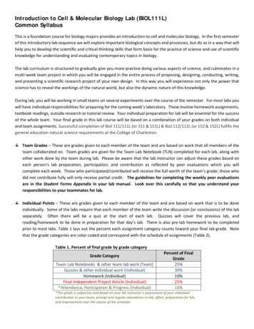
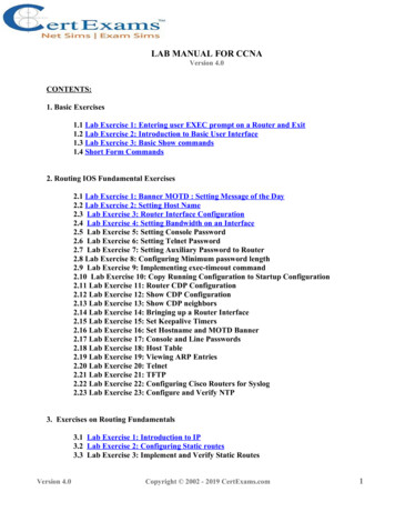
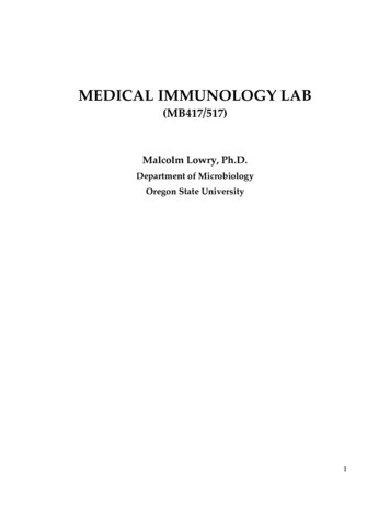
PROPER USE OF EQUIPMENTMICROSCOPE USEThe microscopes are valuable and must be cared for properly. Remove microscope fromcabinet by rolling shelf all the way out, then lifting microscope to the table. Bumping theoculars into the top of the cabinet can damage them.When replacing the microscope after each use:a. Carefully clean oil off the 100X objective by wiping with flat lens paper,followed by flat lens paper saturated with lens cleaner. If lens paper iscrumpled it will scratch the lens. If oil is left on the objective, it will destroythe seal and the lens will need to be replaced ( 300).b. If oil is on the 45X lens, please let the instructor or TA know so it can havespecial cleaning.c. Remove dirt or oil from the stage, condensers or oculars, if needed.d. Store microscope with low power or blank objective in place and close tostage.e. Wrap the cord around the oculars neatly; replace in cabinet when shelf iscompletely rolled out.f. Sign and date the sign-out sheet in the cabinet.g. Have your TA check your microscope and initial sign-out sheet.USE OF PIPETMANPlace sterile tip onto pipetman by carefully opening sterile box of tips and seating plasticshaft down firmly onto tip. Place sterile tip into desired solution and push down to first stop onplunger button. Carefully release. Liquid should have been drawn up into tip. Put tip into thetube you want to dispense liquid into, press plunger button down to first stop and that should doit.Never turn the numbers on the pipetman above the maximum amount. For example, witha P20 never turn dial past 20.0 or you will strip the gears. Place used disposable tips into plasticdiscard container.8
Pipetman Diagram(bold number are in black on pipetman, nonbold numbers are in red on pipetman)P 10P 20Range: 1 µl to 10 µlRange: 2 µl to 20 µl100 10.0 µl 14.5 µl5P 200P 1000Range: 20 µl to 200 µl14514Range: 200 µl to 1000 µl0 145 µl455 450 µl9
Immunology introduction:Functioning immune systems are crucial for the survival of the species but theirimportance is sometimes forgotten when matters of evolution are discussed. All livingplants and animals must be equipped with defense mechanisms against potentialpathogens. For example, the unicellular amoeba produces anti-microbial peptides andmany antibacterial and antifungal compounds have been isolated from terrestrial plantsand algae. Invertebrates rely on innate immune systems whereas vertebrates have anadaptive system as well. The origin of the adaptive system is thought to have occurredwith a serendipitous insertion of a recombination-activating gene (RAG) transposoninto the genome of an ancestor to the jawed fishes before the divergence of cartilaginousfishes. Much research has been conducted on adaptive immune systems, in particularthe mammalian system. However, in recent years more researchers have taken aninterest in the innate system and the interactions that occur between the two differentsystems.The innate immune system.The innate immune system can be divided into first and second lines of defense.The first line takes the form of anatomical and physiological barriers. Examples ofanatomical barriers in mammals are skin and mucosal membranes. Skin, being heavilykeratinized and low in moisture content, is hard for pathogens to penetrate. Thisdifficulty is also enhanced by the low pH of 3-5 that is created by lactic acid and certainfatty acids that are produced in the sebaceous glands. Microbial penetration of mucosalmembranes is also difficult. Abundant mucus secretions can simply wash thepathogens away, and the cilia that line the airways of mammals beat constantly andtransport the trapped pathogens so that they can be cleared.The mucus that is released onto the body surface of teleosts also works as an anatomicaland physiological barrier by making it hard for parasites, bacteria and fungi to enter orto start growing on the skin itself.10
Antimicrobial proteins and peptides.The second line of defense includes a wide range of molecules that inhibit thegrowth and spread of microbes by direct killing, or by facilitation of receptor-mediatedphagocytosis. The skin and mucosal membranes produce antimicrobial proteins andpeptides. Lysozyme, a hydrolytic enzyme that cleaves the peptidoglycan layer in thecell wall of Gram-positive bacteria, is found in tears and in the respiratory mucosa andis also produced in the Paneth cells of the intestinal mucosa of mammals. Lactoferrin,secreted in the mammalian respiratory system, also shows antimicrobial activity. Itsactivity seems to be confined to the first 25 amino acids of the N-terminus and thisfragment gets cleaved off to form the active peptide. Antimicrobial peptides are alsoproduced from the N-terminal of pepsinogenin from the bullfrog, Rana catesbeiana.Other antimicrobial peptides have been isolated, mainly from mammals and Drosophila,and they can be divided into a and b defensins depending on if their secondarystructures mainly consist of an a-helix or a b-sheet.The Drosophila antibacterial and antifungal peptides include cecropins, attacin,diptericin, drosocin and drocomycin. They are secreted from the fat body, analogous tothe liver in mammals. The intracellular signaling that leads to transcription of therelevant genes is conducted by relish, which is homologous to the mammalian NF-kBtranscription factor. This factor is responsible for activation of many of the acute phaseproteins in mammals. A couple of the a-defensins, human defensin-5 (HD-5) and HD6, are constitutively expressed in the Paneth cells of the intestine; HD-5 is also secretedconstitutively into the mucus of the vagina and cervix.Many antimicrobial peptides are cytotoxic to both Gram negative and Grampositive bacteria, and some peptides, such as Metchnikowin from Drosophila, have bothantifungal and antimicrobial activity. While some are expressed constitutively, other11
peptides are inducible. Antimicrobial peptides are also found in neutrophil granulesand recently they have been found to interact with the adaptive immune system byattracting dendritic cells and T-cells to sites of infection.The hepatic (liver) acute phase response.Humoral components of innate immunity are not limited by constitutiveexpression of fixed rates. Indeed increases and decreases in protein synthesis in the liverduring the early phase of an infection or immediately following a traumatic event helpprotect the individual from opportunistic pathogens. These so-called acute phaseproteins (APPs) are secreted into the plasma. Those that increase in quantity areconsidered to be positive APPs whereas those that decrease are negative APPs.More than thirty-five APPs have been identified in mammals. There are threemajor mammalian APPs: C-reactive protein (CRP), Serum Amyloid A (SAP) andSerum Amyloid A (SAA). Others include complement components, serum proteinaseinhibitors, coagulation factors and metal binding proteins.Cells in innate immunityGranulocytes (neutrophils, eosinophils and basophils), dendritic cells,monocytes and tissue macrophages are important components of the secondary line ofdefenses in innate immune systems. Most are phagocytic and can secrete molecules(interleukins) that orchestrate the inflammatory response. Of the granulocytes,neutrophils and eosinophils can phagocytose but basophils cannot. The granules inneutrophils stain with both acidic and basic dyes, eosinophil granules stain with acidicdyes and the granules in basophils, as the name implies, stain with basic dyes.Neutrophils, or polymorphonuclear leukocytes (PMNs) as they are also called,are important in the diagnosis of acute infections in mammals, since their numbersincrease rapidly early in an infection. In mammals, the neutrophil granules containcollagenase and other proteolytic enzymes, lysozyme, lactoferrin, peroxidase, and12
bactericidal/permeability-increasing protein, as well as several a-defensins. Thesegranules fuse with phagosomes and help in the digestion of engulfed particles.Neutrophils also produce different oxygen and nitrogen radicals such as superoxideand nitric oxide that are highly damaging to pathogens.Eosinophils are involved in defense against parasites and also play a role inasthma and allergies. The roles of basophils are not well defined. However, botheosinophils and basophils, by their release of histamines and other vasoactivesubstances, are part of the pathology seen in allergies and asthma.Macrophages and dendritic cells (DC) have many functions. They are capable ofopsonin-dependent and opsonin-independent phagocytosis. Secondly, they producecytokines after they are “activated” by phagocytosed particles and thirdly, they degradecaptured particles and present some of the resulting peptides, in the context of MHC II,on the cell surface. The antigen, held within a grove in the MHC molecule, is thenrecognized by T-helper cells. This event, which involves several signals eventuallyleads to expansion of the specific T-cell clones. These T-cells trigger B-cells to produceantibody. Monocytes circulate in the blood and, when needed outside the vasculature,become activated and migrate into the affected tissue as macrophages.Lymphocytes, B and T-cells, clonal expansion, memory, self-recognition,antibody diversity, primary and secondary responses are hallmarks of the adaptiveimmune system. B-cells, like macrophages and DCs, are antigen-presenting cells(APCs) with the main difference between the cell types being that B-cells candifferentiate into plasma cells that secrete antibodies. A large diversity of antibodyspecificities can be achieved by rearranging immunoglobulin gene segments.13
LAB #1 HISTOLOGY OF LYMPH ORGANSCells of the immune system are located in highly organized compartments inlymphoid tissue. Particularly the spleen and lymph nodes (LN) contain areas wherecertain lymphocytes are found and others are usually not. The advantage to this type ofarrangement allows trafficking of cells to the appropriate sites of the tissue. Inparticular, the cortex is the outer most area of lymph nodes and the inner portion is themedulla. B lymphocytes are mainly organized into follicles and T lymphocytes anddendritic cells are contained in paracortical areas. This type of organization allows Agto be carried into the lymph node by migrating dendritic cells which can then activate Thelper cells. The T helper cells can then interact with B cells and mediate theiractivation. When B cells become activated they will usually develop into plasma cellswhich secrete large quantities of Ig. However, an activated B cell can enter a follicle andbegin to proliferate vigorously as well as somatically mutate their Ig genes. This site isreferred to as a germinal center. Astonishingly, there are documented studies whichshow that a single B cell can clonally expand to encompass an entire germinal center.14
Slides are on display in the microscopes. Please look at the slides and identify some ofthe structures seen.1. Lymph nodes, Peyer’s patch, tonsil and spleen – identify lymphocytes, dendritic cellsand macrophages (can be tricky). Also look for blood vessels.2. Bone marrow - please identify myeloblasts, reticulocytes and band neutrophils.3. Do one drawing of an organ with some cells labeled for your lab report. You canchoose what you want to draw from the slides on display.15
DIFFERENTIAL COUNT1. You will have 3 slides that have been stained with wrights stain of human bloodsmears. One is normal blood from a healthy individual, and will be marked as normal.The other two will be coded as B and C, and are unknown pathologies of human blood.The goal is to determine how the unknown blood samples differ from the normalcontrol. By counting the percentages of white blood cells in each category, you candetermine the likely pathology. Today the unknowns are derived from patients thathave either a viral infection resulting in mononucleosis, or have a nasty case of allergiesor possibly infection with parasites. To figure out which condition you have, you haveto determine what cells would be increased in each type of infection.2. Count 100 “white cells” (lymphocytes and granulocytes, monocytes etc.) andcategorize them into lymphocytes, neutrophils, eosinophils, monocytes and basophils.Do this for the control slide, the B and C slides and then you can make comparisonsbetween them. Each person in the lab pair will need to make the same counts, and thenthe data can be shared in the lab pair. Make a judgment on the cause of infection inyour discussion of your results showing the differential counts you observed.Differential counts are done often in the hospital or at the doctor’s office if any kind ofinfection is suspected. What can a differential count tell you about the status of thepatient? Of course it can tell you if an infection or other condition is affecting theimmune system. What we are doing today is what was done in clinics before the16
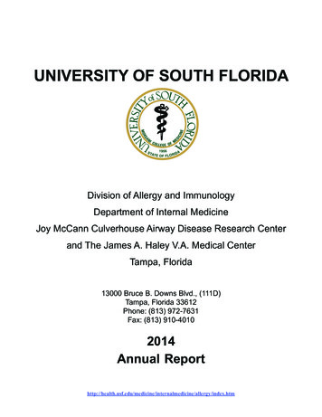
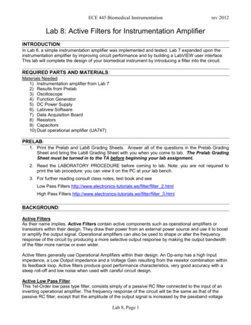
advent of flow cytometry to determine the health status of a patient. It is still doneoften for more detailed analysis if abnormalities are observed.Pictures of a CBC and also the morphology of various white blood cell types follow onthe next few pages.Sample scan of a CBC (cellular blood count) or complete blood count done in a clinic byflow cytometry.The bottom graph is a sedimentation rate calculation on RBC’s. This reflects the activation ofclotting factors and acute phase proteins triggered by inflammation or damage. Thismeasurement is also called “seds” for short in the hospital. You can impress your doctor nexttime by asking what your CBC counts showed and what your “seds” were the next time you get ablood draw.We will be doing flow cytometry later in the quarter.17
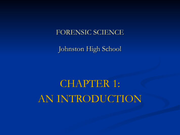
A normal mature lymphocyte is seen on the left compared to a segmented PMN on the right. An RBC isseen to be about 2/3 the size of a normal lymphocyte.Here is a monocyte. It is slightly larger than a lymphocyte and has a folded nucleus. Monocytescan migrate out of the bloodstream and become tissue macrophages under the influence ofcytokines. Note the many small smudgy blue platelets between the RBC's18
In the center of the field is an eosinophil with a bilobed nucleus and numerous reddish granules inthe cytoplasm. Just underneath it is a small lymphocyte. Eosinophils can increase with allergic reactionsand with parasitic infestations.The RBC's here have stacked together in long chains. This is known as "rouleaux formation" andit happens with increased serum proteins, particularly fibrinogen and globulins. Such long chainsof RBC's sediment more readily. This is the mechanism for the sedimentation rate, whichincreases non-specifically with inflammation and increased "acute phase" serum proteins.19
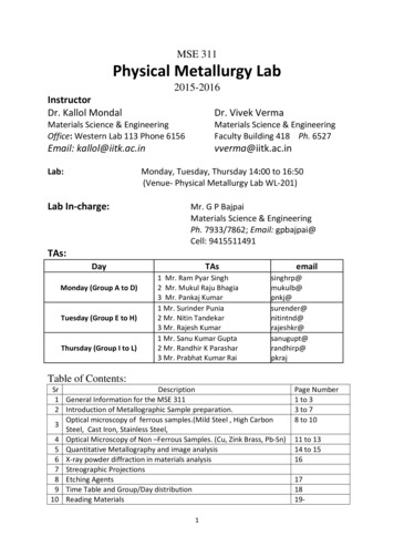
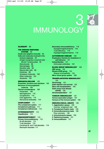
Identify the segmented neutrophil, band neutrophil, lymphocyte, monocyte, eosinophil, basophil, andplatelet in the image above:Answers:Band neutrophil: upper left corner.neutrophil: right middlelymphocyte: left middlemonocyte: lower left cornereosinophil: upper right cornerbasophil: lower right corner20
LAB #2 PHAGOCYTOSISIn this lab we will examine the role of macrophages. In addition to beingimportant in the initiation of a specific immune response (antigen processing andpresentation, secretion of factors), macrophages are also potent effector cells. In theircapacity as phagocytic cells, macrophages actively ingest and digest such variedantigens as whole pathogenic bacteria and parasites as well as injured and dead hostcells. The process of phagocytosis involves several steps. The macrophage is attractedto and moves toward the foreign body by a process called chemotaxis. Followingattachment of the antigen to the macrophage cell membrane, it is engulfed within amembrane-bound structure called a phagosome. As the phagosome moves toward thecell interior it fuses with a structure called a lysosome, which releases a variety ofdestructive agents, including hydrogen peroxide, oxygen free radicals, nitric oxide,peroxidase and lysozyme. Once these compounds have destroyed the contents of thephagolysosome, they are eliminated by exocytosis. Although this process is highlyeffective for dealing with most pathogens, there are certain intracellular pathogenswhich have adapted and can even multiply within phagocytic
3. October 9: Lab 3: Nitric oxide production in response to microbial stimuli. Part 1. October 11: Friday- 1pm Part 2 of Lab 3 Nitric oxide lab. 4. October 16: Lab 4: Macrophage intracellular killing of bacteria Lab report from lab#1 and #2 due 5. October 23: Lab