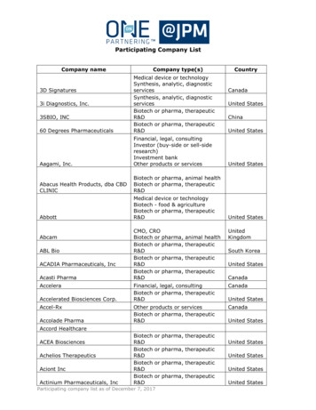
Transcription
ApoptosisTools for cell death series 1
ContentsCell death. . . . . . . . . . . . . . . . . . . . . . . . . . . . . . . . . . . . . . . . . . . . . . . . . . . . . . . . . . . . . . . . 4Apoptosis . . . . . . . . . . . . . . . . . . . . . . . . . . . . . . . . . . . . . . . . . . . . . . . . . . . . . . . . . . . . . . . . 5–– Mechanisms of apoptosis. . . . . . . . . . . . . . . . . . . . . . . . . . . . . . . . . . . . . . . . . . . . . . . . . 5–– Hallmarks of apoptosis . . . . . . . . . . . . . . . . . . . . . . . . . . . . . . . . . . . . . . . . . . . . . . . . . . . 7Detecting apoptosis. . . . . . . . . . . . . . . . . . . . . . . . . . . . . . . . . . . . . . . . . . . . . . . . . . . . . 8–– Activation of pro-apoptotic members of the Bcl-2 family. . . . . . . . . . . . . . . . . . . . . . . 8–– Loss of membrane asymmetry. . . . . . . . . . . . . . . . . . . . . . . . . . . . . . . . . . . . . . . . . . . . . 9–– Caspases . . . . . . . . . . . . . . . . . . . . . . . . . . . . . . . . . . . . . . . . . . . . . . . . . . . . . . . . . . . . . 11–– Antibody-based methods . . . . . . . . . . . . . . . . . . . . . . . . . . . . . . . . . . . . . . . . . . . . . 12–– Substrate-based methods . . . . . . . . . . . . . . . . . . . . . . . . . . . . . . . . . . . . . . . . . . . . . 13–– Calpain and cathepsins. . . . . . . . . . . . . . . . . . . . . . . . . . . . . . . . . . . . . . . . . . . . . . . . . 14–– Mitochondrial transmembrane potential. . . . . . . . . . . . . . . . . . . . . . . . . . . . . . . . . . . 15–– Cytochrome c release . . . . . . . . . . . . . . . . . . . . . . . . . . . . . . . . . . . . . . . . . . . . . . . . . . 17–– Chromatin condensation. . . . . . . . . . . . . . . . . . . . . . . . . . . . . . . . . . . . . . . . . . . . . . . . 19–– Genomic DNA fragmentation . . . . . . . . . . . . . . . . . . . . . . . . . . . . . . . . . . . . . . . . . . . . 19–– Increase of sub G1 population. . . . . . . . . . . . . . . . . . . . . . . . . . . . . . . . . . . . . . . . . . . . 21–– Cell membrane blebbing. . . . . . . . . . . . . . . . . . . . . . . . . . . . . . . . . . . . . . . . . . . . . . . . 22Tips for apoptosis assays . . . . . . . . . . . . . . . . . . . . . . . . . . . . . . . . . . . . . . . . . . . . . . . . 23–– Use of apoptosis inducers and inhibitors. . . . . . . . . . . . . . . . . . . . . . . . . . . . . . . . . . . . 23–– Analyze single cells . . . . . . . . . . . . . . . . . . . . . . . . . . . . . . . . . . . . . . . . . . . . . . . . . . . . . 23–– Be aware of cell line variation. . . . . . . . . . . . . . . . . . . . . . . . . . . . . . . . . . . . . . . . . . . . . 24General considerations . . . . . . . . . . . . . . . . . . . . . . . . . . . . . . . . . . . . . . . . . . . . . . . . . 25References. . . . . . . . . . . . . . . . . . . . . . . . . . . . . . . . . . . . . . . . . . . . . . . . . . . . . . . . . . . . . . 273
Cell deathCell death happens when a cell fails to maintain essential life functions and can benon-programmed, in the case of injury or trauma, or programmed, as in processes likeapoptosis and autophagy.Cell death can be classified according to its morphological appearance (such asapoptotic or necrotic), enzymological criteria (with or without the involvement of distinctproteases), functional aspects (programmed or non-programmed), or immunologicalcharacteristics (immunogenic or non-immunogenic)1.Before studying cell death mechanisms, researchers should ensure cell death hashappened. The Nomenclature Committee on Cell Death (NCCD) has proposed thatresearchers should define a cell as dead when the following features are observed1:1. The cell has lost the plasma membrane integrity2. The cell has undergone complete disintegration3. Whatever is left of the cell has been phagocytosed by the neighboring cells in vivoIn this guide, we aim to provide you with an overview of apoptosis, the most studied andwell-known type of cell death, the most common parameters used to assess apoptosis,and tools you can use to study cell death in your research.4
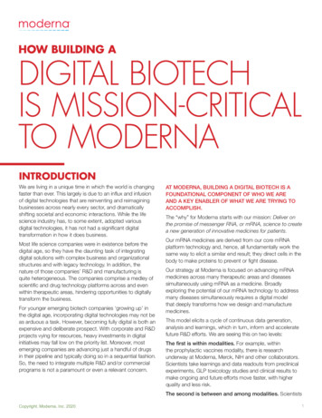
ApoptosisApoptosis is a type of programmed cell death that is critical for numerous normalphysiological processes. Historically, apoptosis has been defined by its morphologicalfeatures, most of which were described in the 1970s by John Kerr2. Apoptoticmorphology includes cell shrinkage, membrane blebbing, chromosome condensation(pyknosis) and nuclear fragmentation (karyorrhexis), DNA laddering, and the eventualengulfment of the cell by phagosomes. In contrast to necrosis, the apoptotic celldoes not provoke an inflammatory response and only individual cells are affected byapoptosis in vivo.Mechanisms of apoptosisApoptosis is characterized by the activation of a family of cysteine-aspartate proteasesknown as caspases, involved in the restricted proteolysis of over 400 proteins. The twomain pathways through which apoptosis is initiated are the intrinsic and extrinsic celldeath pathways, both of these resulting in caspase activation.The intrinsic cell death pathway is governed by the Bcl-2 family of proteins, whichregulate commitment to cell death through mitochondrial permeabilization. Manyintracellular death signals are communicated through the intrinsic cell deathpathway, such as DNA damage, oncogene activation, growth factor deprivation, ERstress, and microtubule disruption. The key step in the intrinsic cell death pathway ispermeabilization of the mitochondrial outer membrane, which has been identified as a‘point of no return’ after which cells are committed to cell death.Following permeabilization, the release of various proteins from the mitochondrialintermembrane space promotes caspase activation and apoptosis. Cytochrome cbinds to apoptosis protease-activating factor-1 (APAF1), inducing its oligomerizationand thereby forming a structure called the apoptosome that recruits and activatesan initiator caspase, caspase-9. Caspase-9 cleaves and activates the executionercaspases, caspase-3 and -7, leading to apoptosis.Activation of the extrinsic cell death pathway occurs following the binding on the cellsurface of “death receptors” to their corresponding ligands such as Fas, TNFR1, or TRAIL.These death receptors have two distinct signaling motifs: death domains (DD) anddeath effector domains (DED) that allow them to interact and recruit other adaptormolecules, such as FAS-associated death domain protein (FADD) and caspase-8, whichcan then directly cleave and activate caspase-3 and caspase-7, leading to apoptosis3.5
BadBimTRAF2FADDRIP1cLAPExtrinsic pathwayTNF, FasL, TRAILTRADDUVTaxolFADDGrowth factorwithdrawalTRADDHypoxia -1FLIPCaspase KKβArtsIntrinsicpathwayCyto cApaf-1NFkBlkBaCaspase 9Caspase 9Caspase 3/6/7Casp 7lkBaNFkBEndo GCalpainCaspase 12LAMINICADROCK[Ca2 ]PARPAIFCAD– Cell shrinkage– Membrane blebbingER stressDNA fragmentationFLIPXIAPNFkBFigure 1. Apoptosis pathway overview. Apoptosis is induced via two main routes involving either themitochondria (intrinsic pathway) or the activation of death receptors (extrinsic pathway), convergingon caspase activation. ANT: adenine nucleotide translocator; CypD: cyclophilin D; MDM: multi-domainmembers; VDAC: voltage-dependent anion channel.Download your apoptosis pathway card6
Hallmarks of apoptosisApoptosis occurs via a complex signaling cascade that is tightly regulated at multiplepoints, providing many opportunities to evaluate the proteins involved (figure 2).LiveDeadParameters of apoptosis1. Loss of membrane asymmetry2. Activation of pro-apoptotic Bcl-2 proteins3. Caspase activation4. iΔΨM and cytochrome c release5. hSub G1 population6. Nuclear condensation7. DNA fragmentation8. Cell membrane blebbingPoint of no returnRelative timeFigure 2. Hallmarks of apoptosis. These events do not happen in a sequential order, andmany of them will overlap and occur at the same time. Loss of membrane asymmetryor initiation of caspase cascade are biochemical features of apoptosis which do notnecessarily lead to cell death. However, other downstream features such as decreaseof the mitochondrial membrane potential (ΔΨm) and concomitant release ofcytochrome c into the cytosol, are generally considered points of no return, after whichit is very unlikely the cell will survive.In the following chapters, we will describe the different tools that you can use to detectapoptosis in your particular samples and to help you identifying the most appropriatemethod for your experimental settings.7
Detecting apoptosisActivation of pro-apoptotic members of the Bcl-2 familyThe Bcl-2 family consists of a number of evolutionarily conserved proteins that shareone or more of theBcl-2 homology (BH) domains. The members of the Bcl-2 family caneither promote or inhibit apoptosis, depending on the BH domains they contain. Theanti-apoptotic members, such as Bcl-2 and Bcl-X(L), conserve all four BH domains; thepro-apoptotic members, such as Bax, Bak or Bad, always contain the BH3 domain andmay have lost other domains.There is a dynamic balance between anti-apoptotic and the pro-apoptotic proteins.When the intrinsic pathway is activated, some of the pro-apoptotic members willdimerize and form pores on the outer mitochondrial membrane, leading to the releaseof APAF1 and other mitochondrial proteins for subsequent activation of downstreamproteins like caspases4.One of the simplest methods to look at activation of pro-apoptotic Bcl-2 proteins is bylooking at the change in protein levels by western blot using a specific antibody. Forexample, figure 3 shows how expression levels of Bax change in spleen and liver frommouse after aspirin treatment.Figure 3. Levels of pro-apoptotic Bax were monitored in rat liver and spleenhomogenates treated with aspirin (ASA) and with or without phyllanthus niruri protein(PNP), a novel antioxidant protein. Bax was detected using anti-Bax antibody [E63](ab32503). Image adapted from Bhattacharyya S. et al 5.However, protein expression levels might not always change in a particular cell type orinjury. In these instances, there are several other techniques that can be used to detectthe activation of Bcl-2 pro-apoptotic members:–– Assess the oligomerization with other pro-apoptotic proteinsThis can be done by immunoprecipitating one of the proteins and detecting itsoligomerization partner by western blot–– Assess the mitochondrial localization of the activated proteinsThis can be done by performing western blot on cell fractions, or by looking atlocalization of the activated proteins in the mitochondria together with mitochondrialdyes by immunofluorescence8
Product highlightAnti-Bcl-2 antibody [E17]Detection of Bcl-2 expression in humancell lysates (20 µg/lane) with anti-Bcl-2antibody[E17] (1:10,000 dilution). Lane 1: Jurkat,lane 2: HeLa, lane 3: SH-SYSY5. Anti-Bcl-2antibody [E17] (ab32124) has beenextensively validated for WB, IP and IHC.Loss of membrane asymmetryThe loss of cellular membrane asymmetry is an early sign of apoptosis, whereinembedded phosphatidylserine (PS) residues in the inner plasma membrane becomeexternalized and signal phagocytosis.Annexin V, a human placental protein that specifically binds to PS in the presence ofcalcium and fluorochrome-conjugated annexin V in particular, is a commonly used toolto detect and quantify the PS exposure characteristic of membrane asymmetry6.The binding of fluorochrome-conjugated annexin V to exposed PS can be detected byflow cytometry or fluorescence microscopy (figure 4). While fluorescence microscopy willallow the visualization of the event, flow cytometry is the most useful method as it allowsfor a quick and accurate quantification of cells with exposed PS.ExEmFigure 4. In a normal healthy cell (left), PS residues are mainly located in the innerplasma membrane. After initiation of apoptosis (right), PS residues relocate to theouter plasma membrane and provide anchorage point for annexin V binding. Uponexcitation, fluorochrome-conjugated annexin V will emit a fluorescent signal.Annexin V detection should be paired with the use of cell viability reagents such aspropidium iodide (PI) or 7-AAD, which are not able to penetrate the plasma membraneand can be used to differentiate between apoptotic and necrotic cells. Table 1 shows aquick representation of how to interpret the data obtained from annexin V and viabilitydye staining.9
Table 1. Stage of cell depending on the annexin V and viability dye staining.Annexin VAnnexin Vviability dye (PI/7-AAD)Viable cellsApoptotic cellsviability dye (PI/7-AAD)Dead cellsLate apoptotic/necrotic cellsIt has recently been described that PS exposure is not exclusive to apoptosis and alsohappens in other types of cell death such as necroptosis7. Therefore, loss of membraneasymmetry should be considered more a confirmatory assay for apoptosis than adefining assay and should be run alongside other apoptosis assays.Annexin V protocol to detect apoptosisA. Incubate cells with annexin V-FITC1. Induce apoptosis by desired method2. Collect 1–5 x 10⁵ cells by centrifugation3. Resuspend cells in 500 μL of 1X binding buffer4. Add 5 μL of annexin V-FITC (ab14085) and 5 μL of propidium iodide (PI) (ab14083))5. Incubate at room temperature for 5 min in the dark6. Proceed to B or C below depending on method of analysisB. Quantify by flow cytometry1. Analyze annexin V-FITC binding by flow cytometry (Ex 488 nm; Em 350 nm) usingFITC signal detector (usually FL1) and PI staining by the phycoerythrin emissionsignal detector (usually FL2).–– For adherent cells, gently trypsinize and wash cells once with serum-containingmedia before incubation with annexin V-FITC (Steps A3–5).C. Detect via fluorescence microscopy1. Place the cell suspension from Step A.5 on a glass slide. Cover the cells with a glasscoverslip. For analyzing adherent cells, grow cells directly on a coverslip.2. Following incubation (Step A5), invert coverslip on a glass slide and visualize cells.The cells can also be washed and fixed in 2% formaldehyde before visualization.–– Cells must be incubated with annexin V-FITC before fixation since any cellmembrane disruption can cause non-specific binding of annexin V to PS on theinner surface of the cell membrane.3. Observe the cells under a fluorescence microscope using a dual filter set for FITCand rhodamine.–– Cells that have bound annexin V-FITC will show green staining in the plasmamembrane.–– Cells that have lost membrane integrity will show red staining (PI) throughoutthe nucleus and a halo of green staining (FITC) on the cell surface (plasmamembrane).10
Product highlightAnnexin V-CF Blue 7-AAD apoptosisdetection kitDetection of PS exposure in Jurkat cells.Jurkat cells were treated with 6 µMcamptothecin for four hours, and PSexposure was detected using AnnexinV-CF Blue 7-AAD Apoptosis DetectionKit (ab214663). The image shows thedifferent gating of live cells, dead cells,and apoptotic cells.Search the right annexin assay for youCaspasesThe caspase family is made up of highly conserved cysteine proteases that playan essential role in apoptosis. Mammalian caspases can be subdivided into threefunctional groups: initiator caspases (caspase-2, -8, -9 and -10), executioner caspases(caspase-3, -6 and -7), and inflammatory caspases (caspase-1, -4, -5, -11 and -12).Initiator caspases initiate the apoptosis signal while the executioner caspases carry outthe mass proteolysis that leads to apoptosis. Inflammatory caspases do not function inapoptosis but are rather involved in inflammatory cytokine signaling and other types ofcell death such as pyroptosis8.Initially synthesized as inactive pro-caspases, caspases become rapidly cleavedand activated in response to granzyme B, death receptors, and apoptosome stimuli.Caspases will then cleave a range of substrates, including downstream caspases,nuclear proteins, plasma membrane proteins, and mitochondrial proteins, ultimatelyleading to cell death.Activation of caspases can be detected using multiple methods (table 2). The answerprovided by different experimental methods will confirm whether one or more specificcaspases are active or inactive. It is always best practice to use more than one methodto confirm specific caspase activation.11
Table 2. Caspase activation detection.Sample typeDetection methodBest to use when you want toProductsLive cells(suspension oradherent)Flow cytometryQuickly detect and quantify howmany cells have active caspasesusing a specific substrate.ab112130Fixed cells(suspension oradherent)FluorescencemicroscopyVisualize which cells have activeab65613caspases. Commonly used whenyou want to visualize other proteinsat the same time.Flow cytometryQuickly detect and quantify howmany cells have active caspasesusing a specific antibody.ab65613Western blotDetect a caspase in its cleavedform as well as in its pro-caspaseform using specific antibodies.ab2324ab32042ab136812Cell or tissuelysates (fresh orfrozen)Absorbance/Quickly detect caspase activation ab39401fluorescence assay in a cell population using a specific ab39383substrate. Easily adaptable for HTPanalysis.Absorbance/Quickly detect caspase activation ab181418fluorescence assay in a cell population using a specific ab168541antibody against the active form.ab119507ab119508Tissue sections(frozen orparaffin)IHCVisualize caspase activation withab32042a specific antibody in discrete cellsin a heterogeneous tissue (patientsample, mouse or rat tissue).Antibody-based methodsThe ability to detect active caspase relies on the specificity of the antibody and wherethe epitope is located. Therefore, it is essential to choose the right antibody for the rightmethod.Figure 5. Detection of caspase-3 by western blot. HeLa cells were left untreated (lanes 1,3, 5, 7) or treated with 1 µM staurosporine for 4 hours (lanes 2, 4, 6, 8). Apoptotic proteins(PARP and caspase-3) and loading control (actin) were detected with Apoptosiswestern blot cocktail (ab136812).12
The antibody against caspase-3 used in figure 5 recognizes an epitope found in bothuncleaved (proenzyme) and cleaved (active form) caspase-3. Western blot analysis isthe recommended assay for such antibodies as it allows differentiation of both forms andalso allows bands to be used as transfection controls (ie there has been no issue in thetransfer process).To determine caspase-3 activation by immunostaining (figure 6), it is more appropriateto use an antibody that only detects the cleaved active form so that only apoptotic cellsare stained.Figure 6. Detection of active caspase-3 by immunofluorescence. HeLa cells were leftuntreated (left) or treated with 1 µM staurosporine for 4 hours (right) and stained withActive Caspase 3 antibody [E83-77] (ab32042) at 1:100 dilution.Substrate-based methodsBiochemical substrates consist of short peptides that contain specific cleavagesequences that are recognized by the caspase and are covalently attached to acolorimetric or fluorogenic detection probe. Upon cleavage of the substrates by thecognate caspase, the colorimetric or fluorogenic compound is liberated, producingan increase in absorbance (colorimetric substrate) or fluorescence light (fluorogenicsubstrate). The resulting signal is proportional to the amount of caspase activity presentin the sample; however, many of the cleavage sites are similar and can be cleaved byother caspases. For example, caspase-3 and -7 have very similar cleavage sites. In thesesituations, the use of specific activators and/or inhibitors of caspase activity is essential toensure determination of the correct activity.More information on which factors to consider when using substrate-basedmethods can be found on our website.Another important consideration when looking at caspase activation is to ensure thatthe specific caspase is activated in the sample you have chosen. For example, thebreast cancer cell line MCF-7 lacks a functional caspase-3, therefore, determination ofcaspase-7 activity would be preferable.13
WebinarCaspase activation webinarin this webinar, Dr Baucher-Hayespresents a new method to look atcaspase activation by FRET usingcaspase substrates linked to fluorescentproteins. This allows the detection ofcaspase activity in live cells.Calpain and cathepsinsCaspases are the main proteases involved in apoptosis. However, other proteases suchas calpains and cathepsins also contribute to regulation of apoptosis in some specificcellular systems.Calpains are non-lysosomal calcium-dependent cysteine proteases composed ofone or two subunits. Calpain cleavage sites are not sequence-specific and tertiarystructure elements rather than primary amino acid sequences seem to be responsiblefor directing cleavage to a specific site. In endothelial cells, activated calpain-1cleaves Bid, leading to cytochrome c release, while in cardiomyocytes, calpain-1 hasbeen shown to activate caspase-3 and PARP following TNF induction, although themechanism by how they regulate apoptosis remain unclear9.Calpain activity can be easily detected in many cell types using a specific calpainsubstrate linked to a colorimetric or fluorogenic detection molecule that will be releasedupon cleavage of the substrate. There are, however, a few considerations to keep inmind when investigating apoptosis-related calpain activity:–– Ensure that there is no contamination from protease-rich lysosomes or other organellesthat would give a false positive–– Consider using inhibitors to lysosomal proteases (such as cathepsins) to limitnonspecific activity–– Use caspase inhibitors (such as z-FA-FMK) in your study in order to differentiatebetween the activities of the different cysteine proteases–– Use cells treated with calpain-specific inhibitors as negative controls to ensure theactivity you are measuring is from calpain–– Calpain is likely to be active at some level in the cell, so use the appropriatebackground controls (untreated cells) to provide a baseline for calpain activation–– Calpain activity itself is not an indication of apoptosis, as calpain has other functionsand is activated during necrosis–– Ensure that you are using appropriate controlsProduct highlightCalpain activity assayCalpain activity was measured in Jurkatcells in the absence (naïve) or presenceof 10 µM Camptothecin (CPT) or 10 µg/mL cycloheximide (CHX) for 4 hours, usingCalpain activity assay (ab65308).14
Cathepsins are proteases found in all animals. While most cathepsins are cysteineproteases (cathepsins B, F, K, L, and S), cathepsin D is an aspartic protease and cathepsinG a serine protease. Cathepsins are generally found in lysosomes. They becomeactivated at the low pH found in this organelle and have been historically associatedwith necrosis. Some cathepsins remain active at neutral pH (in the cytosol)10 and areassociated with apoptosis signals such as caspase-8 activation through TNF alpha11,although cathepsins can cause cell death without the involvement of the mitochondriaor in a caspase-independent manner12.Like caspase and calpain activity, cathepsin activity can be easily detected in manycell types using a specific cathepsin substrate linked to a colorimetric or fluorogenicdetection molecule that will be released upon cleavage of the substrate.Product highlightCathepsin D activity assay kitCathepsin D activity in mouse lysates.Cathepsin D activity was measured in avariety of mouse lysates using CathepsinD activity assay kit (fluorometric)(ab65302).Here are a few things to keep in mind when investigating apoptosis-related cathepsinactivity:–– Use specific cathepsin inhibitors as negative controls to ensure the activity you aremeasuring is cathepsin-specific–– Use caspase inhibitors in your study to differentiate between the activities of thedifferent cysteine proteases–– Unfortunately, some general caspase inhibitors (such as z-FA-FMK) can also inhibitcathepsin activity so be sure you include cathepsin inhibitors as controls–– Cathepsins are also activated during necrosis, and cathepsin activation is not aunique indication of apoptosis so make sure that you are using appropriate controlsand appropriate cell lines to give context to your studyMitochondrial transmembrane potentialA distinctive feature of apoptosis is the disruption of normal mitochondrial function,especially changes that affect the mitochondrial (trans)membrane potential (ΔΨm).DYm is critical for maintaining the physiological function of the respiratory chain togenerate ATP; opening the mitochondrial permeability transition pore (MPTP) leads tothe collapse of the ΔΨm and subsequent release of cytochrome c into the cytosol.Mitochondrial membrane potential is commonly detected using cationic(positively-charged) fluorescent dyes that accumulate in the negatively-chargedmitochondrial matrix. The dye accumulates in inverse proportion to ΔΨm: the morenegative the ΔΨm, the more dye accumulates. This means that a healthy cell willcontain more dye while an apoptotic cell will contain less.These dyes can be used qualitatively with fluorescence microscopy or quantitativelyin flow cytometry or microplate spectrophotometry13. The table below highlights ourmitochondrial membrane potential probes:15
Table 3. Mitochondrial membrane potential TMRMCationic red-orangedye that readilyaccumulates inactive mitochondria.Depolarizedor inactivemitochondriahave decreasedmembrane potentialand fail to sequesterTMRE.Healthy cells:bright orangefluorescenceEx 549 Time-lapseEm 575 fluorescencemicroscopy andimmunofluorescencestainingCationic green-reddye that exhibitspotential-dependentaccumulationin mitochondria.Mitochondrialdepolarization isindicated by adecrease in the red/green fluorescenceintensity ratio.Healthycells: redfluorescenceJC-10, a derivative ofJC-1, has improvedsolubility in aqueousmedia and theability to detectsubtle changesin mitochondrialmembrane potentialloss.Healthycells: orangefluorescenceCationic dye. Innormal cells, the redfluorescence intensityincreases whendye accumulatesin normal cells. Inapoptotic cells,the fluorescenceintensity of the dyedecreases followingthe collapse of theDYmHealthycells: redfluorescence(tetramethylrhodamine, ethyl ester/tetramethylrhodamine, methyl ester)JC-1JC-10MitoOrangedyeMitoNIR dyeApoptoticcells: weakorangefluorescenceEx 530Em 530–570Apoptoticcells: greenfluorescenceBest to use forFlow cytometryor microplatespectrophotometry.Ideal forcomparativemeasurements.Ex 490Em 520–570Apoptoticcells: greenApoptoticcells:weak redfluorescenceHealthycells: nearinfrared (NIR)fluorescenceEx 540 Flow cytometryEm 590 or microplatespectrophotometry.Multiparametricstudy of apoptosisEx 635Em 660Apoptoticcells: weak NIRfluorescenceFCCP (carbonyl cyanide-4-(trifluoromethoxy)phenylhydrazone) is an ionophoreuncoupler of oxidative phosphorylation. Treating cells with FCCP depolarizesmitochondrial membrane potential, which makes FCCP a very good positive control forthese types of studies.16
Product highlightMitochondrial membrane potential assay kitVisualization of mitochondrial membrane potential (ΔΨm). HeLa (A) and Jurkat cells(B) were stained with 200 nM TMRE (TMRE – Mitochondrial membrane potential assaykit (ab113852)) for 20 minutes and immediately imaged.Cytochrome c releaseThe collapse of the ΔΨm is a fairly catastrophic event as it leads to the openingof the MPTP and the subsequent release of cytochrome c into the cytosol. Somestudies suggest that cytochrome c can be released independently of the MPTP13–15,although the importance and mechanism of action of this event is unclear. Once themitochondrial pores are opened and cytochrome c is released, the apoptotic cascadereaches a “point of no return” from which it is very unlikely that the cell can recover,meaning death is the most likely outcome.The most common technique to detect cytochrome c release is through western blot onprotein extracted from different subcellular compartments. It is very important to ensurethat the different subcellular fractions are not contaminated with other fractions. Thiscan be easily checked with specific and reliable subcellular markers:–– Cytoplasmic markers: GAPDH, actin–– Mitochondrial markers: VDAC1, PDH-E1Learn more about organelle controls17
Product highlightApotrack cytochrome c apoptosis WB antibody cocktailtRelease of cytochrome c into the cytosol. Translocation of cytochrome c from themitochondria into the cytosol was detected in untreated and treated cells (STS:staurosporine treatment; FAS: Fas induction). Apotrack cytochrome c apoptosis WBantibody cocktail (ab110415) is a Western blot antibody cocktail that allows detectionof cytochrome c in cytoplasmic and mitochondrial fractions. This product contains aset of organelle control markers to ensure there is no cross-contamination betweencellular fractions. Mito
In this guide, we aim to provide you with an overview of apoptosis, the most studied and . the most common parameters used to assess apoptosis, and tools you can use to study cell death in your research . 5 Apoptosis Apoptosis is a type of programmed cell death that is critical for numerous normal . Casp 7 - Cell shrinkage - Membrane .